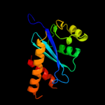
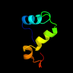
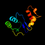
1 d1musa_
98.2
12
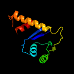
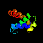
Fold: Ribonuclease H-like motifSuperfamily: Ribonuclease H-likeFamily: Transposase inhibitor (Tn5 transposase)2 d1b7ea_
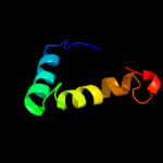
97.5
13
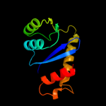
Fold: Ribonuclease H-like motifSuperfamily: Ribonuclease H-likeFamily: Transposase inhibitor (Tn5 transposase)3 d1cxqa_
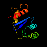
84.2
22
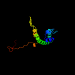
Fold: Ribonuclease H-like motifSuperfamily: Ribonuclease H-likeFamily: Retroviral integrase, catalytic domain4 c3hefB_
79.7
14
PDB header: viral proteinChain: B: PDB Molecule: gene 1 protein;PDBTitle: crystal structure of the bacteriophage sf6 terminase small2 subunit
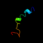
5 d1asua_
70.0
20
Fold: Ribonuclease H-like motifSuperfamily: Ribonuclease H-likeFamily: Retroviral integrase, catalytic domain6 c3nf9A_
47.7
14
PDB header: hydrolase/hydrolase inhibitorChain: A: PDB Molecule: integrase;PDBTitle: structural basis for a new mechanism of inhibition of hiv integrase2 identified by fragment screening and structure based design
7 d1a9xa1
39.7
16
Fold: Carbamoyl phosphate synthetase, large subunit connection domainSuperfamily: Carbamoyl phosphate synthetase, large subunit connection domainFamily: Carbamoyl phosphate synthetase, large subunit connection domain8 d1c0ma2
35.3
15
Fold: Ribonuclease H-like motifSuperfamily: Ribonuclease H-likeFamily: Retroviral integrase, catalytic domain9 c1c0mA_
30.6
19
PDB header: transferaseChain: A: PDB Molecule: protein (integrase);PDBTitle: crystal structure of rsv two-domain integrase
10 d1slma1
29.6
27
Fold: PGBD-likeSuperfamily: PGBD-likeFamily: MMP N-terminal domain11 c3ohwB_
22.4
16
PDB header: protein bindingChain: B: PDB Molecule: phycobilisome lcm core-membrane linker polypeptide;PDBTitle: x-ray structure of phycobilisome lcm core-membrane linker polypeptide2 (fragment 721-860) from synechocystis sp. pcc 6803, northeast3 structural genomics consortium target sgr209e
12 c2ky4A_
22.4
16
PDB header: photosynthesisChain: A: PDB Molecule: phycobilisome linker polypeptide;PDBTitle: solution nmr structure of the pbs linker domain of phycobilisome2 linker polypeptide from anabaena sp. northeast structural genomics3 consortium target nsr123e
13 c2l3wA_
20.8
20
PDB header: photosynthesisChain: A: PDB Molecule: phycobilisome rod linker polypeptide;PDBTitle: solution nmr structure of the pbs linker domain of phycobilisome rod2 linker polypeptide from synechococcus elongatus, northeast structural3 genomics consortium target snr168a
14 d1k78a1
19.8
19
Fold: DNA/RNA-binding 3-helical bundleSuperfamily: Homeodomain-likeFamily: Paired domain15 c3pruD_
19.4
13
PDB header: photosynthesisChain: D: PDB Molecule: phycobilisome 32.1 kda linker polypeptide, phycocyanin-PDBTitle: crystal structure of phycobilisome 32.1 kda linker polypeptide,2 phycocyanin-associated, rod 1 (fragment 14-158) from synechocystis3 sp. pcc 6803, northeast structural genomics consortium target sgr182a
16 c2l06A_
18.7
22
PDB header: protein bindingChain: A: PDB Molecule: phycobilisome lcm core-membrane linker polypeptide;PDBTitle: solution nmr structure of the pbs linker polypeptide domain (fragment2 254-400) of phycobilisome linker protein apce from synechocystis sp.3 pcc 6803. northeast structural genomics consortium target sgr209c
17 d1hyva_
18.2
16
Fold: Ribonuclease H-like motifSuperfamily: Ribonuclease H-likeFamily: Retroviral integrase, catalytic domain18 c1q2iA_
17.8
28
PDB header: antitumor proteinChain: A: PDB Molecule: pnc27;PDBTitle: nmr solution structure of a peptide from the mdm-2 binding2 domain of the p53 protein that is selectively cytotoxic to3 cancer cells
19 d1exqa_
16.0
17
Fold: Ribonuclease H-like motifSuperfamily: Ribonuclease H-likeFamily: Retroviral integrase, catalytic domain20 c3pvpA_
15.9
14
PDB header: dna binding protein/dnaChain: A: PDB Molecule: chromosomal replication initiator protein dnaa;PDBTitle: structure of mycobacterium tuberculosis dnaa-dbd in complex with box22 dna
21 c3f9kV_
not modelled
15.1
14
PDB header: viral protein, recombinationChain: V: PDB Molecule: integrase;PDBTitle: two domain fragment of hiv-2 integrase in complex with ledgf ibd
22 d1su3a1
not modelled
14.8
28
Fold: PGBD-likeSuperfamily: PGBD-likeFamily: MMP N-terminal domain23 c2o8kA_
not modelled
14.2
27
PDB header: transcription/dnaChain: A: PDB Molecule: rna polymerase sigma factor rpon;PDBTitle: nmr structure of the sigma-54 rpon domain bound to the-242 promoter element
24 c3u1nC_
not modelled
14.2
12
PDB header: hydrolaseChain: C: PDB Molecule: sam domain and hd domain-containing protein 1;PDBTitle: structure of the catalytic core of human samhd1
25 c1k6yB_
not modelled
12.7
19
PDB header: transferaseChain: B: PDB Molecule: integrase;PDBTitle: crystal structure of a two-domain fragment of hiv-1 integrase
26 c1x6iB_
not modelled
12.2
13
PDB header: structural genomics, unknown functionChain: B: PDB Molecule: hypothetical protein ygfy;PDBTitle: crystal structure of ygfy from escherichia coli
27 c1bg1A_
not modelled
10.9
11
PDB header: transcription/dnaChain: A: PDB Molecule: protein (transcription factor stat3b);PDBTitle: transcription factor stat3b/dna complex
28 d2fug11
not modelled
9.9
19
Fold: Bromodomain-likeSuperfamily: Nqo1C-terminal domain-likeFamily: Nqo1C-terminal domain-like29 d1nkua_
not modelled
9.1
13
Fold: DNA-glycosylaseSuperfamily: DNA-glycosylaseFamily: 3-Methyladenine DNA glycosylase I (Tag)30 d2d1ha1
not modelled
8.3
17
Fold: DNA/RNA-binding 3-helical bundleSuperfamily: "Winged helix" DNA-binding domainFamily: TrmB-like31 c3iwfA_
not modelled
8.3
4
PDB header: transcription regulatorChain: A: PDB Molecule: transcription regulator rpir family;PDBTitle: the crystal structure of the n-terminal domain of a rpir2 transcriptional regulator from staphylococcus epidermidis to 1.4a
32 c2kvcA_
not modelled
8.2
20
PDB header: unknown functionChain: A: PDB Molecule: putative uncharacterized protein;PDBTitle: solution structure of the mycobacterium tuberculosis protein rv0543c,2 a member of the duf3349 superfamily. seattle structural genomics3 center for infectious disease target mytud.17112.a
33 d1sfka_
not modelled
7.6
19
Fold: Flavivirus capsid protein CSuperfamily: Flavivirus capsid protein CFamily: Flavivirus capsid protein C34 d1s1ma2
not modelled
7.4
24
Fold: P-loop containing nucleoside triphosphate hydrolasesSuperfamily: P-loop containing nucleoside triphosphate hydrolasesFamily: Nitrogenase iron protein-like35 d1c6va_
not modelled
7.3
14
Fold: Ribonuclease H-like motifSuperfamily: Ribonuclease H-likeFamily: Retroviral integrase, catalytic domain36 d1vcoa2
not modelled
6.8
18
Fold: P-loop containing nucleoside triphosphate hydrolasesSuperfamily: P-loop containing nucleoside triphosphate hydrolasesFamily: Nitrogenase iron protein-like37 c1r6rA_
not modelled
5.9
10
PDB header: viral proteinChain: A: PDB Molecule: genome polyprotein;PDBTitle: solution structure of dengue virus capsid protein reveals a2 new fold
38 d1r6ra_
not modelled
5.9
10
Fold: Flavivirus capsid protein CSuperfamily: Flavivirus capsid protein CFamily: Flavivirus capsid protein C39 c1yvlB_
not modelled
5.7
9
PDB header: signaling proteinChain: B: PDB Molecule: signal transducer and activator of transcriptionPDBTitle: structure of unphosphorylated stat1
40 c1ud0B_
not modelled
5.6
20
PDB header: chaperoneChain: B: PDB Molecule: 70 kda heat-shock-like protein;PDBTitle: crystal structure of the c-terminal 10-kda subdomain of hsc70
41 d1h3fa1
not modelled
5.6
9
Fold: Adenine nucleotide alpha hydrolase-likeSuperfamily: Nucleotidylyl transferaseFamily: Class I aminoacyl-tRNA synthetases (RS), catalytic domain42 c2vkpA_
not modelled
5.6
12
PDB header: protein-bindingChain: A: PDB Molecule: btb/poz domain-containing protein 6;PDBTitle: crystal structure of btb domain from btbd6
43 c3d3kD_
not modelled
5.4
12
PDB header: protein bindingChain: D: PDB Molecule: enhancer of mrna-decapping protein 3;PDBTitle: crystal structure of human edc3p
44 c1m6vE_
not modelled
5.4
16
PDB header: ligaseChain: E: PDB Molecule: carbamoyl phosphate synthetase large chain;PDBTitle: crystal structure of the g359f (small subunit) point mutant of2 carbamoyl phosphate synthetase
45 d1j1va_
not modelled
5.3
19
Fold: DNA/RNA-binding 3-helical bundleSuperfamily: TrpR-likeFamily: Chromosomal replication initiation factor DnaA C-terminal domain IV46 c2lkyA_
not modelled
5.2
15
PDB header: structural genomics, unknown functionChain: A: PDB Molecule: uncharacterized protein;PDBTitle: solution structure of msmeg_1053, the second duf3349 annotated protein2 in the genome of mycobacterium smegmatis, seattle structural genomics3 center for infectious disease target mysma.17112.b