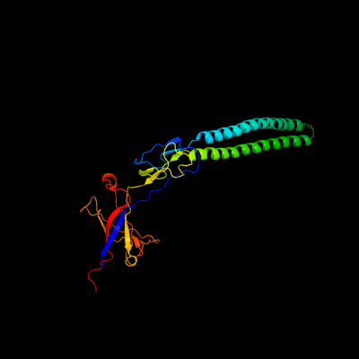
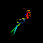
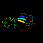
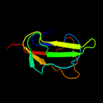
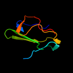
1 c3fppB_
100.0
18
PDB header: membrane proteinChain: B: PDB Molecule: macrolide-specific efflux protein maca;PDBTitle: crystal structure of e.coli maca
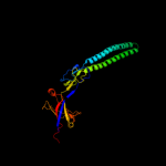
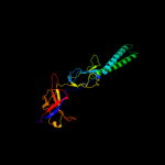
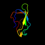
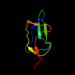
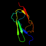
2 c3h9iB_
100.0
15
PDB header: transport proteinChain: B: PDB Molecule: cation efflux system protein cusb;PDBTitle: crystal structure of the membrane fusion protein cusb from escherichia2 coli
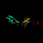
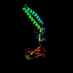
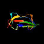
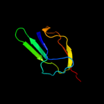
3 c2f1mA_
99.9
23
PDB header: transport proteinChain: A: PDB Molecule: acriflavine resistance protein a;PDBTitle: conformational flexibility in the multidrug efflux system protein acra
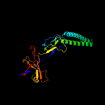
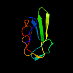
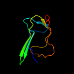
4 c3lnnB_
99.9
21
PDB header: metal transportChain: B: PDB Molecule: membrane fusion protein (mfp) heavy metal cation effluxPDBTitle: crystal structure of zneb from cupriavidus metallidurans
5 c1t5eB_
99.9
21
PDB header: transport proteinChain: B: PDB Molecule: multidrug resistance protein mexa;PDBTitle: the structure of mexa
6 d1vf7a_
99.9
21
Fold: HlyD-like secretion proteinsSuperfamily: HlyD-like secretion proteinsFamily: HlyD-like secretion proteins7 c2k33A_
99.6
27
PDB header: membrane protein, transport proteinChain: A: PDB Molecule: acra;PDBTitle: solution structure of an n-glycosylated protein using in2 vitro glycosylation
8 c2b8gA_
98.6
21
PDB header: biosynthetic proteinChain: A: PDB Molecule: biotin/lipoyl attachment protein;PDBTitle: solution structure of bacillus subtilis blap biotinylated-2 form (energy minimized mean structure)
9 c3n6rK_
98.5
42
PDB header: ligaseChain: K: PDB Molecule: propionyl-coa carboxylase, alpha subunit;PDBTitle: crystal structure of the holoenzyme of propionyl-coa carboxylase (pcc)
10 d1dcza_
98.5
54
Fold: Barrel-sandwich hybridSuperfamily: Single hybrid motifFamily: Biotinyl/lipoyl-carrier proteins and domains11 c2ejgD_
98.4
33
PDB header: ligaseChain: D: PDB Molecule: 149aa long hypothetical methylmalonyl-coa decarboxylasePDBTitle: crystal structure of the biotin protein ligase (mutation r48a) and2 biotin carboxyl carrier protein complex from pyrococcus horikoshii3 ot3
12 d1o78a_
98.3
27
Fold: Barrel-sandwich hybridSuperfamily: Single hybrid motifFamily: Biotinyl/lipoyl-carrier proteins and domains13 c2ejmA_
98.3
48
PDB header: ligaseChain: A: PDB Molecule: methylcrotonoyl-coa carboxylase subunit alpha;PDBTitle: solution structure of ruh-072, an apo-biotnyl domain form2 human acetyl coenzyme a carboxylase
14 c2dn8A_
98.1
13
PDB header: ligaseChain: A: PDB Molecule: acetyl-coa carboxylase 2;PDBTitle: solution structure of rsgi ruh-053, an apo-biotin carboxy2 carrier protein from human transcarboxylase
15 c2kccA_
98.0
19
PDB header: ligaseChain: A: PDB Molecule: acetyl-coa carboxylase 2;PDBTitle: solution structure of biotinoyl domain from human acetyl-2 coa carboxylase 2
16 d1qjoa_
98.0
23
Fold: Barrel-sandwich hybridSuperfamily: Single hybrid motifFamily: Biotinyl/lipoyl-carrier proteins and domains17 d1iyua_
97.9
34
Fold: Barrel-sandwich hybridSuperfamily: Single hybrid motifFamily: Biotinyl/lipoyl-carrier proteins and domains18 d1k8ma_
97.9
26
Fold: Barrel-sandwich hybridSuperfamily: Single hybrid motifFamily: Biotinyl/lipoyl-carrier proteins and domains19 d1bdoa_
97.9
18
Fold: Barrel-sandwich hybridSuperfamily: Single hybrid motifFamily: Biotinyl/lipoyl-carrier proteins and domains20 d1ghja_
97.9
29
Fold: Barrel-sandwich hybridSuperfamily: Single hybrid motifFamily: Biotinyl/lipoyl-carrier proteins and domains21 c2q8iB_
not modelled
97.8
52
PDB header: transferaseChain: B: PDB Molecule: dihydrolipoyllysine-residue acetyltransferase component ofPDBTitle: pyruvate dehydrogenase kinase isoform 3 in complex with antitumor drug2 radicicol
22 c2l5tA_
not modelled
97.7
38
PDB header: transferaseChain: A: PDB Molecule: lipoamide acyltransferase;PDBTitle: solution nmr structure of e2 lipoyl domain from thermoplasma2 acidophilum
23 d1y8ob1
not modelled
97.7
52
Fold: Barrel-sandwich hybridSuperfamily: Single hybrid motifFamily: Biotinyl/lipoyl-carrier proteins and domains24 c2dncA_
not modelled
97.6
46
PDB header: transferaseChain: A: PDB Molecule: pyruvate dehydrogenase protein x component;PDBTitle: solution structure of rsgi ruh-054, a lipoyl domain from2 human 2-oxoacid dehydrogenase
25 d1laba_
not modelled
97.5
37
Fold: Barrel-sandwich hybridSuperfamily: Single hybrid motifFamily: Biotinyl/lipoyl-carrier proteins and domains26 d1gjxa_
not modelled
97.4
34
Fold: Barrel-sandwich hybridSuperfamily: Single hybrid motifFamily: Biotinyl/lipoyl-carrier proteins and domains27 d1pmra_
not modelled
97.3
26
Fold: Barrel-sandwich hybridSuperfamily: Single hybrid motifFamily: Biotinyl/lipoyl-carrier proteins and domains28 c2dneA_
not modelled
97.1
48
PDB header: transferaseChain: A: PDB Molecule: dihydrolipoyllysine-residue acetyltransferasePDBTitle: solution structure of rsgi ruh-058, a lipoyl domain of2 human 2-oxoacid dehydrogenase
29 d1glaf_
not modelled
95.9
24
Fold: Barrel-sandwich hybridSuperfamily: Duplicated hybrid motifFamily: Glucose permease-like30 d2gpra_
not modelled
95.7
32
Fold: Barrel-sandwich hybridSuperfamily: Duplicated hybrid motifFamily: Glucose permease-like31 d2f3ga_
not modelled
95.6
20
Fold: Barrel-sandwich hybridSuperfamily: Duplicated hybrid motifFamily: Glucose permease-like32 d1gpra_
not modelled
95.4
18
Fold: Barrel-sandwich hybridSuperfamily: Duplicated hybrid motifFamily: Glucose permease-like33 d2pnrc1
not modelled
95.2
21
Fold: Barrel-sandwich hybridSuperfamily: Single hybrid motifFamily: Biotinyl/lipoyl-carrier proteins and domains34 c2qf7A_
not modelled
95.0
23
PDB header: ligaseChain: A: PDB Molecule: pyruvate carboxylase protein;PDBTitle: crystal structure of a complete multifunctional pyruvate carboxylase2 from rhizobium etli
35 c2jkuA_
not modelled
94.7
20
PDB header: ligaseChain: A: PDB Molecule: propionyl-coa carboxylase alpha chain,PDBTitle: crystal structure of the n-terminal region of the biotin2 acceptor domain of human propionyl-coa carboxylase
36 d1uoua3
not modelled
93.3
25
Fold: alpha/beta-HammerheadSuperfamily: Pyrimidine nucleoside phosphorylase C-terminal domainFamily: Pyrimidine nucleoside phosphorylase C-terminal domain37 d1brwa3
not modelled
93.0
31
Fold: alpha/beta-HammerheadSuperfamily: Pyrimidine nucleoside phosphorylase C-terminal domainFamily: Pyrimidine nucleoside phosphorylase C-terminal domain38 d2tpta3
not modelled
92.0
25
Fold: alpha/beta-HammerheadSuperfamily: Pyrimidine nucleoside phosphorylase C-terminal domainFamily: Pyrimidine nucleoside phosphorylase C-terminal domain39 c2dsjA_
not modelled
91.1
36
PDB header: transferaseChain: A: PDB Molecule: pyrimidine-nucleoside (thymidine) phosphorylase;PDBTitle: crystal structure of project id tt0128 from thermus thermophilus hb8
40 c1otpA_
not modelled
90.3
24
PDB header: phosphorylaseChain: A: PDB Molecule: thymidine phosphorylase;PDBTitle: structural and theoretical studies suggest domain movement produces an2 active conformation of thymidine phosphorylase
41 c3h5qA_
not modelled
90.2
41
PDB header: transferaseChain: A: PDB Molecule: pyrimidine-nucleoside phosphorylase;PDBTitle: crystal structure of a putative pyrimidine-nucleoside phosphorylase2 from staphylococcus aureus
42 c2j0fC_
not modelled
90.2
24
PDB header: transferaseChain: C: PDB Molecule: thymidine phosphorylase;PDBTitle: structural basis for non-competitive product inhibition in2 human thymidine phosphorylase: implication for drug design
43 c2gu1A_
not modelled
90.2
21
PDB header: hydrolaseChain: A: PDB Molecule: zinc peptidase;PDBTitle: crystal structure of a zinc containing peptidase from2 vibrio cholerae
44 c1brwB_
not modelled
88.8
27
PDB header: transferaseChain: B: PDB Molecule: protein (pyrimidine nucleoside phosphorylase);PDBTitle: the crystal structure of pyrimidine nucleoside2 phosphorylase in a closed conformation
45 c3fmcC_
88.7
17
PDB header: hydrolaseChain: C: PDB Molecule: putative succinylglutamate desuccinylase / aspartoacylase;PDBTitle: crystal structure of a putative succinylglutamate desuccinylase /2 aspartoacylase family protein (sama_0604) from shewanella amazonensis3 sb2b at 1.80 a resolution
46 c2qj8B_
not modelled
87.9
19
PDB header: hydrolaseChain: B: PDB Molecule: mlr6093 protein;PDBTitle: crystal structure of an aspartoacylase family protein (mlr6093) from2 mesorhizobium loti maff303099 at 2.00 a resolution
47 c2hsiB_
not modelled
86.1
17
PDB header: structural genomics, unknown functionChain: B: PDB Molecule: putative peptidase m23;PDBTitle: crystal structure of putative peptidase m23 from2 pseudomonas aeruginosa, new york structural genomics3 consortium
48 c2aukA_
not modelled
83.5
20
PDB header: transferaseChain: A: PDB Molecule: dna-directed rna polymerase beta' chain;PDBTitle: structure of e. coli rna polymerase beta' g/g' insert
49 c3cdxB_
not modelled
82.4
19
PDB header: hydrolaseChain: B: PDB Molecule: succinylglutamatedesuccinylase/aspartoacylase;PDBTitle: crystal structure of2 succinylglutamatedesuccinylase/aspartoacylase from3 rhodobacter sphaeroides
50 d1qwya_
not modelled
82.4
13
Fold: Barrel-sandwich hybridSuperfamily: Duplicated hybrid motifFamily: Peptidoglycan hydrolase LytM51 c3na6A_
not modelled
81.8
16
PDB header: hydrolaseChain: A: PDB Molecule: succinylglutamate desuccinylase/aspartoacylase;PDBTitle: crystal structure of a succinylglutamate desuccinylase (tm1040_2694)2 from silicibacter sp. tm1040 at 2.00 a resolution
52 d1qpoa2
not modelled
79.2
22
Fold: alpha/beta-HammerheadSuperfamily: Nicotinate/Quinolinate PRTase N-terminal domain-likeFamily: NadC N-terminal domain-like53 c2xhaB_
not modelled
75.8
17
PDB header: transcriptionChain: B: PDB Molecule: transcription antitermination protein nusg;PDBTitle: crystal structure of domain 2 of thermotoga maritima n-utilization2 substance g (nusg)
54 c1tqqC_
not modelled
75.5
11
PDB header: transport proteinChain: C: PDB Molecule: outer membrane protein tolc;PDBTitle: structure of tolc in complex with hexamminecobalt
55 d1e2wa2
not modelled
75.2
19
Fold: Barrel-sandwich hybridSuperfamily: Rudiment single hybrid motifFamily: Cytochrome f, small domain56 c2b44A_
not modelled
75.0
13
PDB header: hydrolaseChain: A: PDB Molecule: glycyl-glycine endopeptidase lytm;PDBTitle: truncated s. aureus lytm, p 32 2 1 crystal form
57 d1wp1a_
not modelled
74.7
11
Fold: Outer membrane efflux proteins (OEP)Superfamily: Outer membrane efflux proteins (OEP)Family: Outer membrane efflux proteins (OEP)58 d1o4ua2
not modelled
74.4
15
Fold: alpha/beta-HammerheadSuperfamily: Nicotinate/Quinolinate PRTase N-terminal domain-likeFamily: NadC N-terminal domain-like59 d1ci3m2
not modelled
73.9
19
Fold: Barrel-sandwich hybridSuperfamily: Rudiment single hybrid motifFamily: Cytochrome f, small domain60 c1yc9A_
not modelled
73.7
9
PDB header: membrane proteinChain: A: PDB Molecule: multidrug resistance protein;PDBTitle: the crystal structure of the outer membrane protein vcec from the2 bacterial pathogen vibrio cholerae at 1.8 resolution
61 d1tova_
not modelled
73.3
16
Fold: SH3-like barrelSuperfamily: Cap-Gly domainFamily: Cap-Gly domain62 d1ek9a_
not modelled
73.1
11
Fold: Outer membrane efflux proteins (OEP)Superfamily: Outer membrane efflux proteins (OEP)Family: Outer membrane efflux proteins (OEP)63 c3dtpA_
not modelled
73.0
11
PDB header: contractile proteinChain: A: PDB Molecule: myosin 2 heavy chain chimera of smooth andPDBTitle: tarantula heavy meromyosin obtained by flexible docking to2 tarantula muscle thick filament cryo-em 3d-map
64 c3nyyA_
not modelled
72.8
22
PDB header: hydrolaseChain: A: PDB Molecule: putative glycyl-glycine endopeptidase lytm;PDBTitle: crystal structure of a putative glycyl-glycine endopeptidase lytm2 (rumgna_02482) from ruminococcus gnavus atcc 29149 at 1.60 a3 resolution
65 d1qapa2
not modelled
72.1
10
Fold: alpha/beta-HammerheadSuperfamily: Nicotinate/Quinolinate PRTase N-terminal domain-likeFamily: NadC N-terminal domain-like66 c3pikA_
not modelled
71.8
9
PDB header: transport proteinChain: A: PDB Molecule: cation efflux system protein cusc;PDBTitle: outer membrane protein cusc
67 c3it5B_
not modelled
70.6
10
PDB header: hydrolaseChain: B: PDB Molecule: protease lasa;PDBTitle: crystal structure of the lasa virulence factor from pseudomonas2 aeruginosa
68 c2xhcA_
not modelled
68.8
17
PDB header: transcriptionChain: A: PDB Molecule: transcription antitermination protein nusg;PDBTitle: crystal structure of thermotoga maritima n-utilization substance g2 (nusg)
69 c2aujD_
not modelled
68.6
31
PDB header: transferaseChain: D: PDB Molecule: dna-directed rna polymerase beta' chain;PDBTitle: structure of thermus aquaticus rna polymerase beta'-subunit2 insert
70 d1ixda_
not modelled
68.5
19
Fold: SH3-like barrelSuperfamily: Cap-Gly domainFamily: Cap-Gly domain71 c3d4rE_
not modelled
67.8
18
PDB header: unknown functionChain: E: PDB Molecule: domain of unknown function from the pfam-b_34464 family;PDBTitle: crystal structure of a duf2118 family protein (mmp0046) from2 methanococcus maripaludis at 2.20 a resolution
72 d1whla_
not modelled
67.6
23
Fold: SH3-like barrelSuperfamily: Cap-Gly domainFamily: Cap-Gly domain73 d2cp2a1
not modelled
66.7
19
Fold: SH3-like barrelSuperfamily: Cap-Gly domainFamily: Cap-Gly domain74 d2coya1
not modelled
66.7
22
Fold: SH3-like barrelSuperfamily: Cap-Gly domainFamily: Cap-Gly domain75 d2cp6a1
not modelled
66.4
21
Fold: SH3-like barrelSuperfamily: Cap-Gly domainFamily: Cap-Gly domain76 d1whma_
not modelled
65.2
17
Fold: SH3-like barrelSuperfamily: Cap-Gly domainFamily: Cap-Gly domain77 d1whga_
not modelled
64.5
16
Fold: SH3-like barrelSuperfamily: Cap-Gly domainFamily: Cap-Gly domain78 c1y4cA_
not modelled
63.4
13
PDB header: de novo proteinChain: A: PDB Molecule: maltose binding protein fused with designedPDBTitle: designed helical protein fusion mbp
79 c3gnnA_
not modelled
62.6
8
PDB header: transferaseChain: A: PDB Molecule: nicotinate-nucleotide pyrophosphorylase;PDBTitle: crystal structure of nicotinate-nucleotide2 pyrophosphorylase from burkholderi pseudomallei
80 c3ipkA_
not modelled
62.5
14
PDB header: cell adhesionChain: A: PDB Molecule: agi/ii;PDBTitle: crystal structure of a3vp1 of agi/ii of streptococcus mutans
81 c1h9mB_
not modelled
62.0
22
PDB header: binding proteinChain: B: PDB Molecule: molybdenum-binding-protein;PDBTitle: two crystal structures of the cytoplasmic molybdate-binding2 protein modg suggest a novel cooperative binding mechanism3 and provide insights into ligand-binding specificity.4 peg-grown form with molybdate bound
82 c3e1yG_
not modelled
61.8
15
PDB header: translationChain: G: PDB Molecule: eukaryotic peptide chain release factor gtp-binding subunitPDBTitle: crystal structure of human erf1/erf3 complex
83 c2qqsB_
not modelled
61.5
22
PDB header: oxidoreductaseChain: B: PDB Molecule: jmjc domain-containing histone demethylationPDBTitle: jmjd2a tandem tudor domains in complex with a trimethylated2 histone h4-k20 peptide
84 c1o4uA_
not modelled
61.3
15
PDB header: transferaseChain: A: PDB Molecule: type ii quinolic acid phosphoribosyltransferase;PDBTitle: crystal structure of a nicotinate nucleotide pyrophosphorylase2 (tm1645) from thermotoga maritima at 2.50 a resolution
85 c3ghgK_
not modelled
58.4
6
PDB header: blood clottingChain: K: PDB Molecule: fibrinogen beta chain;PDBTitle: crystal structure of human fibrinogen
86 d2cp3a1
not modelled
58.2
17
Fold: SH3-like barrelSuperfamily: Cap-Gly domainFamily: Cap-Gly domain87 c1h9sA_
not modelled
58.1
20
PDB header: transcription regulatorChain: A: PDB Molecule: molybdenum transport protein mode;PDBTitle: molybdate bound complex of dimop domain of mode from e.coli
88 c3dlmA_
not modelled
57.9
19
PDB header: transferaseChain: A: PDB Molecule: histone-lysine n-methyltransferase setdb1;PDBTitle: crystal structure of tudor domain of human histone-lysine n-2 methyltransferase setdb1
89 d2ix0a1
not modelled
57.9
12
Fold: OB-foldSuperfamily: Nucleic acid-binding proteinsFamily: Cold shock DNA-binding domain-like90 c3pajA_
not modelled
57.2
15
PDB header: transferaseChain: A: PDB Molecule: nicotinate-nucleotide pyrophosphorylase, carboxylating;PDBTitle: 2.00 angstrom resolution crystal structure of a quinolinate2 phosphoribosyltransferase from vibrio cholerae o1 biovar eltor str.3 n16961
91 c1qapA_
not modelled
57.1
10
PDB header: glycosyltransferaseChain: A: PDB Molecule: quinolinic acid phosphoribosyltransferase;PDBTitle: quinolinic acid phosphoribosyltransferase with bound2 quinolinic acid
92 c1deqF_
not modelled
57.0
9
PDB header: PDB COMPND: 93 d2cp0a1
not modelled
56.6
16
Fold: SH3-like barrelSuperfamily: Cap-Gly domainFamily: Cap-Gly domain94 c1e2vB_
not modelled
56.5
19
PDB header: electron transport proteinsChain: B: PDB Molecule: cytochrome f;PDBTitle: n153q mutant of cytochrome f from chlamydomonas reinhardtii
95 c3m9bK_
not modelled
56.3
12
PDB header: chaperoneChain: K: PDB Molecule: proteasome-associated atpase;PDBTitle: crystal structure of the amino terminal coiled coil domain and the2 inter domain of the mycobacterium tuberculosis proteasomal atpase mpa
96 c3e20A_
not modelled
56.0
17
PDB header: translationChain: A: PDB Molecule: eukaryotic peptide chain release factor gtp-bindingPDBTitle: crystal structure of s.pombe erf1/erf3 complex
97 c1ctmA_
not modelled
56.0
6
PDB header: electron transport(cytochrome)Chain: A: PDB Molecule: cytochrome f;PDBTitle: crystal structure of chloroplast cytochrome f reveals a2 novel cytochrome fold and unexpected heme ligation
98 d1onla_
not modelled
54.6
20
Fold: Barrel-sandwich hybridSuperfamily: Single hybrid motifFamily: Biotinyl/lipoyl-carrier proteins and domains99 c2z0wA_
not modelled
54.1
11
PDB header: protein bindingChain: A: PDB Molecule: cap-gly domain-containing linker protein 4;PDBTitle: crystal structure of the 2nd cap-gly domain in human restin-2 like protein 2 reveals a swapped-dimer
100 c2jxmB_
not modelled
53.8
19
PDB header: electron transportChain: B: PDB Molecule: cytochrome f;PDBTitle: ensemble of twenty structures of the prochlorothrix2 hollandica plastocyanin- cytochrome f complex
101 c3csqC_
not modelled
53.8
12
PDB header: hydrolaseChain: C: PDB Molecule: morphogenesis protein 1;PDBTitle: crystal and cryoem structural studies of a cell wall2 degrading enzyme in the bacteriophage phi29 tail
102 c2jbmA_
not modelled
53.4
0
PDB header: transferaseChain: A: PDB Molecule: nicotinate-nucleotide pyrophosphorylase;PDBTitle: qprtase structure from human
103 c1zeqX_
not modelled
51.8
15
PDB header: metal binding proteinChain: X: PDB Molecule: cation efflux system protein cusf;PDBTitle: 1.5 a structure of apo-cusf residues 6-88 from escherichia2 coli
104 c2edgA_
not modelled
51.8
16
PDB header: biosynthetic proteinChain: A: PDB Molecule: glycine cleavage system h protein;PDBTitle: solution structure of the gcv_h domain from mouse glycine
105 c2xdpA_
not modelled
51.6
24
PDB header: oxidoreductaseChain: A: PDB Molecule: lysine-specific demethylase 4c;PDBTitle: crystal structure of the tudor domain of human jmjd2c
106 c3tqvA_
not modelled
51.0
12
PDB header: transferaseChain: A: PDB Molecule: nicotinate-nucleotide pyrophosphorylase;PDBTitle: structure of the nicotinate-nucleotide pyrophosphorylase from2 francisella tularensis.
107 d2e3ha1
not modelled
50.8
18
Fold: SH3-like barrelSuperfamily: Cap-Gly domainFamily: Cap-Gly domain108 c1tu2B_
not modelled
50.5
25
PDB header: electron transportChain: B: PDB Molecule: apocytochrome f;PDBTitle: the complex of nostoc cytochrome f and plastocyanin determin with2 paramagnetic nmr. based on the structures of cytochrome f and3 plastocyanin, 10 structures
109 c2e4hA_
not modelled
49.5
18
PDB header: structural proteinChain: A: PDB Molecule: restin;PDBTitle: solution structure of cytoskeletal protein in complex with2 tubulin tail
110 c2b7pA_
not modelled
48.4
25
PDB header: transferaseChain: A: PDB Molecule: probable nicotinate-nucleotide pyrophosphorylase;PDBTitle: crystal structure of quinolinic acid phosphoribosyltransferase from2 helicobacter pylori
111 c1ei3E_
not modelled
46.4
8
PDB header: PDB COMPND: 112 c3l0gD_
not modelled
46.3
20
PDB header: transferaseChain: D: PDB Molecule: nicotinate-nucleotide pyrophosphorylase;PDBTitle: crystal structure of nicotinate-nucleotide pyrophosphorylase from2 ehrlichia chaffeensis at 2.05a resolution
113 c2e75C_
not modelled
46.2
19
PDB header: photosynthesisChain: C: PDB Molecule: apocytochrome f;PDBTitle: crystal structure of the cytochrome b6f complex with 2-nonyl-4-2 hydroxyquinoline n-oxide (nqno) from m.laminosus
114 d1tu2b2
not modelled
45.5
25
Fold: Barrel-sandwich hybridSuperfamily: Rudiment single hybrid motifFamily: Cytochrome f, small domain115 c2d3eD_
not modelled
45.0
7
PDB header: contractile proteinChain: D: PDB Molecule: general control protein gcn4 and tropomyosin 1PDBTitle: crystal structure of the c-terminal fragment of rabbit2 skeletal alpha-tropomyosin
116 c3ojaB_
not modelled
43.1
8
PDB header: protein bindingChain: B: PDB Molecule: anopheles plasmodium-responsive leucine-rich repeat proteinPDBTitle: crystal structure of lrim1/apl1c complex
117 c2jvvA_
not modelled
42.9
26
PDB header: transcriptionChain: A: PDB Molecule: transcription antitermination protein nusg;PDBTitle: solution structure of e. coli nusg carboxyterminal domain
118 c2kvqG_
not modelled
42.9
26
PDB header: transcriptionChain: G: PDB Molecule: transcription antitermination protein nusg;PDBTitle: solution structure of nuse:nusg-ctd complex
119 c1qpoA_
not modelled
41.8
22
PDB header: transferaseChain: A: PDB Molecule: quinolinate acid phosphoribosyl transferase;PDBTitle: quinolinate phosphoribosyl transferase (qaprtase) apo-enzyme from2 mycobacterium tuberculosis
120 d2hqha1
not modelled
41.3
17
Fold: SH3-like barrelSuperfamily: Cap-Gly domainFamily: Cap-Gly domain