1 c3nreB_
100.0
20
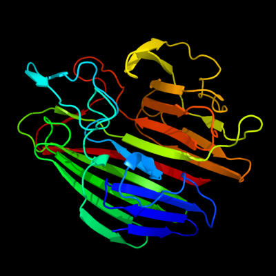
PDB header: isomeraseChain: B: PDB Molecule: aldose 1-epimerase;PDBTitle: crystal structure of a putative aldose 1-epimerase (b2544) from2 escherichia coli k12 at 1.59 a resolution
2 d1z45a1
100.0
23
Fold: SupersandwichSuperfamily: Galactose mutarotase-likeFamily: Aldose 1-epimerase (mutarotase)3 d1so0a_
100.0
23
Fold: SupersandwichSuperfamily: Galactose mutarotase-likeFamily: Aldose 1-epimerase (mutarotase)4 c3imhB_
100.0
19
PDB header: isomeraseChain: B: PDB Molecule: galactose-1-epimerase;PDBTitle: crystal structure of galactose 1-epimerase from lactobacillus2 acidophilus ncfm
5 c1ygaA_
100.0
22
PDB header: isomeraseChain: A: PDB Molecule: hypothetical 37.9 kda protein in bio3-hxt17PDBTitle: crystal structure of saccharomyces cerevisiae yn9a protein,2 new york structural genomics consortium
6 c1z45A_
100.0
22
PDB header: isomeraseChain: A: PDB Molecule: gal10 bifunctional protein;PDBTitle: crystal structure of the gal10 fusion protein galactose2 mutarotase/udp-galactose 4-epimerase from saccharomyces3 cerevisiae complexed with nad, udp-glucose, and galactose
7 d1lura_
100.0
22
Fold: SupersandwichSuperfamily: Galactose mutarotase-likeFamily: Aldose 1-epimerase (mutarotase)8 d1nsza_
100.0
20
Fold: SupersandwichSuperfamily: Galactose mutarotase-likeFamily: Aldose 1-epimerase (mutarotase)9 c3os7B_
100.0
19
PDB header: isomeraseChain: B: PDB Molecule: galactose mutarotase-like protein;PDBTitle: crystal structure of a galactose mutarotase-like protein (ca_c0697)2 from clostridium acetobutylicum at 1.80 a resolution
10 c3os7D_
100.0
19
PDB header: isomeraseChain: D: PDB Molecule: galactose mutarotase-like protein;PDBTitle: crystal structure of a galactose mutarotase-like protein (ca_c0697)2 from clostridium acetobutylicum at 1.80 a resolution
11 c3dcdA_
100.0
16
PDB header: structural genomics, unknown functionChain: A: PDB Molecule: galactose mutarotase related enzyme;PDBTitle: x-ray structure of the galactose mutarotase related enzyme q5fkd7 from2 lactobacillus acidophilus at the resolution 1.9a. northeast3 structural genomics consortium target lar33.
12 c3q1nA_
100.0
20
PDB header: isomeraseChain: A: PDB Molecule: galactose mutarotase related enzyme;PDBTitle: crystal structure of a galactose mutarotase-like protein (lsei_2598)2 from lactobacillus casei atcc 334 at 1.61 a resolution
13 c3mwxA_
100.0
22
PDB header: isomeraseChain: A: PDB Molecule: aldose 1-epimerase;PDBTitle: crystal structure of a putative galactose mutarotase (bsu18360) from2 bacillus subtilis at 1.45 a resolution
14 c3k25B_
100.0
20
PDB header: structural genomics, unknown functionChain: B: PDB Molecule: slr1438 protein;PDBTitle: crystal structure of slr1438 protein from synechocystis sp. pcc 6803,2 northeast structural genomics consortium target sgr112
15 c2cisA_
100.0
13
PDB header: isomeraseChain: A: PDB Molecule: hexose-6-phosphate mutarotase;PDBTitle: structure-based functional annotation: yeast ymr099c codes2 for a d-hexose-6-phosphate mutarotase. complex with3 tagatose-6-phosphate
16 c2htbB_
100.0
15
PDB header: isomeraseChain: B: PDB Molecule: putative enzyme related to aldose 1-epimerase;PDBTitle: crystal structure of a putative mutarotase (yead) from2 salmonella typhimurium in monoclinic form
17 d1jova_
100.0
18
Fold: SupersandwichSuperfamily: Galactose mutarotase-likeFamily: Hypothetical protein HI131718 c3ty1B_
100.0
11
PDB header: isomeraseChain: B: PDB Molecule: hypothetical aldose 1-epimerase;PDBTitle: crystal structure of a hypothetical aldose 1-epimerase (kpn_04629)2 from klebsiella pneumoniae subsp. pneumoniae mgh 78578 at 1.90 a3 resolution
19 c3blcB_
98.1
12
PDB header: chaperone,protein transportChain: B: PDB Molecule: inner membrane protein oxaa;PDBTitle: crystal structure of the periplasmic domain of the escherichia coli2 yidc
20 c3bs6B_
98.1
10
PDB header: membrane protein, protein transportChain: B: PDB Molecule: inner membrane protein oxaa;PDBTitle: 1.8 angstrom crystal structure of the periplasmic domain of2 the membrane insertase yidc
21 d1jz8a4
not modelled
93.2
12
Fold: SupersandwichSuperfamily: Galactose mutarotase-likeFamily: beta-Galactosidase, domain 522 c2yfnA_
not modelled
89.8
13
PDB header: hydrolaseChain: A: PDB Molecule: alpha-galactosidase-sucrose kinase agask;PDBTitle: galactosidase domain of alpha-galactosidase-sucrose kinase,2 agask
23 c2xn1B_
not modelled
89.0
10
PDB header: hydrolaseChain: B: PDB Molecule: alpha-galactosidase;PDBTitle: structure of alpha-galactosidase from lactobacillus acidophilus ncfm2 with tris
24 c3rgbA_
not modelled
86.3
14
PDB header: oxidoreductaseChain: A: PDB Molecule: methane monooxygenase subunit b2;PDBTitle: crystal structure of particulate methane monooxygenase from2 methylococcus capsulatus (bath)
25 c1yewI_
not modelled
86.3
14
PDB header: oxidoreductase, membrane proteinChain: I: PDB Molecule: particulate methane monooxygenase, b subunit;PDBTitle: crystal structure of particulate methane monooxygenase
26 c3mi6A_
not modelled
83.2
11
PDB header: hydrolaseChain: A: PDB Molecule: alpha-galactosidase;PDBTitle: crystal structure of the alpha-galactosidase from lactobacillus2 brevis, northeast structural genomics consortium target lbr11.
27 c3rfrI_
not modelled
82.2
17
PDB header: oxidoreductaseChain: I: PDB Molecule: pmob;PDBTitle: crystal structure of particulate methane monooxygenase (pmmo) from2 methylocystis sp. strain m
28 c3bgaB_
not modelled
75.3
17
PDB header: hydrolaseChain: B: PDB Molecule: beta-galactosidase;PDBTitle: crystal structure of beta-galactosidase from bacteroides2 thetaiotaomicron vpi-5482
29 c1jz6C_
not modelled
74.5
14
PDB header: hydrolaseChain: C: PDB Molecule: beta-galactosidase;PDBTitle: e. coli (lacz) beta-galactosidase in complex with galacto-2 tetrazole
30 c3mv14_
not modelled
71.1
14
PDB header: hydrolaseChain: 4: PDB Molecule: beta-galactosidase;PDBTitle: e.coli (lacz) beta-galactosidase (r599a) in complex with guanidinium
31 c1so9A_
not modelled
65.5
10
PDB header: metal transportChain: A: PDB Molecule: cytochrome c oxidase assembly protein ctag;PDBTitle: solution structure of apocox11, 30 structures
32 d1so9a_
not modelled
65.5
10
Fold: Ctag/Cox11Superfamily: Ctag/Cox11Family: Ctag/Cox1133 d1ejxb_
not modelled
63.0
17
Fold: beta-clipSuperfamily: Urease, beta-subunitFamily: Urease, beta-subunit34 d4ubpb_
not modelled
62.3
26
Fold: beta-clipSuperfamily: Urease, beta-subunitFamily: Urease, beta-subunit35 d1e9ya1
not modelled
52.4
22
Fold: beta-clipSuperfamily: Urease, beta-subunitFamily: Urease, beta-subunit36 c3qgaD_
not modelled
50.7
22
PDB header: hydrolaseChain: D: PDB Molecule: fusion of urease beta and gamma subunits;PDBTitle: 3.0 a model of iron containing urease urea2b2 from helicobacter2 mustelae
37 d1ji1a2
not modelled
49.2
8
Fold: Glycosyl hydrolase domainSuperfamily: Glycosyl hydrolase domainFamily: alpha-Amylases, C-terminal beta-sheet domain38 c3qbtH_
not modelled
47.2
26
PDB header: protein transport/hydrolaseChain: H: PDB Molecule: inositol polyphosphate 5-phosphatase ocrl-1;PDBTitle: crystal structure of ocrl1 540-678 in complex with rab8a:gppnhp
39 c1e9zA_
not modelled
44.9
22
PDB header: hydrolaseChain: A: PDB Molecule: urease subunit alpha;PDBTitle: crystal structure of helicobacter pylori urease
40 c3obaA_
not modelled
39.7
9
PDB header: hydrolaseChain: A: PDB Molecule: beta-galactosidase;PDBTitle: structure of the beta-galactosidase from kluyveromyces lactis
41 d1nkga3
not modelled
38.0
11
Fold: SupersandwichSuperfamily: Galactose mutarotase-likeFamily: Rhamnogalacturonase B, RhgB, N-terminal domain42 c1nkgA_
not modelled
32.3
11
PDB header: lyaseChain: A: PDB Molecule: rhamnogalacturonase b;PDBTitle: rhamnogalacturonan lyase from aspergillus aculeatus
43 d1wzla2
not modelled
25.0
17
Fold: Glycosyl hydrolase domainSuperfamily: Glycosyl hydrolase domainFamily: alpha-Amylases, C-terminal beta-sheet domain44 c3isyA_
not modelled
24.4
6
PDB header: protein bindingChain: A: PDB Molecule: intracellular proteinase inhibitor;PDBTitle: crystal structure of an intracellular proteinase inhibitor (ipi,2 bsu11130) from bacillus subtilis at 2.61 a resolution
45 c2r39A_
not modelled
19.8
14
PDB header: structural genomics, unknown functionChain: A: PDB Molecule: fixg-related protein;PDBTitle: crystal structure of fixg-related protein from vibrio parahaemolyticus
46 c2x3bB_
not modelled
19.6
13
PDB header: hydrolaseChain: B: PDB Molecule: toxic extracellular endopeptidase;PDBTitle: asap1 inactive mutant e294a, an extracellular toxic zinc2 metalloendopeptidase
47 c3ac0B_
not modelled
16.7
20
PDB header: hydrolaseChain: B: PDB Molecule: beta-glucosidase i;PDBTitle: crystal structure of beta-glucosidase from kluyveromyces marxianus in2 complex with glucose
48 c2cqtA_
not modelled
16.2
9
PDB header: transferaseChain: A: PDB Molecule: cellobiose phosphorylase;PDBTitle: crystal structure of cellvibrio gilvus cellobiose phosphorylase2 crystallized from sodium/potassium phosphate
49 d2je8a2
not modelled
15.1
12
Fold: Immunoglobulin-like beta-sandwichSuperfamily: beta-Galactosidase/glucuronidase domainFamily: beta-Galactosidase/glucuronidase domain50 c2zf9D_
not modelled
12.6
15
PDB header: structural proteinChain: D: PDB Molecule: scae cell-surface anchored scaffoldin protein;PDBTitle: crystal structure of a type iii cohesin module from the cellulosomal2 scae cell-surface anchoring scaffoldin of ruminococcus flavefaciens
51 c2kl8A_
not modelled
11.5
31
PDB header: de novo proteinChain: A: PDB Molecule: or15;PDBTitle: solution nmr structure of de novo designed ferredoxin-like2 fold protein, northeast structural genomics consortium3 target or15
52 d1cc3a_
not modelled
10.7
20
Fold: Cupredoxin-likeSuperfamily: CupredoxinsFamily: Plastocyanin/azurin-like53 d1g94a1
not modelled
10.4
21
Fold: Glycosyl hydrolase domainSuperfamily: Glycosyl hydrolase domainFamily: alpha-Amylases, C-terminal beta-sheet domain54 d1cx1a_
not modelled
9.8
18
Fold: Galactose-binding domain-likeSuperfamily: Galactose-binding domain-likeFamily: CBM4/955 d1nbca_
not modelled
9.0
15
Fold: Common fold of diphtheria toxin/transcription factors/cytochrome fSuperfamily: Carbohydrate-binding domainFamily: Cellulose-binding domain family III56 c3cfuA_
not modelled
8.7
33
PDB header: lipoproteinChain: A: PDB Molecule: uncharacterized lipoprotein yjha;PDBTitle: crystal structure of the yjha protein from bacillus2 subtilis. northeast structural genomics consortium target3 sr562
57 c1v7wA_
not modelled
8.6
11
PDB header: transferaseChain: A: PDB Molecule: chitobiose phosphorylase;PDBTitle: crystal structure of vibrio proteolyticus chitobiose phosphorylase in2 complex with glcnac
58 d1gu3a_
not modelled
8.1
18
Fold: Galactose-binding domain-likeSuperfamily: Galactose-binding domain-likeFamily: CBM4/959 d1v7wa2
not modelled
8.1
12
Fold: SupersandwichSuperfamily: Galactose mutarotase-likeFamily: Glycosyltransferase family 36 N-terminal domain60 d1ktba1
not modelled
8.0
14
Fold: Glycosyl hydrolase domainSuperfamily: Glycosyl hydrolase domainFamily: alpha-Amylases, C-terminal beta-sheet domain61 d1g43a_
not modelled
7.9
5
Fold: Common fold of diphtheria toxin/transcription factors/cytochrome fSuperfamily: Carbohydrate-binding domainFamily: Cellulose-binding domain family III62 c3zqwA_
not modelled
7.7
15
PDB header: carbohydrate-binding proteinChain: A: PDB Molecule: cellulosomal scaffoldin;PDBTitle: structure of cbm3b of major scaffoldin subunit scaa from2 acetivibrio cellulolyticus
63 c2l8aA_
not modelled
7.6
9
PDB header: hydrolaseChain: A: PDB Molecule: endoglucanase;PDBTitle: structure of a novel cbm3 lacking the calcium-binding site
64 d1dkza2
not modelled
7.4
23
Fold: Heat shock protein 70kD (HSP70), peptide-binding domainSuperfamily: Heat shock protein 70kD (HSP70), peptide-binding domainFamily: Heat shock protein 70kD (HSP70), peptide-binding domain65 d2ccwa1
not modelled
7.3
17
Fold: Cupredoxin-likeSuperfamily: CupredoxinsFamily: Plastocyanin/azurin-like66 c3dqgC_
not modelled
7.0
13
PDB header: chaperoneChain: C: PDB Molecule: heat shock 70 kda protein f;PDBTitle: peptide-binding domain of heat shock 70 kda protein f, mitochondrial2 precursor, from caenorhabditis elegans.
67 d1u00a2
not modelled
6.7
20
Fold: Heat shock protein 70kD (HSP70), peptide-binding domainSuperfamily: Heat shock protein 70kD (HSP70), peptide-binding domainFamily: Heat shock protein 70kD (HSP70), peptide-binding domain68 c3qfgA_
not modelled
6.6
7
PDB header: structural genomics, unknown functionChain: A: PDB Molecule: uncharacterized protein;PDBTitle: structure of a putative lipoprotein from staphylococcus aureus subsp.2 aureus nctc 8325
69 d1cuoa_
not modelled
6.4
17
Fold: Cupredoxin-likeSuperfamily: CupredoxinsFamily: Plastocyanin/azurin-like70 d3dhpa1
not modelled
6.4
19
Fold: Glycosyl hydrolase domainSuperfamily: Glycosyl hydrolase domainFamily: alpha-Amylases, C-terminal beta-sheet domain71 d1mspa_
not modelled
6.4
22
Fold: Immunoglobulin-like beta-sandwichSuperfamily: PapD-likeFamily: MSP-like72 c2wo4A_
not modelled
6.3
19
PDB header: hydrolaseChain: A: PDB Molecule: glycoside hydrolase, family 9;PDBTitle: 3b' carbohydrate-binding module from the cel9v glycoside2 hydrolase from clostridium thermocellum, in-house data
73 d2vzsa2
not modelled
6.2
16
Fold: Immunoglobulin-like beta-sandwichSuperfamily: beta-Galactosidase/glucuronidase domainFamily: beta-Galactosidase/glucuronidase domain74 d3es6b1
not modelled
6.0
8
Fold: Immunoglobulin-like beta-sandwichSuperfamily: E set domainsFamily: SVA-like75 d2j5wa3
not modelled
5.9
0
Fold: Cupredoxin-likeSuperfamily: CupredoxinsFamily: Multidomain cupredoxins76 d1hfua2
not modelled
5.8
13
Fold: Cupredoxin-likeSuperfamily: CupredoxinsFamily: Multidomain cupredoxins77 d2f2ha2
not modelled
5.8
8
Fold: SupersandwichSuperfamily: Galactose mutarotase-likeFamily: YicI N-terminal domain-like78 d1azca_
not modelled
5.6
17
Fold: Cupredoxin-likeSuperfamily: CupredoxinsFamily: Plastocyanin/azurin-like79 c3tdqB_
not modelled
5.6
19
PDB header: cell adhesionChain: B: PDB Molecule: pily2 protein;PDBTitle: crystal structure of a fimbrial biogenesis protein pily22 (pily2_pa4555) from pseudomonas aeruginosa pao1 at 2.10 a resolution
80 d1jaea1
not modelled
5.2
16
Fold: Glycosyl hydrolase domainSuperfamily: Glycosyl hydrolase domainFamily: alpha-Amylases, C-terminal beta-sheet domain81 c2aenH_
not modelled
5.2
12
PDB header: viral proteinChain: H: PDB Molecule: outer capsid protein vp4, vp8* core;PDBTitle: crystal structure of the rotavirus strain ds-1 vp8* core