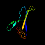
1 c2iiiA_
100.0
28
PDB header: lyaseChain: A: PDB Molecule: s-adenosylmethionine decarboxylase proenzyme;PDBTitle: crystal structure of the adenosylmethionine decarboxylase (aq_254)2 from aquifex aeolicus vf5
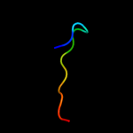
2 d1vr7a1
100.0
35
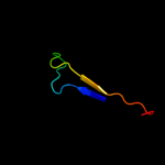
Fold: S-adenosylmethionine decarboxylaseSuperfamily: S-adenosylmethionine decarboxylaseFamily: Bacterial S-adenosylmethionine decarboxylase3 c1vr7A_
100.0
35
PDB header: lyaseChain: A: PDB Molecule: s-adenosylmethionine decarboxylase proenzyme;PDBTitle: crystal structure of s-adenosylmethionine decarboxylase proenzyme2 (tm0655) from thermotoga maritima at 1.2 a resolution
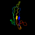
4 c3iwdC_
99.7
39
PDB header: lyaseChain: C: PDB Molecule: s-adenosylmethionine decarboxylase;PDBTitle: t. maritima adometdc complex with 5'-deoxy-5'-dimethyl2 thioadenosine
5 c3iwcD_
98.9
22
PDB header: lyaseChain: D: PDB Molecule: s-adenosylmethionine decarboxylase;PDBTitle: t. maritima adometdc complex with s-adenosylmethionine2 methyl ester
6 c1mhmA_
81.8
24
PDB header: lyaseChain: A: PDB Molecule: s-adenosylmethionine decarboxylase;PDBTitle: crystal structure of s-adenosylmethionine decarboxylase2 from potato
7 c3ep3A_
79.9
26
PDB header: lyaseChain: A: PDB Molecule: s-adenosylmethionine decarboxylase alpha chain;PDBTitle: human adometdc d174n mutant with no putrescine bound
8 c3somO_
58.8
30
PDB header: oxidoreductaseChain: O: PDB Molecule: methylmalonic aciduria and homocystinuria type c protein;PDBTitle: crystal structure of human mmachc
9 c1msvB_
54.9
26
PDB header: lyaseChain: B: PDB Molecule: s-adenosylmethionine decarboxylase proenzyme;PDBTitle: the s68a s-adenosylmethionine decarboxylase proenzyme2 processing mutant.
10 d1jl0a_
50.2
28
Fold: S-adenosylmethionine decarboxylaseSuperfamily: S-adenosylmethionine decarboxylaseFamily: S-adenosylmethionine decarboxylase11 c2lb0A_
41.7
17
PDB header: signaling protein/transcriptionChain: A: PDB Molecule: e3 ubiquitin-protein ligase smurf1;PDBTitle: structure of the first ww domain of human smurf1 in complex with a di-2 phosphorylated human smad1 derived peptide
12 d1u58a1
39.9
19
Fold: Immunoglobulin-like beta-sandwichSuperfamily: ImmunoglobulinFamily: C1 set domains (antibody constant domain-like)13 c2lazA_
39.4
17
PDB header: signaling protein/transcriptionChain: A: PDB Molecule: e3 ubiquitin-protein ligase smurf1;PDBTitle: structure of the first ww domain of human smurf1 in complex with a2 mono-phosphorylated human smad1 derived peptide
14 c3c6kC_
34.5
32
PDB header: transferaseChain: C: PDB Molecule: spermine synthase;PDBTitle: crystal structure of human spermine synthase in complex2 with spermidine and 5-methylthioadenosine
15 c1d9kD_
32.8
21
PDB header: immune systemChain: D: PDB Molecule: mhc i-ak b chain (beta chain);PDBTitle: crystal structure of complex between d10 tcr and pmhc i-ak/ca
16 c3k1hA_
31.8
19
PDB header: chaperoneChain: A: PDB Molecule: putative uncharacterized protein;PDBTitle: crystal structure of hp1076 from h.pylori
17 c2l4jA_
31.7
25
PDB header: transcriptionChain: A: PDB Molecule: yes-associated protein 2 (yap2);PDBTitle: yap ww2
18 d1mh5h2
29.2
22
Fold: Immunoglobulin-like beta-sandwichSuperfamily: ImmunoglobulinFamily: C1 set domains (antibody constant domain-like)19 d2f21a1
26.9
17
Fold: WW domain-likeSuperfamily: WW domainFamily: WW domain20 c2kl5A_
26.3
23
PDB header: structural genomics, unknown functionChain: A: PDB Molecule: uncharacterized protein yutd;PDBTitle: solution nmr structure of protein yutd from b.subtilis, northeast2 structural genomics consortium target sr232
21 c1iaoB_
not modelled
26.2
21
PDB header: mhc iiChain: B: PDB Molecule: mhc class ii i-ad;PDBTitle: class ii mhc i-ad in complex with ovalbumin peptide 323-339
22 c1iebD_
not modelled
23.8
24
PDB header: histocompatibility antigenChain: D: PDB Molecule: mhc class ii i-ek;PDBTitle: histocompatibility antigen
23 c2q6wD_
not modelled
23.3
19
PDB header: immune systemChain: D: PDB Molecule: hla class ii histocompatibility antigen, drPDBTitle: the structure of hla-dra, drb3*0101 (dr52a) with bound2 platelet integrin peptide associated with fetal and3 neonatal alloimmune thrombocytopenia
24 c2kq0A_
not modelled
22.3
50
PDB header: ligaseChain: A: PDB Molecule: e3 ubiquitin-protein ligase nedd4;PDBTitle: human nedd4 3rd ww domain complex with ebola zaire virus matrix2 protein vp40 derived peptide ilptappeymea
25 c1pqzA_
not modelled
21.6
19
PDB header: viral protein/immune systemChain: A: PDB Molecule: mcmv m144;PDBTitle: murine cytomegalovirus immunomodulatory protein m144
26 c2p24A_
not modelled
20.0
16
PDB header: immune systemChain: A: PDB Molecule: h-2 class ii histocompatibility antigen, a-u alpha chain;PDBTitle: i-au/mbp125-135
27 c3lqzA_
not modelled
18.7
22
PDB header: immune systemChain: A: PDB Molecule: hla class ii histocompatibility antigen, dp alpha 1 chain;PDBTitle: crystal structure of hla-dp2
28 d2jmfa1
not modelled
17.9
31
Fold: WW domain-likeSuperfamily: WW domainFamily: WW domain29 c2jmfA_
not modelled
17.6
33
PDB header: ligase/signaling proteinChain: A: PDB Molecule: e3 ubiquitin-protein ligase suppressor of deltex;PDBTitle: solution structure of the su(dx) ww4- notch py peptide2 complex
30 c2xm5A_
not modelled
17.1
29
PDB header: transferaseChain: A: PDB Molecule: cloq;PDBTitle: structural and mechanistic analysis of the magnesium-2 independent aromatic prenyltransferase cloq from the3 clorobiocin biosynthetic pathway
31 c2p24B_
not modelled
16.7
21
PDB header: immune systemChain: B: PDB Molecule: h-2 class ii histocompatibility antigen, a-u beta chain;PDBTitle: i-au/mbp125-135
32 c1ik9C_
not modelled
16.2
33
PDB header: gene regulation/ligaseChain: C: PDB Molecule: dna ligase iv;PDBTitle: crystal structure of a xrcc4-dna ligase iv complex
33 c1wr4A_
not modelled
16.0
23
PDB header: ligaseChain: A: PDB Molecule: ubiquitin-protein ligase nedd4-2;PDBTitle: solution structure of the second ww domain of nedd4-2
34 c1ymzA_
not modelled
15.8
15
PDB header: unknown functionChain: A: PDB Molecule: cc45;PDBTitle: cc45, an artificial ww domain designed using statistical2 coupling analysis
35 c1zhbJ_
not modelled
15.8
17
PDB header: immune systemChain: J: PDB Molecule: h-2 class i histocompatibility antigen, d-bPDBTitle: crystal structure of the murine class i major2 histocompatibility complex of h-2db, b2-microglobulin, and3 a 9-residue peptide derived from rat dopamine beta-4 monooxigenase
36 c2ez5W_
not modelled
15.5
40
PDB header: signalling protein,ligaseChain: W: PDB Molecule: e3 ubiquitin-protein ligase nedd4;PDBTitle: solution structure of the dnedd4 ww3* domain- comm lpsy2 peptide complex
37 c2ysbA_
not modelled
15.0
38
PDB header: protein bindingChain: A: PDB Molecule: salvador homolog 1 protein;PDBTitle: solution structure of the first ww domain from the mouse2 salvador homolog 1 protein (sav1)
38 c1e0mA_
not modelled
15.0
23
PDB header: de novo proteinChain: A: PDB Molecule: wwprototype;PDBTitle: prototype ww domain
39 d1i5hw_
not modelled
14.8
27
Fold: WW domain-likeSuperfamily: WW domainFamily: WW domain40 c2zajA_
not modelled
14.7
25
PDB header: protein bindingChain: A: PDB Molecule: membrane-associated guanylate kinase, ww and pdzPDBTitle: solution structure of the short-isoform of the second ww2 domain from the human membrane-associated guanylate kinase,3 ww and pdz domain-containing protein 1 (magi-1)
41 c2yshA_
not modelled
14.6
8
PDB header: protein bindingChain: A: PDB Molecule: growth-arrest-specific protein 7;PDBTitle: solution structure of the ww domain from the human growth-2 arrest-specific protein 7, gas-7
42 c1yiuA_
not modelled
14.6
42
PDB header: ligaseChain: A: PDB Molecule: itchy e3 ubiquitin protein ligase;PDBTitle: itch e3 ubiquitin ligase ww3 domain
43 c1v1cA_
not modelled
14.0
29
PDB header: sh3-domainChain: A: PDB Molecule: obscurin;PDBTitle: solution structure of the sh3 domain of obscurin
44 d1tk7a2
not modelled
14.0
33
Fold: WW domain-likeSuperfamily: WW domainFamily: WW domain45 c1wmvA_
not modelled
13.0
46
PDB header: oxidoreductase, apoptosisChain: A: PDB Molecule: ww domain containing oxidoreductase;PDBTitle: solution structure of the second ww domain of wwox
46 c3l4hA_
not modelled
13.0
38
PDB header: protein bindingChain: A: PDB Molecule: e3 ubiquitin-protein ligase hecw1;PDBTitle: helical box domain and second ww domain of the human e3 ubiquitin-2 protein ligase hecw1
47 c2clvA_
not modelled
12.9
16
PDB header: immune systemChain: A: PDB Molecule: h-2 class i histocompatibility antigen, k-bPDBTitle: mhc class i natural mutant h-2kbm8 heavy chain complexed2 with beta-2 microglobulin and pbm8 peptide
48 d1k9ra_
not modelled
12.9
25
Fold: WW domain-likeSuperfamily: WW domainFamily: WW domain49 c2ysgA_
not modelled
12.7
42
PDB header: protein bindingChain: A: PDB Molecule: syntaxin-binding protein 4;PDBTitle: solution structure of the ww domain from the human syntaxin-2 binding protein 4
50 c2dmvA_
not modelled
12.1
31
PDB header: ligaseChain: A: PDB Molecule: itchy homolog e3 ubiquitin protein ligase;PDBTitle: solution structure of the second ww domain of itchy homolog2 e3 ubiquitin protein ligase (itch)
51 c2djyA_
not modelled
11.7
27
PDB header: ligase/signaling proteinChain: A: PDB Molecule: smad ubiquitination regulatory factor 2;PDBTitle: solution structure of smurf2 ww3 domain-smad7 py peptide2 complex
52 c2lawA_
not modelled
11.6
23
PDB header: signaling protein/transcriptionChain: A: PDB Molecule: yorkie homolog;PDBTitle: structure of the second ww domain from human yap in complex with a2 human smad1 derived peptide
53 c3zr8X_
not modelled
10.9
16
PDB header: protein bindingChain: X: PDB Molecule: avr3a11;PDBTitle: crystal structure of rxlr effector avr3a11 from phytophthora capsici
54 d2nnab1
not modelled
10.9
20
Fold: Immunoglobulin-like beta-sandwichSuperfamily: ImmunoglobulinFamily: C1 set domains (antibody constant domain-like)55 c1wr7A_
not modelled
10.8
43
PDB header: ligaseChain: A: PDB Molecule: nedd4-2;PDBTitle: solution structure of the third ww domain of nedd4-2
56 d1tk7a1
not modelled
10.7
17
Fold: WW domain-likeSuperfamily: WW domainFamily: WW domain57 c2yscA_
not modelled
10.6
9
PDB header: protein bindingChain: A: PDB Molecule: amyloid beta a4 precursor protein-binding familyPDBTitle: solution structure of the ww domain from the human amyloid2 beta a4 precursor protein-binding family b member 3, apbb3
58 c2dwvB_
not modelled
10.5
17
PDB header: protein bindingChain: B: PDB Molecule: salvador homolog 1 protein;PDBTitle: solution structure of the second ww domain from mouse2 salvador homolog 1 protein (mww45)
59 c3zv0A_
not modelled
10.5
19
PDB header: cell cycleChain: A: PDB Molecule: protein shq1;PDBTitle: structure of the shq1p-cbf5p complex
60 c2yf6A_
not modelled
10.2
19
PDB header: immune systemChain: A: PDB Molecule: major histocompatibility complex class i glycoproteinPDBTitle: complex of a b21 chicken mhc class i molecule and a 10mer2 chicken peptide
61 c3bzeA_
not modelled
10.2
17
PDB header: immune systemChain: A: PDB Molecule: hla class i histocompatibility antigen, alpha chain e;PDBTitle: the human non-classical major histocompatibility complex molecule hla-2 e
62 d1ynja1
not modelled
10.1
37
Fold: DCoH-likeSuperfamily: RBP11-like subunits of RNA polymeraseFamily: RNA polymerase alpha subunit dimerisation domain63 c2ysfA_
not modelled
9.8
29
PDB header: protein bindingChain: A: PDB Molecule: e3 ubiquitin-protein ligase itchy homolog;PDBTitle: solution structure of the fourth ww domain from the human2 e3 ubiquitin-protein ligase itchy homolog, itch
64 c1zt7C_
not modelled
9.5
16
PDB header: immune systemChain: C: PDB Molecule: h-2 class i histocompatibility antigen, k-kPDBTitle: crystal structure of class i mhc h-2kk in complex with a2 nonapeptide
65 c3ssbJ_
not modelled
9.1
41
PDB header: hydrolase/hydrolase inhibitorChain: J: PDB Molecule: inducible metalloproteinase inhibitor protein;PDBTitle: structure of insect metalloproteinase inhibitor in complex with2 thermolysin
66 c1sebB_
not modelled
8.7
21
PDB header: complex (mhc ii/peptide/toxin)Chain: B: PDB Molecule: hla class ii histocompatibility antigen;PDBTitle: complex of the human mhc class ii glycoprotein hla-dr1 and2 the bacterial superantigen seb
67 c1ypzA_
not modelled
8.3
17
PDB header: immune systemChain: A: PDB Molecule: h2-t22 protein;PDBTitle: immune receptor
68 c2kykA_
not modelled
8.3
31
PDB header: ligaseChain: A: PDB Molecule: e3 ubiquitin-protein ligase itchy homolog;PDBTitle: the sandwich region between two lmp2a py motif regulates the2 interaction between aip4ww2domain and py motif
69 d1twfj_
not modelled
8.3
14
Fold: DNA/RNA-binding 3-helical bundleSuperfamily: RNA polymerase subunit RPB10Family: RNA polymerase subunit RPB1070 c3g27A_
not modelled
7.7
58
PDB header: protein bindingChain: A: PDB Molecule: 82 prophage-derived uncharacterized protein ybco;PDBTitle: structure of a putative bacteriophage protein from escherichia coli2 str. k-12 substr. mg1655
71 c2le2B_
not modelled
7.7
26
PDB header: hydrolase inhibitorChain: B: PDB Molecule: p56;PDBTitle: novel dimeric structure of phage phi29-encoded protein p56: insights2 into uracil-dna glycosylase inhibition
72 c3lqzB_
not modelled
7.6
19
PDB header: immune systemChain: B: PDB Molecule: hla-dp2 beta chain linked with dra peptide;PDBTitle: crystal structure of hla-dp2
73 c1s9vA_
not modelled
7.3
21
PDB header: immune systemChain: A: PDB Molecule: hla class ii histocompatibility antigen, dq(3)PDBTitle: crystal structure of hla-dq2 complexed with deamidated2 gliadin peptide
74 c1f3jD_
not modelled
7.1
17
PDB header: immune systemChain: D: PDB Molecule: h-2 class ii histocompatibility antigen;PDBTitle: histocompatibility antigen i-ag7
75 c2ysdA_
not modelled
6.8
40
PDB header: protein bindingChain: A: PDB Molecule: membrane-associated guanylate kinase, ww and pdzPDBTitle: solution structure of the first ww domain from the human2 membrane-associated guanylate kinase, ww and pdz domain-3 containing protein 1. magi-1
76 d1xoda1
not modelled
6.8
11
Fold: PH domain-like barrelSuperfamily: PH domain-likeFamily: Enabled/VASP homology 1 domain (EVH1 domain)77 c2kxqA_
not modelled
6.8
33
PDB header: protein bindingChain: A: PDB Molecule: e3 ubiquitin-protein ligase smurf2;PDBTitle: solution structure of smurf2 ww2 and ww3 bound to smad7 py motif2 containing peptide
78 c1p4lA_
not modelled
6.8
16
PDB header: immune systemChain: A: PDB Molecule: mhc class i h-2kb heavy chain;PDBTitle: crystal structure of nk receptor ly49c mutant with its mhc2 class i ligand h-2kb
79 c1cd1C_
not modelled
6.7
18
PDB header: cd1Chain: C: PDB Molecule: cd1;PDBTitle: cd1(mouse) antigen presenting molecule
80 d1kn6a_
not modelled
6.4
24
Fold: Ferredoxin-likeSuperfamily: Protease propeptides/inhibitorsFamily: Prohormone convertase 1 pro-domain81 c2l2lA_
not modelled
6.3
29
PDB header: transferaseChain: A: PDB Molecule: transcriptional repressor p66-alpha;PDBTitle: solution structure of the coiled-coil complex between mbd2 and2 p66alpha
82 c2fwoA_
not modelled
6.3
16
PDB header: immune system/viral proteinChain: A: PDB Molecule: h-2 class i histocompatibility antigen, k-dPDBTitle: mhc class i h-2kd heavy chain in complex with beta-2 2microglobulin and peptide derived from influenza3 nucleoprotein
83 c2j42A_
not modelled
6.2
22
PDB header: toxinChain: A: PDB Molecule: c2 toxin component-ii;PDBTitle: low quality crystal structure of the transport component c2-2 ii of the c2-toxin from clostridium botulinum
84 d1bdfa1
not modelled
6.1
37
Fold: DCoH-likeSuperfamily: RBP11-like subunits of RNA polymeraseFamily: RNA polymerase alpha subunit dimerisation domain85 d1mr1c_
not modelled
5.8
20
Fold: SAND domain-likeSuperfamily: SAND domain-likeFamily: SMAD4-binding domain of oncoprotein Ski86 c1xz0A_
not modelled
5.7
18
PDB header: immune systemChain: A: PDB Molecule: t-cell surface glycoprotein cd1a;PDBTitle: crystal structure of cd1a in complex with a synthetic mycobactin2 lipopeptide
87 c2ysiA_
not modelled
5.6
8
PDB header: protein bindingChain: A: PDB Molecule: transcription elongation regulator 1;PDBTitle: solution structure of the first ww domain from the mouse2 transcription elongation regulator 1, transcription factor3 ca150
88 c2f0cC_
not modelled
5.4
20
PDB header: virus/viral proteinChain: C: PDB Molecule: phage tp901-1 orf49 (bpp);PDBTitle: structure of the receptor binding protein (orf49, bbp) from lactophage2 tp901-1
89 c1mwaH_
not modelled
5.2
16
PDB header: immune systemChain: H: PDB Molecule: h-2kbm3 mhc class i molecule heavy chain;PDBTitle: 2c/h-2kbm3/dev8 allogeneic complex
90 c1mwaI_
not modelled
5.2
16
PDB header: immune systemChain: I: PDB Molecule: h-2kbm3 mhc class i molecule heavy chain;PDBTitle: 2c/h-2kbm3/dev8 allogeneic complex
91 c1zt4C_
not modelled
5.2
18
PDB header: immune systemChain: C: PDB Molecule: t-cell surface glycoprotein cd1d;PDBTitle: the crystal structure of human cd1d with and without alpha-2 galactosylceramide
92 d1smya1
not modelled
5.2
37
Fold: DCoH-likeSuperfamily: RBP11-like subunits of RNA polymeraseFamily: RNA polymerase alpha subunit dimerisation domain93 d1d5mb1
not modelled
5.2
21
Fold: Immunoglobulin-like beta-sandwichSuperfamily: ImmunoglobulinFamily: C1 set domains (antibody constant domain-like)