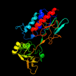
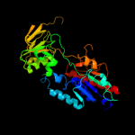
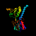
1 c1f6mF_
100.0
100
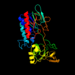
PDB header: oxidoreductaseChain: F: PDB Molecule: thioredoxin reductase;PDBTitle: crystal structure of a complex between thioredoxin2 reductase, thioredoxin, and the nadp+ analog, aadp+
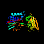
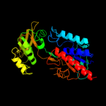
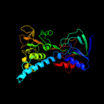
2 c1hyuA_
100.0
34
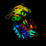
PDB header: oxidoreductaseChain: A: PDB Molecule: alkyl hydroperoxide reductase subunit f;PDBTitle: crystal structure of intact ahpf
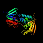
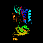
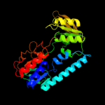
3 c3r9uA_
100.0
36
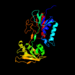
PDB header: oxidoreductaseChain: A: PDB Molecule: thioredoxin reductase;PDBTitle: thioredoxin-disulfide reductase from campylobacter jejuni.
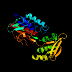
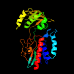
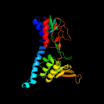
4 c2q7vA_
100.0
42
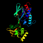
PDB header: oxidoreductaseChain: A: PDB Molecule: thioredoxin reductase;PDBTitle: crystal structure of deinococcus radiodurans thioredoxin2 reductase
5 c2v6oA_
100.0
17
PDB header: oxidoreductaseChain: A: PDB Molecule: thioredoxin glutathione reductase;PDBTitle: structure of schistosoma mansoni thioredoxin-glutathione2 reductase (smtgr)
6 c1vdcA_
100.0
45
PDB header: oxidoreductaseChain: A: PDB Molecule: nadph dependent thioredoxin reductase;PDBTitle: structure of nadph dependent thioredoxin reductase
7 c3ctyA_
100.0
28
PDB header: oxidoreductaseChain: A: PDB Molecule: thioredoxin reductase;PDBTitle: crystal structure of t. acidophilum thioredoxin reductase
8 c1fl2A_
100.0
32
PDB header: oxidoreductaseChain: A: PDB Molecule: alkyl hydroperoxide reductase subunit f;PDBTitle: catalytic core component of the alkylhydroperoxide reductase ahpf from2 e.coli
9 c2a87A_
100.0
47
PDB header: oxidoreductaseChain: A: PDB Molecule: thioredoxin reductase;PDBTitle: crystal structure of m. tuberculosis thioredoxin reductase
10 c2zbwA_
100.0
25
PDB header: oxidoreductaseChain: A: PDB Molecule: thioredoxin reductase;PDBTitle: crystal structure of thioredoxin reductase-like protein from thermus2 thermophilus hb8
11 c1gthD_
100.0
18
PDB header: oxidoreductaseChain: D: PDB Molecule: dihydropyrimidine dehydrogenase;PDBTitle: dihydropyrimidine dehydrogenase (dpd) from pig, ternary2 complex with nadph and 5-iodouracil
12 c3d8xB_
100.0
51
PDB header: oxidoreductaseChain: B: PDB Molecule: thioredoxin reductase 1;PDBTitle: crystal structure of saccharomyces cerevisiae nadph dependent2 thioredoxin reductase 1
13 c3f8rD_
100.0
35
PDB header: oxidoreductaseChain: D: PDB Molecule: thioredoxin reductase (trxb-3);PDBTitle: crystal structure of sulfolobus solfataricus thioredoxin2 reductase b3 in complex with two nadp molecules
14 c2q0lA_
100.0
33
PDB header: oxidoreductaseChain: A: PDB Molecule: thioredoxin reductase;PDBTitle: helicobacter pylori thioredoxin reductase reduced by sodium dithionite2 in complex with nadp+
15 c2r9zB_
100.0
22
PDB header: oxidoreductaseChain: B: PDB Molecule: glutathione amide reductase;PDBTitle: glutathione amide reductase from chromatium gracile
16 c1ojtA_
100.0
20
PDB header: oxidoreductaseChain: A: PDB Molecule: surface protein;PDBTitle: structure of dihydrolipoamide dehydrogenase
17 c1geuA_
100.0
19
PDB header: oxidoreductase(flavoenzyme)Chain: A: PDB Molecule: glutathione reductase;PDBTitle: anatomy of an engineered nad-binding site
18 c2a8xA_
100.0
21
PDB header: oxidoreductaseChain: A: PDB Molecule: dihydrolipoyl dehydrogenase;PDBTitle: crystal structure of lipoamide dehydrogenase from2 mycobacterium tuberculosis
19 c3dgzA_
100.0
17
PDB header: oxidoreductaseChain: A: PDB Molecule: thioredoxin reductase 2;PDBTitle: crystal structure of mouse mitochondrial thioredoxin reductase, c-2 terminal 3-residue truncation
20 c1ebdB_
100.0
21
PDB header: complex (oxidoreductase/transferase)Chain: B: PDB Molecule: dihydrolipoamide dehydrogenase;PDBTitle: dihydrolipoamide dehydrogenase complexed with the binding2 domain of the dihydrolipoamide acetylase
21 c2nvkX_
not modelled
100.0
18
PDB header: oxidoreductaseChain: X: PDB Molecule: thioredoxin reductase;PDBTitle: crystal structure of thioredoxin reductase from drosophila2 melanogaster
22 c3urhB_
not modelled
100.0
17
PDB header: oxidoreductaseChain: B: PDB Molecule: dihydrolipoyl dehydrogenase;PDBTitle: crystal structure of a dihydrolipoamide dehydrogenase from2 sinorhizobium meliloti 1021
23 c2eq8E_
not modelled
100.0
20
PDB header: oxidoreductaseChain: E: PDB Molecule: pyruvate dehydrogenase complex, dihydrolipoamidePDBTitle: crystal structure of lipoamide dehydrogenase from thermus thermophilus2 hb8 with psbdp
24 c3lzxB_
not modelled
100.0
25
PDB header: oxidoreductaseChain: B: PDB Molecule: ferredoxin--nadp reductase 2;PDBTitle: crystal structure of ferredoxin-nadp+ oxidoreductase from bacillus2 subtilis (form ii)
25 c1zkqA_
not modelled
100.0
17
PDB header: oxidoreductaseChain: A: PDB Molecule: thioredoxin reductase 2, mitochondrial;PDBTitle: crystal structure of mouse thioredoxin reductase type 2
26 c1bwcA_
not modelled
100.0
16
PDB header: oxidoreductaseChain: A: PDB Molecule: protein (glutathione reductase);PDBTitle: structure of human glutathione reductase complexed with ajoene2 inhibitor and subversive substrate
27 c3ic9D_
not modelled
100.0
18
PDB header: oxidoreductaseChain: D: PDB Molecule: dihydrolipoamide dehydrogenase;PDBTitle: the structure of dihydrolipoamide dehydrogenase from colwellia2 psychrerythraea 34h.
28 c3lxdA_
not modelled
100.0
20
PDB header: oxidoreductaseChain: A: PDB Molecule: fad-dependent pyridine nucleotide-disulphidePDBTitle: crystal structure of ferredoxin reductase arr from novosphingobium2 aromaticivorans
29 c2hqmB_
not modelled
100.0
18
PDB header: oxidoreductaseChain: B: PDB Molecule: glutathione reductase;PDBTitle: crystal structure of glutathione reductase glr1 from the yeast2 saccharomyces cerevisiae
30 c1lpfB_
not modelled
100.0
21
PDB header: oxidoreductaseChain: B: PDB Molecule: dihydrolipoamide dehydrogenase;PDBTitle: three-dimensional structure of lipoamide dehydrogenase from2 pseudomonas fluorescens at 2.8 angstroms resolution.3 analysis of redox and thermostability properties
31 c2c3dB_
not modelled
100.0
18
PDB header: oxidoreductaseChain: B: PDB Molecule: 2-oxopropyl-com reductase;PDBTitle: 2.15 angstrom crystal structure of 2-ketopropyl coenzyme m2 oxidoreductase carboxylase with a coenzyme m disulfide3 bound at the active site
32 c3o0hA_
not modelled
100.0
17
PDB header: oxidoreductaseChain: A: PDB Molecule: glutathione reductase;PDBTitle: crystal structure of glutathione reductase from bartonella henselae
33 c3ab1B_
not modelled
100.0
20
PDB header: oxidoreductaseChain: B: PDB Molecule: ferredoxin--nadp reductase;PDBTitle: crystal structure of ferredoxin nadp+ oxidoreductase
34 c1v59B_
not modelled
100.0
17
PDB header: oxidoreductaseChain: B: PDB Molecule: dihydrolipoamide dehydrogenase;PDBTitle: crystal structure of yeast lipoamide dehydrogenase2 complexed with nad+
35 c3l8kB_
not modelled
100.0
15
PDB header: oxidoreductaseChain: B: PDB Molecule: dihydrolipoyl dehydrogenase;PDBTitle: crystal structure of a dihydrolipoyl dehydrogenase from2 sulfolobus solfataricus
36 c1lvlA_
not modelled
100.0
22
PDB header: oxidoreductaseChain: A: PDB Molecule: dihydrolipoamide dehydrogenase;PDBTitle: the refined structure of pseudomonas putida lipoamide dehydrogenase2 complexed with nad+ at 2.45 angstroms resolution
37 c1zx9A_
not modelled
100.0
21
PDB header: oxidoreductaseChain: A: PDB Molecule: mercuric reductase;PDBTitle: crystal structure of tn501 mera
38 c3ntaA_
not modelled
100.0
20
PDB header: oxidoreductaseChain: A: PDB Molecule: fad-dependent pyridine nucleotide-disulphidePDBTitle: structure of the shewanella loihica pv-4 nadh-dependent persulfide2 reductase
39 c2eq7B_
not modelled
100.0
19
PDB header: oxidoreductaseChain: B: PDB Molecule: 2-oxoglutarate dehydrogenase e3 component;PDBTitle: crystal structure of lipoamide dehydrogenase from thermus thermophilus2 hb8 with psbdo
40 c2bcpA_
not modelled
100.0
25
PDB header: oxidoreductaseChain: A: PDB Molecule: nadh oxidase;PDBTitle: structural analysis of streptococcus pyogenes nadh oxidase:2 c44s nox with azide
41 c2w0hA_
not modelled
100.0
17
PDB header: oxidoreductaseChain: A: PDB Molecule: trypanothione reductase;PDBTitle: x ray structure of leishmania infantum trypanothione2 reductase in complex with antimony and nadph
42 c1zmcG_
not modelled
100.0
18
PDB header: oxidoreductaseChain: G: PDB Molecule: dihydrolipoyl dehydrogenase;PDBTitle: crystal structure of human dihydrolipoamide dehydrogenase2 complexed to nad+
43 c3oc4A_
not modelled
100.0
22
PDB header: oxidoreductaseChain: A: PDB Molecule: oxidoreductase, pyridine nucleotide-disulfide family;PDBTitle: crystal structure of a pyridine nucleotide-disulfide family2 oxidoreductase from the enterococcus faecalis v583
44 c1q1wA_
not modelled
100.0
16
PDB header: oxidoreductaseChain: A: PDB Molecule: putidaredoxin reductase;PDBTitle: crystal structure of putidaredoxin reductase from2 pseudomonas putida
45 c2qaeA_
not modelled
100.0
21
PDB header: oxidoreductaseChain: A: PDB Molecule: dihydrolipoyl dehydrogenase;PDBTitle: crystal structure analysis of trypanosoma cruzi lipoamide2 dehydrogenase
46 c1dxlC_
not modelled
100.0
16
PDB header: oxidoreductaseChain: C: PDB Molecule: dihydrolipoamide dehydrogenase;PDBTitle: dihydrolipoamide dehydrogenase of glycine decarboxylase2 from pisum sativum
47 c1gv4A_
not modelled
100.0
19
PDB header: oxidoreductaseChain: A: PDB Molecule: programed cell death protein 8;PDBTitle: murine apoptosis-inducing factor (aif)
48 c1tytA_
not modelled
100.0
18
PDB header: oxidoreductaseChain: A: PDB Molecule: trypanothione reductase, oxidized form;PDBTitle: crystal and molecular structure of crithidia fasciculata2 trypanothione reductase at 2.6 angstroms resolution
49 c2cfyB_
not modelled
100.0
20
PDB header: oxidoreductaseChain: B: PDB Molecule: thioredoxin reductase 1;PDBTitle: crystal structure of human thioredoxin reductase 1
50 c3fg2P_
not modelled
100.0
20
PDB header: oxidoreductaseChain: P: PDB Molecule: putative rubredoxin reductase;PDBTitle: crystal structure of ferredoxin reductase for the cyp199a2 system from2 rhodopseudomonas palustris
51 c3icrA_
not modelled
100.0
19
PDB header: oxidoreductaseChain: A: PDB Molecule: coenzyme a-disulfide reductase;PDBTitle: crystal structure of oxidized bacillus anthracis coadr-rhd
52 c1onfA_
not modelled
100.0
18
PDB header: oxidoreductaseChain: A: PDB Molecule: glutathione reductase;PDBTitle: crystal structure of plasmodium falciparum glutathione reductase
53 c3fbsB_
not modelled
100.0
19
PDB header: oxidoreductaseChain: B: PDB Molecule: oxidoreductase;PDBTitle: the crystal structure of the oxidoreductase from agrobacterium2 tumefaciens
54 c1ndaD_
not modelled
100.0
17
PDB header: oxidoreductaseChain: D: PDB Molecule: trypanothione oxidoreductase;PDBTitle: the structure of trypanosoma cruzi trypanothione reductase2 in the oxidized and nadph reduced state
55 c1xdiA_
not modelled
100.0
19
PDB header: unknown functionChain: A: PDB Molecule: rv3303c-lpda;PDBTitle: crystal structure of lpda (rv3303c) from mycobacterium tuberculosis
56 c1yqzA_
not modelled
100.0
20
PDB header: oxidoreductaseChain: A: PDB Molecule: coenzyme a disulfide reductase;PDBTitle: structure of coenzyme a-disulfide reductase from2 staphylococcus aureus refined at 1.54 angstrom resolution
57 c2vdcI_
not modelled
100.0
19
PDB header: oxidoreductaseChain: I: PDB Molecule: glutamate synthase [nadph] small chain;PDBTitle: the 9.5 a resolution structure of glutamate synthase from2 cryo-electron microscopy and its oligomerization behavior3 in solution: functional implications.
58 c2gr2A_
not modelled
100.0
17
PDB header: oxidoreductaseChain: A: PDB Molecule: ferredoxin reductase;PDBTitle: crystal structure of ferredoxin reductase, bpha4 (oxidized form)
59 c3ef6A_
not modelled
100.0
16
PDB header: oxidoreductaseChain: A: PDB Molecule: toluene 1,2-dioxygenase system ferredoxin--nad(+)PDBTitle: crystal structure of toluene 2,3-dioxygenase reductase
60 c2cduB_
not modelled
100.0
15
PDB header: oxidoreductaseChain: B: PDB Molecule: nadph oxidase;PDBTitle: the crystal structure of water-forming nad(p)h oxidase from2 lactobacillus sanfranciscensis
61 c1m6iA_
not modelled
100.0
17
PDB header: oxidoreductaseChain: A: PDB Molecule: programmed cell death protein 8;PDBTitle: crystal structure of apoptosis inducing factor (aif)
62 c3kd9B_
not modelled
100.0
21
PDB header: oxidoreductaseChain: B: PDB Molecule: coenzyme a disulfide reductase;PDBTitle: crystal structure of pyridine nucleotide disulfide oxidoreductase from2 pyrococcus horikoshii
63 c1nhqA_
not modelled
100.0
24
PDB header: oxidoreductase (h2o2(a))Chain: A: PDB Molecule: nadh peroxidase;PDBTitle: crystallographic analyses of nadh peroxidase cys42ala and cys42ser2 mutants: active site structure, mechanistic implications, and an3 unusual environment of arg303
64 c2v3aA_
not modelled
100.0
17
PDB header: oxidoreductaseChain: A: PDB Molecule: rubredoxin reductase;PDBTitle: crystal structure of rubredoxin reductase from pseudomonas2 aeruginosa.
65 c3iwaA_
not modelled
99.9
21
PDB header: oxidoreductaseChain: A: PDB Molecule: fad-dependent pyridine nucleotide-disulphidePDBTitle: crystal structure of a fad-dependent pyridine nucleotide-disulphide2 oxidoreductase from desulfovibrio vulgaris
66 c1xhcA_
not modelled
99.9
18
PDB header: oxidoreductaseChain: A: PDB Molecule: nadh oxidase /nitrite reductase;PDBTitle: nadh oxidase /nitrite reductase from pyrococcus furiosus pfu-1140779-2 001
67 c3cgdB_
not modelled
99.9
21
PDB header: oxidoreductaseChain: B: PDB Molecule: pyridine nucleotide-disulfide oxidoreductase, class i;PDBTitle: pyridine nucleotide complexes with bacillus anthracis coenzyme a-2 disulfide reductase: a structural analysis of dual nad(p)h3 specificity
68 c3kpgA_
not modelled
99.9
17
PDB header: oxidoreductaseChain: A: PDB Molecule: sulfide-quinone reductase, putative;PDBTitle: crystal structure of sulfide:quinone oxidoreductase from2 acidithiobacillus ferrooxidans in complex with decylubiquinone
69 c3kljA_
not modelled
99.9
20
PDB header: oxidoreductaseChain: A: PDB Molecule: nad(fad)-dependent dehydrogenase, nirb-family (n-terminalPDBTitle: crystal structure of nadh:rubredoxin oxidoreductase from clostridium2 acetobutylicum
70 c3h8lA_
not modelled
99.9
16
PDB header: oxidoreductaseChain: A: PDB Molecule: nadh oxidase;PDBTitle: the first x-ray structure of a sulfide:quinone2 oxidoreductase: insights into sulfide oxidation mechanism
71 c1djnB_
not modelled
99.9
17
PDB header: oxidoreductaseChain: B: PDB Molecule: trimethylamine dehydrogenase;PDBTitle: structural and biochemical characterization of recombinant wild type2 trimethylamine dehydrogenase from methylophilus methylotrophus (sp.3 w3a1)
72 c3hyxC_
not modelled
99.9
12
PDB header: oxidoreductaseChain: C: PDB Molecule: sulfide-quinone reductase;PDBTitle: 3-d x-ray structure of the sulfide:quinone oxidoreductase from aquifex2 aeolicus in complex with aurachin c
73 c1fcdB_
not modelled
99.9
16
PDB header: electron transport(flavocytochrome)Chain: B: PDB Molecule: flavocytochrome c sulfide dehydrogenase (flavin-PDBTitle: the structure of flavocytochrome c sulfide dehydrogenase2 from a purple phototrophic bacterium chromatium vinosum at3 2.5 angstroms resolution
74 c3k30B_
not modelled
99.9
18
PDB header: oxidoreductaseChain: B: PDB Molecule: histamine dehydrogenase;PDBTitle: histamine dehydrogenase from nocardiodes simplex
75 c3d1cA_
not modelled
99.9
15
PDB header: oxidoreductaseChain: A: PDB Molecule: flavin-containing putative monooxygenase;PDBTitle: crystal structure of flavin-containing putative monooxygenase2 (np_373108.1) from staphylococcus aureus mu50 at 2.40 a resolution
76 c1ps9A_
not modelled
99.9
20
PDB header: oxidoreductaseChain: A: PDB Molecule: 2,4-dienoyl-coa reductase;PDBTitle: the crystal structure and reaction mechanism of e. coli 2,4-2 dienoyl coa reductase
77 c1x31A_
not modelled
99.9
17
PDB header: oxidoreductaseChain: A: PDB Molecule: sarcosine oxidase alpha subunit;PDBTitle: crystal structure of heterotetrameric sarcosine oxidase from2 corynebacterium sp. u-96
78 c1cjcA_
not modelled
99.9
14
PDB header: oxidoreductaseChain: A: PDB Molecule: protein (adrenodoxin reductase);PDBTitle: structure of adrenodoxin reductase of mitochondrial p4502 systems
79 c1lqtB_
not modelled
99.9
17
PDB header: oxidoreductaseChain: B: PDB Molecule: fpra;PDBTitle: a covalent modification of nadp+ revealed by the atomic resolution2 structure of fpra, a mycobacterium tuberculosis oxidoreductase
80 c1y56A_
not modelled
99.9
18
PDB header: oxidoreductaseChain: A: PDB Molecule: hypothetical protein ph1363;PDBTitle: crystal structure of l-proline dehydrogenase from p.horikoshii
81 d3grsa1
not modelled
99.9
17
Fold: FAD/NAD(P)-binding domainSuperfamily: FAD/NAD(P)-binding domainFamily: FAD/NAD-linked reductases, N-terminal and central domains82 d1trba1
not modelled
99.9
98
Fold: FAD/NAD(P)-binding domainSuperfamily: FAD/NAD(P)-binding domainFamily: FAD/NAD-linked reductases, N-terminal and central domains83 d3lada1
not modelled
99.9
22
Fold: FAD/NAD(P)-binding domainSuperfamily: FAD/NAD(P)-binding domainFamily: FAD/NAD-linked reductases, N-terminal and central domains84 d1ojta1
not modelled
99.9
22
Fold: FAD/NAD(P)-binding domainSuperfamily: FAD/NAD(P)-binding domainFamily: FAD/NAD-linked reductases, N-terminal and central domains85 d1lvla1
not modelled
99.9
18
Fold: FAD/NAD(P)-binding domainSuperfamily: FAD/NAD(P)-binding domainFamily: FAD/NAD-linked reductases, N-terminal and central domains86 d1h6va1
not modelled
99.9
16
Fold: FAD/NAD(P)-binding domainSuperfamily: FAD/NAD(P)-binding domainFamily: FAD/NAD-linked reductases, N-terminal and central domains87 d1d4ca2
not modelled
99.9
21
Fold: FAD/NAD(P)-binding domainSuperfamily: FAD/NAD(P)-binding domainFamily: Succinate dehydrogenase/fumarate reductase flavoprotein N-terminal domain88 d1ebda1
not modelled
99.9
19
Fold: FAD/NAD(P)-binding domainSuperfamily: FAD/NAD(P)-binding domainFamily: FAD/NAD-linked reductases, N-terminal and central domains89 d1lpfa1
not modelled
99.9
24
Fold: FAD/NAD(P)-binding domainSuperfamily: FAD/NAD(P)-binding domainFamily: FAD/NAD-linked reductases, N-terminal and central domains90 c3s5wB_
not modelled
99.8
16
PDB header: oxidoreductaseChain: B: PDB Molecule: l-ornithine 5-monooxygenase;PDBTitle: ornithine hydroxylase (pvda) from pseudomonas aeruginosa
91 d1qo8a2
not modelled
99.8
15
Fold: FAD/NAD(P)-binding domainSuperfamily: FAD/NAD(P)-binding domainFamily: Succinate dehydrogenase/fumarate reductase flavoprotein N-terminal domain92 d1v59a1
not modelled
99.8
19
Fold: FAD/NAD(P)-binding domainSuperfamily: FAD/NAD(P)-binding domainFamily: FAD/NAD-linked reductases, N-terminal and central domains93 d1aoga1
not modelled
99.8
13
Fold: FAD/NAD(P)-binding domainSuperfamily: FAD/NAD(P)-binding domainFamily: FAD/NAD-linked reductases, N-terminal and central domains94 d1y0pa2
not modelled
99.8
20
Fold: FAD/NAD(P)-binding domainSuperfamily: FAD/NAD(P)-binding domainFamily: Succinate dehydrogenase/fumarate reductase flavoprotein N-terminal domain95 d1dxla1
not modelled
99.8
21
Fold: FAD/NAD(P)-binding domainSuperfamily: FAD/NAD(P)-binding domainFamily: FAD/NAD-linked reductases, N-terminal and central domains96 d1feca1
not modelled
99.8
18
Fold: FAD/NAD(P)-binding domainSuperfamily: FAD/NAD(P)-binding domainFamily: FAD/NAD-linked reductases, N-terminal and central domains97 d2bs2a2
not modelled
99.8
12
Fold: FAD/NAD(P)-binding domainSuperfamily: FAD/NAD(P)-binding domainFamily: Succinate dehydrogenase/fumarate reductase flavoprotein N-terminal domain98 d1vdca1
not modelled
99.8
37
Fold: FAD/NAD(P)-binding domainSuperfamily: FAD/NAD(P)-binding domainFamily: FAD/NAD-linked reductases, N-terminal and central domains99 d1seza1
not modelled
99.8
12
Fold: FAD/NAD(P)-binding domainSuperfamily: FAD/NAD(P)-binding domainFamily: FAD-linked reductases, N-terminal domain100 d1jnra2
not modelled
99.8
19
Fold: FAD/NAD(P)-binding domainSuperfamily: FAD/NAD(P)-binding domainFamily: Succinate dehydrogenase/fumarate reductase flavoprotein N-terminal domain101 c3cesB_
not modelled
99.8
17
PDB header: rna binding proteinChain: B: PDB Molecule: trna uridine 5-carboxymethylaminomethyl modification enzymePDBTitle: crystal structure of e.coli mnmg (gida), a highly-conserved trna2 modifying enzyme
102 c3g05B_
not modelled
99.8
14
PDB header: rna binding proteinChain: B: PDB Molecule: trna uridine 5-carboxymethylaminomethyl modification enzymePDBTitle: crystal structure of n-terminal domain (2-550) of e.coli mnmg
103 d1kf6a2
not modelled
99.8
18
Fold: FAD/NAD(P)-binding domainSuperfamily: FAD/NAD(P)-binding domainFamily: Succinate dehydrogenase/fumarate reductase flavoprotein N-terminal domain104 c1w4xA_
not modelled
99.8
20
PDB header: oxygenaseChain: A: PDB Molecule: phenylacetone monooxygenase;PDBTitle: phenylacetone monooxygenase, a baeyer-villiger2 monooxygenase
105 c3gwdA_
not modelled
99.8
19
PDB header: oxidoreductaseChain: A: PDB Molecule: cyclohexanone monooxygenase;PDBTitle: closed crystal structure of cyclohexanone monooxygenase
106 d2gmha1
not modelled
99.8
15
Fold: FAD/NAD(P)-binding domainSuperfamily: FAD/NAD(P)-binding domainFamily: FAD-linked reductases, N-terminal domain107 c2vq7B_
not modelled
99.8
16
PDB header: oxidoreductaseChain: B: PDB Molecule: flavin-containing monooxygenase;PDBTitle: bacterial flavin-containing monooxygenase in complex with2 nadp: native data
108 d1mo9a1
not modelled
99.7
22
Fold: FAD/NAD(P)-binding domainSuperfamily: FAD/NAD(P)-binding domainFamily: FAD/NAD-linked reductases, N-terminal and central domains109 d2gqfa1
not modelled
99.7
15
Fold: FAD/NAD(P)-binding domainSuperfamily: FAD/NAD(P)-binding domainFamily: HI0933 N-terminal domain-like110 d2iida1
not modelled
99.7
12
Fold: FAD/NAD(P)-binding domainSuperfamily: FAD/NAD(P)-binding domainFamily: FAD-linked reductases, N-terminal domain111 d1reoa1
not modelled
99.7
12
Fold: FAD/NAD(P)-binding domainSuperfamily: FAD/NAD(P)-binding domainFamily: FAD-linked reductases, N-terminal domain112 c2zxiC_
not modelled
99.7
15
PDB header: fad-binding proteinChain: C: PDB Molecule: trna uridine 5-carboxymethylaminomethylPDBTitle: structure of aquifex aeolicus gida in the form ii crystal
113 d1neka2
not modelled
99.7
15
Fold: FAD/NAD(P)-binding domainSuperfamily: FAD/NAD(P)-binding domainFamily: Succinate dehydrogenase/fumarate reductase flavoprotein N-terminal domain114 c1vqwB_
not modelled
99.7
16
PDB header: structural genomics, unknown functionChain: B: PDB Molecule: protein with similarity to flavin-containingPDBTitle: crystal structure of a protein with similarity to flavin-2 containing monooxygenases and to mammalian dimethylalanine3 monooxygenases
115 c3cp8C_
not modelled
99.7
16
PDB header: oxidoreductaseChain: C: PDB Molecule: trna uridine 5-carboxymethylaminomethylPDBTitle: crystal structure of gida from chlorobium tepidum
116 c1chuA_
not modelled
99.7
12
PDB header: flavoenzymeChain: A: PDB Molecule: protein (l-aspartate oxidase);PDBTitle: structure of l-aspartate oxidase: implications for the2 succinate dehydrogenase/ fumarate reducatse family
117 c2gmhA_
not modelled
99.7
13
PDB header: oxidoreductaseChain: A: PDB Molecule: electron transfer flavoprotein-ubiquinonePDBTitle: structure of porcine electron transfer flavoprotein-2 ubiquinone oxidoreductase in complexed with ubiquinone
118 d1gv4a1
not modelled
99.7
17
Fold: FAD/NAD(P)-binding domainSuperfamily: FAD/NAD(P)-binding domainFamily: FAD/NAD-linked reductases, N-terminal and central domains119 c1yvvB_
not modelled
99.7
14
PDB header: oxidoreductaseChain: B: PDB Molecule: amine oxidase, flavin-containing;PDBTitle: x-ray structurure of p. syringae q888a4 oxidoreductase at2 resolution 2.5a. northeast structural genomics consortium3 target psr10.
120 c3atrA_
not modelled
99.7
16
PDB header: oxidoreductaseChain: A: PDB Molecule: conserved archaeal protein;PDBTitle: geranylgeranyl reductase (ggr) from sulfolobus acidocaldarius co-2 crystallized with its ligand