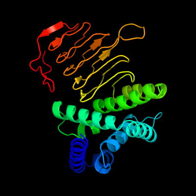
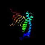
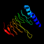
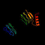
| 1 | d1t3da_
|
|
 |
100.0 |
97 |
Fold:Single-stranded left-handed beta-helix
Superfamily:Trimeric LpxA-like enzymes
Family:Serine acetyltransferase |
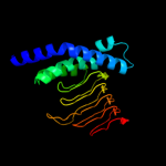
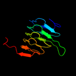
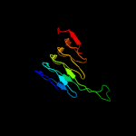
| 2 | c1t3dB_
|
|
 |
100.0 |
97 |
PDB header:transferase
Chain: B: PDB Molecule:serine acetyltransferase;
PDBTitle: crystal structure of serine acetyltransferase from e.coli at 2.2a
|
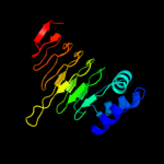
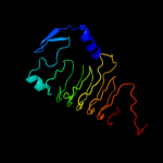
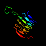
| 3 | c3mc4A_
|
|
 |
100.0 |
54 |
PDB header:transferase
Chain: A: PDB Molecule:ww/rsp5/wwp domain:bacterial transferase
PDBTitle: crystal structure of ww/rsp5/wwp domain: bacterial2 transferase hexapeptide repeat: serine o-acetyltransferase3 from brucella melitensis
|
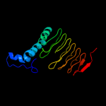
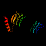
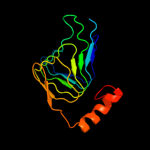
| 4 | d1ssqa_
|
|
 |
100.0 |
75 |
Fold:Single-stranded left-handed beta-helix
Superfamily:Trimeric LpxA-like enzymes
Family:Serine acetyltransferase |
| 5 | c3f1xA_
|
|
 |
100.0 |
29 |
PDB header:transferase
Chain: A: PDB Molecule:serine acetyltransferase;
PDBTitle: three dimensional structure of the serine acetyltransferase from2 bacteroides vulgatus, northeast structural genomics consortium target3 bvr62.
|
| 6 | c3q1xA_
|
|
 |
100.0 |
37 |
PDB header:transferase
Chain: A: PDB Molecule:serine acetyltransferase;
PDBTitle: crystal structure of entamoeba histolytica serine acetyltransferase 12 in complex with l-serine
|
| 7 | d1ocxa_
|
|
 |
99.9 |
19 |
Fold:Single-stranded left-handed beta-helix
Superfamily:Trimeric LpxA-like enzymes
Family:Galactoside acetyltransferase-like |
| 8 | c3ectA_
|
|
 |
99.9 |
22 |
PDB header:transferase
Chain: A: PDB Molecule:hexapeptide-repeat containing-acetyltransferase;
PDBTitle: crystal structure of the hexapeptide-repeat containing-2 acetyltransferase vca0836 from vibrio cholerae
|
| 9 | c2ic7A_
|
|
 |
99.9 |
19 |
PDB header:transferase
Chain: A: PDB Molecule:maltose transacetylase;
PDBTitle: crystal structure of maltose transacetylase from2 geobacillus kaustophilus
|
| 10 | d1krra_
|
|
 |
99.9 |
30 |
Fold:Single-stranded left-handed beta-helix
Superfamily:Trimeric LpxA-like enzymes
Family:Galactoside acetyltransferase-like |
| 11 | d3tdta_
|
|
 |
99.9 |
20 |
Fold:Single-stranded left-handed beta-helix
Superfamily:Trimeric LpxA-like enzymes
Family:Tetrahydrodipicolinate-N-succinlytransferase, THDP-succinlytransferase, DapD |
| 12 | c3r0sA_
|
|
 |
99.9 |
26 |
PDB header:transferase
Chain: A: PDB Molecule:acyl-[acyl-carrier-protein]--udp-n-acetylglucosamine o-
PDBTitle: udp-n-acetylglucosamine acyltransferase from campylobacter jejuni
|
| 13 | c3i3aC_
|
|
 |
99.9 |
25 |
PDB header:transferase
Chain: C: PDB Molecule:acyl-[acyl-carrier-protein]--udp-n-
PDBTitle: structural basis for the sugar nucleotide and acyl chain2 selectivity of leptospira interrogans lpxa
|
| 14 | c3srtB_
|
|
 |
99.9 |
26 |
PDB header:transferase
Chain: B: PDB Molecule:maltose o-acetyltransferase;
PDBTitle: the crystal structure of a maltose o-acetyltransferase from2 clostridium difficile 630
|
| 15 | c3fttA_
|
|
 |
99.9 |
23 |
PDB header:transferase
Chain: A: PDB Molecule:putative acetyltransferase sacol2570;
PDBTitle: crystal structure of the galactoside o-acetyltransferase2 from staphylococcus aureus
|
| 16 | d1mr7a_
|
|
 |
99.9 |
24 |
Fold:Single-stranded left-handed beta-helix
Superfamily:Trimeric LpxA-like enzymes
Family:Galactoside acetyltransferase-like |
| 17 | d1j2za_
|
|
 |
99.9 |
33 |
Fold:Single-stranded left-handed beta-helix
Superfamily:Trimeric LpxA-like enzymes
Family:UDP N-acetylglucosamine acyltransferase |
| 18 | d1xata_
|
|
 |
99.9 |
26 |
Fold:Single-stranded left-handed beta-helix
Superfamily:Trimeric LpxA-like enzymes
Family:Galactoside acetyltransferase-like |
| 19 | d2jf2a1
|
|
 |
99.9 |
27 |
Fold:Single-stranded left-handed beta-helix
Superfamily:Trimeric LpxA-like enzymes
Family:UDP N-acetylglucosamine acyltransferase |
| 20 | d3bswa1
|
|
 |
99.9 |
21 |
Fold:Single-stranded left-handed beta-helix
Superfamily:Trimeric LpxA-like enzymes
Family:PglD-like |
| 21 | c3jqyB_ |
|
not modelled |
99.9 |
30 |
PDB header:transferase
Chain: B: PDB Molecule:polysialic acid o-acetyltransferase;
PDBTitle: crystal strucutre of the polysia specific acetyltransferase neuo
|
| 22 | c3cj8B_ |
|
not modelled |
99.9 |
34 |
PDB header:transferase
Chain: B: PDB Molecule:2,3,4,5-tetrahydropyridine-2-carboxylate n-
PDBTitle: crystal structure of 2,3,4,5-tetrahydropyridine-2-carboxylate n-2 succinyltransferase from enterococcus faecalis v583
|
| 23 | c2iu9C_ |
|
not modelled |
99.9 |
23 |
PDB header:transferase
Chain: C: PDB Molecule:udp-3-o-[3-hydroxymyristoyl] glucosamine
PDBTitle: chlamydia trachomatis lpxd with 100mm udpglcnac (complex ii)
|
| 24 | c3fsbB_ |
|
not modelled |
99.9 |
27 |
PDB header:transferase
Chain: B: PDB Molecule:qdtc;
PDBTitle: crystal structure of qdtc, the dtdp-3-amino-3,6-dideoxy-d-2 glucose n-acetyl transferase from thermoanaerobacterium3 thermosaccharolyticum in complex with coa and dtdp-3-amino-4 quinovose
|
| 25 | c3eevC_ |
|
not modelled |
99.8 |
22 |
PDB header:transferase
Chain: C: PDB Molecule:chloramphenicol acetyltransferase;
PDBTitle: crystal structure of chloramphenicol acetyltransferase vca0300 from2 vibrio cholerae o1 biovar eltor
|
| 26 | c3pmoA_ |
|
not modelled |
99.8 |
16 |
PDB header:transferase
Chain: A: PDB Molecule:udp-3-o-[3-hydroxymyristoyl] glucosamine n-acyltransferase;
PDBTitle: the structure of lpxd from pseudomonas aeruginosa at 1.3 a resolution
|
| 27 | c3mqhD_ |
|
not modelled |
99.8 |
34 |
PDB header:transferase
Chain: D: PDB Molecule:lipopolysaccharides biosynthesis acetyltransferase;
PDBTitle: crystal structure of the 3-n-acetyl transferase wlbb from bordetella2 petrii in complex with coa and udp-3-amino-2-acetamido-2,3-dideoxy3 glucuronic acid
|
| 28 | c3eg4A_ |
|
not modelled |
99.8 |
20 |
PDB header:transferase
Chain: A: PDB Molecule:2,3,4,5-tetrahydropyridine-2,6-dicarboxylate n-
PDBTitle: crystal structure of 2,3,4,5-tetrahydropyridine-2-2 carboxylate n-succinyltransferase from brucella melitensis3 biovar abortus 2308
|
| 29 | d1g97a1 |
|
not modelled |
99.8 |
19 |
Fold:Single-stranded left-handed beta-helix
Superfamily:Trimeric LpxA-like enzymes
Family:GlmU C-terminal domain-like |
| 30 | c3eh0C_ |
|
not modelled |
99.8 |
22 |
PDB header:transferase
Chain: C: PDB Molecule:udp-3-o-[3-hydroxymyristoyl] glucosamine n-
PDBTitle: crystal structure of lpxd from escherichia coli
|
| 31 | c2wlgA_ |
|
not modelled |
99.8 |
19 |
PDB header:transferase
Chain: A: PDB Molecule:polysialic acid o-acetyltransferase;
PDBTitle: crystallographic analysis of the polysialic acid o-2 acetyltransferase oatwy
|
| 32 | c1hm8A_ |
|
not modelled |
99.8 |
19 |
PDB header:transferase
Chain: A: PDB Molecule:udp-n-acetylglucosamine-1-phosphate uridyltransferase;
PDBTitle: crystal structure of s.pneumoniae n-acetylglucosamine-1-phosphate2 uridyltransferase, glmu, bound to acetyl coenzyme a
|
| 33 | d2oi6a1 |
|
not modelled |
99.7 |
21 |
Fold:Single-stranded left-handed beta-helix
Superfamily:Trimeric LpxA-like enzymes
Family:GlmU C-terminal domain-like |
| 34 | d1v3wa_ |
|
not modelled |
99.7 |
26 |
Fold:Single-stranded left-handed beta-helix
Superfamily:Trimeric LpxA-like enzymes
Family:gamma-carbonic anhydrase-like |
| 35 | c2oi6A_ |
|
not modelled |
99.7 |
19 |
PDB header:transferase
Chain: A: PDB Molecule:bifunctional protein glmu;
PDBTitle: e. coli glmu- complex with udp-glcnac, coa and glcn-1-po4
|
| 36 | c2v0hA_ |
|
not modelled |
99.7 |
23 |
PDB header:transferase
Chain: A: PDB Molecule:bifunctional protein glmu;
PDBTitle: characterization of substrate binding and catalysis of the2 potential antibacterial target n-acetylglucosamine-1-3 phosphate uridyltransferase (glmu)
|
| 37 | c3ixcA_ |
|
not modelled |
99.7 |
18 |
PDB header:transferase
Chain: A: PDB Molecule:hexapeptide transferase family protein;
PDBTitle: crystal structure of hexapeptide transferase family protein from2 anaplasma phagocytophilum
|
| 38 | c3r3rA_ |
|
not modelled |
99.7 |
21 |
PDB header:transferase
Chain: A: PDB Molecule:ferripyochelin binding protein;
PDBTitle: structure of the yrda ferripyochelin binding protein from salmonella2 enterica
|
| 39 | d1xhda_ |
|
not modelled |
99.7 |
26 |
Fold:Single-stranded left-handed beta-helix
Superfamily:Trimeric LpxA-like enzymes
Family:gamma-carbonic anhydrase-like |
| 40 | c3fsyC_ |
|
not modelled |
99.6 |
20 |
PDB header:transferase
Chain: C: PDB Molecule:tetrahydrodipicolinate n-succinyltransferase;
PDBTitle: structure of tetrahydrodipicolinate n-succinyltransferase2 (rv1201c;dapd) in complex with succinyl-coa from mycobacterium3 tuberculosis
|
| 41 | c3r1wA_ |
|
not modelled |
99.6 |
24 |
PDB header:lyase
Chain: A: PDB Molecule:carbonic anhydrase;
PDBTitle: crystal structure of a carbonic anhydrase from a crude oil degrading2 psychrophilic library
|
| 42 | c2ggqA_ |
|
not modelled |
99.5 |
20 |
PDB header:transferase
Chain: A: PDB Molecule:401aa long hypothetical glucose-1-phosphate
PDBTitle: complex of hypothetical glucose-1-phosphate thymidylyltransferase from2 sulfolobus tokodaii
|
| 43 | c3c8vA_ |
|
not modelled |
99.5 |
13 |
PDB header:transferase
Chain: A: PDB Molecule:putative acetyltransferase;
PDBTitle: crystal structure of putative acetyltransferase (yp_390128.1) from2 desulfovibrio desulfuricans g20 at 2.28 a resolution
|
| 44 | c2rijA_ |
|
not modelled |
99.3 |
15 |
PDB header:transferase
Chain: A: PDB Molecule:putative 2,3,4,5-tetrahydropyridine-2-carboxylate n-
PDBTitle: crystal structure of a putative 2,3,4,5-tetrahydropyridine-2-2 carboxylate n-succinyltransferase (cj1605c, dapd) from campylobacter3 jejuni at 1.90 a resolution
|
| 45 | c1qreA_ |
|
not modelled |
99.2 |
12 |
PDB header:lyase
Chain: A: PDB Molecule:carbonic anhydrase;
PDBTitle: a closer look at the active site of gamma-carbonic anhydrases: high2 resolution crystallographic studies of the carbonic anhydrase from3 methanosarcina thermophila
|
| 46 | d1qrea_ |
|
not modelled |
99.2 |
12 |
Fold:Single-stranded left-handed beta-helix
Superfamily:Trimeric LpxA-like enzymes
Family:gamma-carbonic anhydrase-like |
| 47 | d2f9ca1 |
|
not modelled |
99.0 |
17 |
Fold:Single-stranded left-handed beta-helix
Superfamily:Trimeric LpxA-like enzymes
Family:YdcK-like |
| 48 | c2qkxA_ |
|
not modelled |
98.8 |
16 |
PDB header:transferase
Chain: A: PDB Molecule:bifunctional protein glmu;
PDBTitle: n-acetyl glucosamine 1-phosphate uridyltransferase from mycobacterium2 tuberculosis complex with n-acetyl glucosamine 1-phosphate
|
| 49 | d1fxja1 |
|
not modelled |
98.8 |
14 |
Fold:Single-stranded left-handed beta-helix
Superfamily:Trimeric LpxA-like enzymes
Family:GlmU C-terminal domain-like |
| 50 | c3kwdA_ |
|
not modelled |
98.8 |
22 |
PDB header:lyase, protein binding, photosynthesis
Chain: A: PDB Molecule:carbon dioxide concentrating mechanism protein;
PDBTitle: inactive truncation of the beta-carboxysomal gamma-carbonic anhydrase,2 ccmm, form 1
|
| 51 | d1yp2a1 |
|
not modelled |
98.8 |
14 |
Fold:Single-stranded left-handed beta-helix
Superfamily:Trimeric LpxA-like enzymes
Family:GlmU C-terminal domain-like |
| 52 | c1yp3C_ |
|
not modelled |
98.7 |
16 |
PDB header:transferase
Chain: C: PDB Molecule:glucose-1-phosphate adenylyltransferase small
PDBTitle: crystal structure of potato tuber adp-glucose2 pyrophosphorylase in complex with atp
|
| 53 | c3d98A_ |
|
not modelled |
98.7 |
16 |
PDB header:transferase
Chain: A: PDB Molecule:bifunctional protein glmu;
PDBTitle: crystal structure of glmu from mycobacterium tuberculosis, ligand-free2 form
|
| 54 | c3brkX_ |
|
not modelled |
98.6 |
21 |
PDB header:transferase
Chain: X: PDB Molecule:glucose-1-phosphate adenylyltransferase;
PDBTitle: crystal structure of adp-glucose pyrophosphorylase from2 agrobacterium tumefaciens
|
| 55 | c1fwyA_ |
|
not modelled |
98.5 |
11 |
PDB header:transferase
Chain: A: PDB Molecule:udp-n-acetylglucosamine pyrophosphorylase;
PDBTitle: crystal structure of n-acetylglucosamine 1-phosphate2 uridyltransferase bound to udp-glcnac
|
| 56 | d2icya1 |
|
not modelled |
19.4 |
26 |
Fold:Single-stranded left-handed beta-helix
Superfamily:Trimeric LpxA-like enzymes
Family:GlmU C-terminal domain-like |
| 57 | d1o57a2 |
|
not modelled |
18.4 |
20 |
Fold:PRTase-like
Superfamily:PRTase-like
Family:Phosphoribosyltransferases (PRTases) |
| 58 | c1b0nB_ |
|
not modelled |
17.0 |
31 |
PDB header:transcription regulator
Chain: B: PDB Molecule:protein (sini protein);
PDBTitle: sinr protein/sini protein complex
|
| 59 | d1b0nb_ |
|
not modelled |
17.0 |
31 |
Fold:Dimerisation interlock
Superfamily:SinR repressor dimerisation domain-like
Family:SinR repressor dimerisation domain-like |
| 60 | c2ioaA_ |
|
not modelled |
15.7 |
13 |
PDB header:ligase, hydrolase
Chain: A: PDB Molecule:bifunctional glutathionylspermidine
PDBTitle: e. coli bifunctional glutathionylspermidine2 synthetase/amidase incomplex with mg2+ and adp and3 phosphinate inhibitor
|
| 61 | c3qxyA_ |
|
not modelled |
15.4 |
25 |
PDB header:transferase
Chain: A: PDB Molecule:n-lysine methyltransferase setd6;
PDBTitle: human setd6 in complex with rela lys310
|
| 62 | c3oqvA_ |
|
not modelled |
13.5 |
13 |
PDB header:protein binding
Chain: A: PDB Molecule:albc;
PDBTitle: albc, a cyclodipeptide synthase from streptomyces noursei
|
| 63 | d2ivxa2 |
|
not modelled |
9.0 |
19 |
Fold:Cyclin-like
Superfamily:Cyclin-like
Family:Cyclin |
| 64 | c3smtA_ |
|
not modelled |
9.0 |
11 |
PDB header:transferase
Chain: A: PDB Molecule:histone-lysine n-methyltransferase setd3;
PDBTitle: crystal structure of human set domain-containing protein3
|
| 65 | c3bitA_ |
|
not modelled |
8.7 |
14 |
PDB header:transcription
Chain: A: PDB Molecule:fact complex subunit spt16;
PDBTitle: crystal structure of yeast spt16 n-terminal domain
|
| 66 | c2vjjA_ |
|
not modelled |
8.5 |
11 |
PDB header:viral protein
Chain: A: PDB Molecule:tailspike protein;
PDBTitle: tailspike protein of e.coli bacteriophage hk620 in complex2 with hexasaccharide
|
| 67 | c2d7cD_ |
|
not modelled |
8.5 |
11 |
PDB header:protein transport
Chain: D: PDB Molecule:rab11 family-interacting protein 3;
PDBTitle: crystal structure of human rab11 in complex with fip3 rab-2 binding domain
|
| 68 | c3gt2A_ |
|
not modelled |
8.1 |
15 |
PDB header:unknown function
Chain: A: PDB Molecule:putative uncharacterized protein;
PDBTitle: crystal structure of the p60 domain from m. avium2 paratuberculosis antigen map1272c
|
| 69 | d3blhb1 |
|
not modelled |
7.8 |
18 |
Fold:Cyclin-like
Superfamily:Cyclin-like
Family:Cyclin |
| 70 | c2hv8D_ |
|
not modelled |
7.7 |
19 |
PDB header:protein transport
Chain: D: PDB Molecule:rab11 family-interacting protein 3;
PDBTitle: crystal structure of gtp-bound rab11 in complex with fip3
|
| 71 | c2z9fC_ |
|
not modelled |
7.5 |
21 |
PDB header:biosynthetic protein
Chain: C: PDB Molecule:cellulose synthase operon protein d;
PDBTitle: crystal structure of axcesd protein from acetobacter xylinum
|
| 72 | c2zkrq_ |
|
not modelled |
6.9 |
19 |
PDB header:ribosomal protein/rna
Chain: Q: PDB Molecule:rna expansion segment es31 part ii;
PDBTitle: structure of a mammalian ribosomal 60s subunit within an2 80s complex obtained by docking homology models of the rna3 and proteins into an 8.7 a cryo-em map
|
| 73 | c3oo2A_ |
|
not modelled |
6.6 |
15 |
PDB header:isomerase
Chain: A: PDB Molecule:alanine racemase 1;
PDBTitle: 2.37 angstrom resolution crystal structure of an alanine racemase2 (alr) from staphylococcus aureus subsp. aureus col
|
| 74 | c2zuuA_ |
|
not modelled |
6.5 |
8 |
PDB header:transferase
Chain: A: PDB Molecule:lacto-n-biose phosphorylase;
PDBTitle: crystal structure of galacto-n-biose/lacto-n-biose i phosphorylase in2 complex with glcnac
|
| 75 | d1ex4a1 |
|
not modelled |
6.4 |
27 |
Fold:SH3-like barrel
Superfamily:DNA-binding domain of retroviral integrase
Family:DNA-binding domain of retroviral integrase |
| 76 | c4a1aP_ |
|
not modelled |
6.2 |
16 |
PDB header:ribosome
Chain: P: PDB Molecule:60s ribosomal protein l21;
PDBTitle: t.thermophila 60s ribosomal subunit in complex with2 initiation factor 6. this file contains 5s rrna,3 5.8s rrna and proteins of molecule 3.
|
| 77 | d1ihwa_ |
|
not modelled |
5.9 |
27 |
Fold:SH3-like barrel
Superfamily:DNA-binding domain of retroviral integrase
Family:DNA-binding domain of retroviral integrase |
| 78 | c3iz5U_ |
|
not modelled |
5.8 |
19 |
PDB header:ribosome
Chain: U: PDB Molecule:60s ribosomal protein l21 (l21e);
PDBTitle: localization of the large subunit ribosomal proteins into a 5.5 a2 cryo-em map of triticum aestivum translating 80s ribosome
|
| 79 | c3izcU_ |
|
not modelled |
5.8 |
16 |
PDB header:ribosome
Chain: U: PDB Molecule:60s ribosomal protein rpl21 (l21e);
PDBTitle: localization of the large subunit ribosomal proteins into a 6.1 a2 cryo-em map of saccharomyces cerevisiae translating 80s ribosome
|
| 80 | d1hska2 |
|
not modelled |
5.8 |
22 |
Fold:Uridine diphospho-N-Acetylenolpyruvylglucosamine reductase, MurB, C-terminal domain
Superfamily:Uridine diphospho-N-Acetylenolpyruvylglucosamine reductase, MurB, C-terminal domain
Family:Uridine diphospho-N-Acetylenolpyruvylglucosamine reductase, MurB, C-terminal domain |
| 81 | c2x9qA_ |
|
not modelled |
5.6 |
15 |
PDB header:ligase
Chain: A: PDB Molecule:cyclodipeptide synthetase;
PDBTitle: structure of the mycobacterium tuberculosis protein, rv2275,2 demonstrates that cyclodipeptide synthetases are related3 to type i trna-synthetases.
|
| 82 | d1uxya2 |
|
not modelled |
5.3 |
26 |
Fold:Uridine diphospho-N-Acetylenolpyruvylglucosamine reductase, MurB, C-terminal domain
Superfamily:Uridine diphospho-N-Acetylenolpyruvylglucosamine reductase, MurB, C-terminal domain
Family:Uridine diphospho-N-Acetylenolpyruvylglucosamine reductase, MurB, C-terminal domain |
| 83 | c2cu2A_ |
|
not modelled |
5.3 |
21 |
PDB header:transferase
Chain: A: PDB Molecule:putative mannose-1-phosphate guanylyl transferase;
PDBTitle: crystal structure of mannose-1-phosphate geranyltransferase from2 thermus thermophilus hb8
|
| 84 | d1ugpa_ |
|
not modelled |
5.3 |
16 |
Fold:Nitrile hydratase alpha chain
Superfamily:Nitrile hydratase alpha chain
Family:Nitrile hydratase alpha chain |
| 85 | c3qwvA_ |
|
not modelled |
5.3 |
25 |
PDB header:transferase
Chain: A: PDB Molecule:set and mynd domain-containing protein 2;
PDBTitle: crystal structure of histone lysine methyltransferase smyd2 in complex2 with the cofactor product adohcy
|
| 86 | d2g46a1 |
|
not modelled |
5.2 |
8 |
Fold:beta-clip
Superfamily:SET domain
Family:Viral histone H3 Lysine 27 Methyltransferase |
| 87 | d2z1ca1 |
|
not modelled |
5.2 |
13 |
Fold:OB-fold
Superfamily:HupF/HypC-like
Family:HupF/HypC-like |
| 88 | c1ds5F_ |
|
not modelled |
5.2 |
33 |
PDB header:transferase
Chain: F: PDB Molecule:casein kinase, beta chain;
PDBTitle: dimeric crystal structure of the alpha subunit in complex2 with two beta peptides mimicking the architecture of the3 tetrameric protein kinase ck2 holoenzyme.
|
| 89 | d2ar0a1 |
|
not modelled |
5.1 |
14 |
Fold:S-adenosyl-L-methionine-dependent methyltransferases
Superfamily:S-adenosyl-L-methionine-dependent methyltransferases
Family:N-6 DNA Methylase-like |