| 1 | c2a5hC_
|
|
 |
100.0 |
32 |
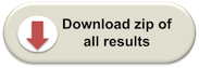
PDB header:isomerase
Chain: C: PDB Molecule:l-lysine 2,3-aminomutase;
PDBTitle: 2.1 angstrom x-ray crystal structure of lysine-2,3-aminomutase from2 clostridium subterminale sb4, with michaelis analog (l-alpha-lysine3 external aldimine form of pyridoxal-5'-phosphate).
|
| 2 | c3rfaA_
|
|
 |
99.9 |
15 |
PDB header:oxidoreductase
Chain: A: PDB Molecule:ribosomal rna large subunit methyltransferase n;
PDBTitle: x-ray structure of rlmn from escherichia coli in complex with s-2 adenosylmethionine
|
| 3 | d1tv8a_
|
|
 |
99.8 |
15 |
Fold:TIM beta/alpha-barrel
Superfamily:Radical SAM enzymes
Family:MoCo biosynthesis proteins |
| 4 | c2yx0A_
|
|
 |
99.7 |
14 |
PDB header:metal binding protein
Chain: A: PDB Molecule:radical sam enzyme;
PDBTitle: crystal structure of p. horikoshii tyw1
|
| 5 | c3c8fA_
|
|
 |
99.6 |
12 |
PDB header:oxidoreductase
Chain: A: PDB Molecule:pyruvate formate-lyase 1-activating enzyme;
PDBTitle: 4fe-4s-pyruvate formate-lyase activating enzyme with2 partially disordered adomet
|
| 6 | c3cixA_
|
|
 |
99.6 |
12 |
PDB header:adomet binding protein
Chain: A: PDB Molecule:fefe-hydrogenase maturase;
PDBTitle: x-ray structure of the [fefe]-hydrogenase maturase hyde from2 thermotoga maritima in complex with thiocyanate
|
| 7 | c3t7vA_
|
|
 |
99.5 |
9 |
PDB header:transferase
Chain: A: PDB Molecule:methylornithine synthase pylb;
PDBTitle: crystal structure of methylornithine synthase (pylb)
|
| 8 | c1r30A_
|
|
 |
99.5 |
15 |
PDB header:transferase
Chain: A: PDB Molecule:biotin synthase;
PDBTitle: the crystal structure of biotin synthase, an s-2 adenosylmethionine-dependent radical enzyme
|
| 9 | d1r30a_
|
|
 |
99.5 |
15 |
Fold:TIM beta/alpha-barrel
Superfamily:Radical SAM enzymes
Family:Biotin synthase |
| 10 | c2qgqF_
|
|
 |
99.5 |
12 |
PDB header:structural genomics, unknown function
Chain: F: PDB Molecule:protein tm_1862;
PDBTitle: crystal structure of tm_1862 from thermotoga maritima.2 northeast structural genomics consortium target vr77
|
| 11 | d1olta_
|
|
 |
99.4 |
12 |
Fold:TIM beta/alpha-barrel
Superfamily:Radical SAM enzymes
Family:Oxygen-independent coproporphyrinogen III oxidase HemN |
| 12 | c2z2uA_
|
|
 |
99.1 |
18 |
PDB header:metal binding protein
Chain: A: PDB Molecule:upf0026 protein mj0257;
PDBTitle: crystal structure of archaeal tyw1
|
| 13 | c3canA_
|
|
 |
99.0 |
15 |
PDB header:lyase activator
Chain: A: PDB Molecule:pyruvate-formate lyase-activating enzyme;
PDBTitle: crystal structure of a domain of pyruvate-formate lyase-activating2 enzyme from bacteroides vulgatus atcc 8482
|
| 14 | c2f06B_
|
|
 |
92.4 |
13 |
PDB header:structural genomics, unknown function
Chain: B: PDB Molecule:conserved hypothetical protein;
PDBTitle: crystal structure of protein bt0572 from bacteroides thetaiotaomicron
|
| 15 | d2nu7b1
|
|
 |
87.0 |
16 |
Fold:Flavodoxin-like
Superfamily:Succinyl-CoA synthetase domains
Family:Succinyl-CoA synthetase domains |
| 16 | c2rfgB_
|
|
 |
82.2 |
10 |
PDB header:lyase
Chain: B: PDB Molecule:dihydrodipicolinate synthase;
PDBTitle: crystal structure of dihydrodipicolinate synthase from hahella2 chejuensis at 1.5a resolution
|
| 17 | c2r8wB_
|
|
 |
79.0 |
13 |
PDB header:lyase
Chain: B: PDB Molecule:agr_c_1641p;
PDBTitle: the crystal structure of dihydrodipicolinate synthase (atu0899) from2 agrobacterium tumefaciens str. c58
|
| 18 | c3daqB_
|
|
 |
78.6 |
10 |
PDB header:lyase
Chain: B: PDB Molecule:dihydrodipicolinate synthase;
PDBTitle: crystal structure of dihydrodipicolinate synthase from methicillin-2 resistant staphylococcus aureus
|
| 19 | d1f74a_
|
|
 |
76.3 |
10 |
Fold:TIM beta/alpha-barrel
Superfamily:Aldolase
Family:Class I aldolase |
| 20 | c2vc6A_
|
|
 |
75.7 |
11 |
PDB header:lyase
Chain: A: PDB Molecule:dihydrodipicolinate synthase;
PDBTitle: structure of mosa from s. meliloti with pyruvate bound
|
| 21 | c2nu9E_ |
|
not modelled |
74.2 |
15 |
PDB header:ligase
Chain: E: PDB Molecule:succinyl-coa synthetase beta chain;
PDBTitle: c123at mutant of e. coli succinyl-coa synthetase2 orthorhombic crystal form
|
| 22 | c3lerA_ |
|
not modelled |
74.1 |
8 |
PDB header:lyase
Chain: A: PDB Molecule:dihydrodipicolinate synthase;
PDBTitle: crystal structure of dihydrodipicolinate synthase from2 campylobacter jejuni subsp. jejuni nctc 11168
|
| 23 | d1hl2a_ |
|
not modelled |
73.5 |
10 |
Fold:TIM beta/alpha-barrel
Superfamily:Aldolase
Family:Class I aldolase |
| 24 | c3lciA_ |
|
not modelled |
73.4 |
13 |
PDB header:lyase
Chain: A: PDB Molecule:n-acetylneuraminate lyase;
PDBTitle: the d-sialic acid aldolase mutant v251w
|
| 25 | c3na8A_ |
|
not modelled |
72.5 |
10 |
PDB header:lyase
Chain: A: PDB Molecule:putative dihydrodipicolinate synthetase;
PDBTitle: crystal structure of a putative dihydrodipicolinate synthetase from2 pseudomonas aeruginosa
|
| 26 | c3si9B_ |
|
not modelled |
71.1 |
7 |
PDB header:lyase
Chain: B: PDB Molecule:dihydrodipicolinate synthase;
PDBTitle: crystal structure of dihydrodipicolinate synthase from bartonella2 henselae
|
| 27 | c3e96B_ |
|
not modelled |
70.4 |
10 |
PDB header:lyase
Chain: B: PDB Molecule:dihydrodipicolinate synthase;
PDBTitle: crystal structure of dihydrodipicolinate synthase from2 bacillus clausii
|
| 28 | c3h5dD_ |
|
not modelled |
70.3 |
13 |
PDB header:lyase
Chain: D: PDB Molecule:dihydrodipicolinate synthase;
PDBTitle: dihydrodipicolinate synthase from drug-resistant streptococcus2 pneumoniae
|
| 29 | c2yxgD_ |
|
not modelled |
69.1 |
9 |
PDB header:lyase
Chain: D: PDB Molecule:dihydrodipicolinate synthase;
PDBTitle: crystal structure of dihyrodipicolinate synthase (dapa)
|
| 30 | c3fkkA_ |
|
not modelled |
68.2 |
10 |
PDB header:lyase
Chain: A: PDB Molecule:l-2-keto-3-deoxyarabonate dehydratase;
PDBTitle: structure of l-2-keto-3-deoxyarabonate dehydratase
|
| 31 | c3dz1A_ |
|
not modelled |
67.7 |
10 |
PDB header:lyase
Chain: A: PDB Molecule:dihydrodipicolinate synthase;
PDBTitle: crystal structure of dihydrodipicolinate synthase from2 rhodopseudomonas palustris at 1.87a resolution
|
| 32 | c3n2xB_ |
|
not modelled |
66.9 |
13 |
PDB header:lyase
Chain: B: PDB Molecule:uncharacterized protein yage;
PDBTitle: crystal structure of yage, a prophage protein belonging to the2 dihydrodipicolinic acid synthase family from e. coli k12 in complex3 with pyruvate
|
| 33 | c3eb2A_ |
|
not modelled |
64.6 |
9 |
PDB header:lyase
Chain: A: PDB Molecule:putative dihydrodipicolinate synthetase;
PDBTitle: crystal structure of dihydrodipicolinate synthase from2 rhodopseudomonas palustris at 2.0a resolution
|
| 34 | c3bi8A_ |
|
not modelled |
63.6 |
13 |
PDB header:lyase
Chain: A: PDB Molecule:dihydrodipicolinate synthase;
PDBTitle: structure of dihydrodipicolinate synthase from clostridium2 botulinum
|
| 35 | c2ehhE_ |
|
not modelled |
63.0 |
11 |
PDB header:lyase
Chain: E: PDB Molecule:dihydrodipicolinate synthase;
PDBTitle: crystal structure of dihydrodipicolinate synthase from2 aquifex aeolicus
|
| 36 | c2kboA_ |
|
not modelled |
61.7 |
8 |
PDB header:hydrolase
Chain: A: PDB Molecule:dna dc->du-editing enzyme apobec-3g;
PDBTitle: structure, interaction, and real-time monitoring of the2 enzymatic reaction of wild type apobec3g
|
| 37 | d2hiya1 |
|
not modelled |
61.1 |
22 |
Fold:SP0830-like
Superfamily:SP0830-like
Family:SP0830-like |
| 38 | c3pueA_ |
|
not modelled |
59.5 |
11 |
PDB header:lyase
Chain: A: PDB Molecule:dihydrodipicolinate synthase;
PDBTitle: crystal structure of the complex of dhydrodipicolinate synthase from2 acinetobacter baumannii with lysine at 2.6a resolution
|
| 39 | c3g0sA_ |
|
not modelled |
58.8 |
12 |
PDB header:lyase
Chain: A: PDB Molecule:dihydrodipicolinate synthase;
PDBTitle: dihydrodipicolinate synthase from salmonella typhimurium lt2
|
| 40 | c3cprB_ |
|
not modelled |
57.2 |
15 |
PDB header:lyase
Chain: B: PDB Molecule:dihydrodipicolinate synthetase;
PDBTitle: the crystal structure of corynebacterium glutamicum2 dihydrodipicolinate synthase to 2.2 a resolution
|
| 41 | d2a6na1 |
|
not modelled |
56.4 |
10 |
Fold:TIM beta/alpha-barrel
Superfamily:Aldolase
Family:Class I aldolase |
| 42 | c3ewbX_ |
|
not modelled |
56.2 |
15 |
PDB header:transferase
Chain: X: PDB Molecule:2-isopropylmalate synthase;
PDBTitle: crystal structure of n-terminal domain of putative 2-2 isopropylmalate synthase from listeria monocytogenes
|
| 43 | d1m1ha2 |
|
not modelled |
55.6 |
23 |
Fold:Ferredoxin-like
Superfamily:N-utilization substance G protein NusG, N-terminal domain
Family:N-utilization substance G protein NusG, N-terminal domain |
| 44 | c2fmoA_ |
|
not modelled |
55.4 |
17 |
PDB header:oxidoreductase
Chain: A: PDB Molecule:5,10-methylenetetrahydrofolate reductase;
PDBTitle: ala177val mutant of e. coli methylenetetrahydrofolate2 reductase
|
| 45 | c3noeA_ |
|
not modelled |
54.1 |
11 |
PDB header:lyase
Chain: A: PDB Molecule:dihydrodipicolinate synthase;
PDBTitle: crystal structure of dihydrodipicolinate synthase from pseudomonas2 aeruginosa
|
| 46 | d1eucb1 |
|
not modelled |
54.0 |
15 |
Fold:Flavodoxin-like
Superfamily:Succinyl-CoA synthetase domains
Family:Succinyl-CoA synthetase domains |
| 47 | c2v9dB_ |
|
not modelled |
49.0 |
12 |
PDB header:lyase
Chain: B: PDB Molecule:yage;
PDBTitle: crystal structure of yage, a prophage protein belonging to2 the dihydrodipicolinic acid synthase family from e. coli3 k12
|
| 48 | c3lkeA_ |
|
not modelled |
48.9 |
7 |
PDB header:lyase
Chain: A: PDB Molecule:enoyl-coa hydratase;
PDBTitle: crystal structure of enoyl-coa hydratase from bacillus2 halodurans
|
| 49 | d1qwga_ |
|
not modelled |
47.6 |
14 |
Fold:TIM beta/alpha-barrel
Superfamily:(2r)-phospho-3-sulfolactate synthase ComA
Family:(2r)-phospho-3-sulfolactate synthase ComA |
| 50 | c3b4uB_ |
|
not modelled |
47.2 |
10 |
PDB header:lyase
Chain: B: PDB Molecule:dihydrodipicolinate synthase;
PDBTitle: crystal structure of dihydrodipicolinate synthase from agrobacterium2 tumefaciens str. c58
|
| 51 | c1eucB_ |
|
not modelled |
46.2 |
19 |
PDB header:ligase
Chain: B: PDB Molecule:succinyl-coa synthetase, beta chain;
PDBTitle: crystal structure of dephosphorylated pig heart, gtp-2 specific succinyl-coa synthetase
|
| 52 | c2pg8C_ |
|
not modelled |
46.0 |
21 |
PDB header:ligand binding protein
Chain: C: PDB Molecule:dpgc;
PDBTitle: crystal structure of r254k mutanat of dpgc with bound substrate analog
|
| 53 | c1zcoA_ |
|
not modelled |
44.2 |
12 |
PDB header:lyase
Chain: A: PDB Molecule:2-dehydro-3-deoxyphosphoheptonate aldolase;
PDBTitle: crystal structure of pyrococcus furiosus 3-deoxy-d-arabino-2 heptulosonate 7-phosphate synthase
|
| 54 | c3fluD_ |
|
not modelled |
43.7 |
10 |
PDB header:lyase
Chain: D: PDB Molecule:dihydrodipicolinate synthase;
PDBTitle: crystal structure of dihydrodipicolinate synthase from the pathogen2 neisseria meningitidis
|
| 55 | d1o5ka_ |
|
not modelled |
43.7 |
13 |
Fold:TIM beta/alpha-barrel
Superfamily:Aldolase
Family:Class I aldolase |
| 56 | c3c52B_ |
|
not modelled |
43.4 |
10 |
PDB header:lyase
Chain: B: PDB Molecule:fructose-bisphosphate aldolase;
PDBTitle: class ii fructose-1,6-bisphosphate aldolase from2 helicobacter pylori in complex with3 phosphoglycolohydroxamic acid, a competitive inhibitor
|
| 57 | c2nx9B_ |
|
not modelled |
42.1 |
14 |
PDB header:lyase
Chain: B: PDB Molecule:oxaloacetate decarboxylase 2, subunit alpha;
PDBTitle: crystal structure of the carboxyltransferase domain of the2 oxaloacetate decarboxylase na+ pump from vibrio cholerae
|
| 58 | c3ndcB_ |
|
not modelled |
41.1 |
23 |
PDB header:transferase
Chain: B: PDB Molecule:precorrin-4 c(11)-methyltransferase;
PDBTitle: crystal structure of precorrin-4 c11-methyltransferase from2 rhodobacter capsulatus
|
| 59 | c3kwpA_ |
|
not modelled |
40.6 |
10 |
PDB header:transferase
Chain: A: PDB Molecule:predicted methyltransferase;
PDBTitle: crystal structure of putative methyltransferase from lactobacillus2 brevis
|
| 60 | c2nytB_ |
|
not modelled |
40.5 |
13 |
PDB header:hydrolase
Chain: B: PDB Molecule:probable c->u-editing enzyme apobec-2;
PDBTitle: the apobec2 crystal structure and functional implications2 for aid
|
| 61 | c2zf8A_ |
|
not modelled |
40.1 |
11 |
PDB header:structural protein
Chain: A: PDB Molecule:component of sodium-driven polar flagellar motor;
PDBTitle: crystal structure of moty
|
| 62 | c3d0cB_ |
|
not modelled |
40.0 |
10 |
PDB header:lyase
Chain: B: PDB Molecule:dihydrodipicolinate synthase;
PDBTitle: crystal structure of dihydrodipicolinate synthase from2 oceanobacillus iheyensis at 1.9 a resolution
|
| 63 | d1va0a1 |
|
not modelled |
39.9 |
23 |
Fold:Tetrapyrrole methylase
Superfamily:Tetrapyrrole methylase
Family:Tetrapyrrole methylase |
| 64 | d1xxxa1 |
|
not modelled |
38.7 |
14 |
Fold:TIM beta/alpha-barrel
Superfamily:Aldolase
Family:Class I aldolase |
| 65 | d1nvma2 |
|
not modelled |
35.9 |
10 |
Fold:TIM beta/alpha-barrel
Superfamily:Aldolase
Family:HMGL-like |
| 66 | c1vs1B_ |
|
not modelled |
35.8 |
10 |
PDB header:transferase
Chain: B: PDB Molecule:3-deoxy-7-phosphoheptulonate synthase;
PDBTitle: crystal structure of 3-deoxy-d-arabino-heptulosonate-7-2 phosphate synthase (dahp synthase) from aeropyrum pernix3 in complex with mn2+ and pep
|
| 67 | d1xkya1 |
|
not modelled |
35.6 |
10 |
Fold:TIM beta/alpha-barrel
Superfamily:Aldolase
Family:Class I aldolase |
| 68 | c2r94B_ |
|
not modelled |
35.4 |
10 |
PDB header:lyase
Chain: B: PDB Molecule:2-keto-3-deoxy-(6-phospho-)gluconate aldolase;
PDBTitle: crystal structure of kd(p)ga from t.tenax
|
| 69 | c3rrvC_ |
|
not modelled |
34.1 |
7 |
PDB header:isomerase
Chain: C: PDB Molecule:enoyl-coa hydratase/isomerase;
PDBTitle: crystal structure of an enoyl-coa hydratase/isomerase from2 mycobacterium paratuberculosis
|
| 70 | c3nvtA_ |
|
not modelled |
33.3 |
11 |
PDB header:transferase/isomerase
Chain: A: PDB Molecule:3-deoxy-d-arabino-heptulosonate 7-phosphate synthase;
PDBTitle: 1.95 angstrom crystal structure of a bifunctional 3-deoxy-7-2 phosphoheptulonate synthase/chorismate mutase (aroa) from listeria3 monocytogenes egd-e
|
| 71 | c2nuxB_ |
|
not modelled |
32.6 |
12 |
PDB header:lyase
Chain: B: PDB Molecule:2-keto-3-deoxygluconate/2-keto-3-deoxy-6-phospho gluconate
PDBTitle: 2-keto-3-deoxygluconate aldolase from sulfolobus acidocaldarius,2 native structure in p6522 at 2.5 a resolution
|
| 72 | c1xuzA_ |
|
not modelled |
32.6 |
16 |
PDB header:biosynthetic protein
Chain: A: PDB Molecule:polysialic acid capsule biosynthesis protein siac;
PDBTitle: crystal structure analysis of sialic acid synthase (neub)from2 neisseria meningitidis, bound to mn2+, phosphoenolpyruvate, and n-3 acetyl mannosaminitol
|
| 73 | c2higA_ |
|
not modelled |
31.9 |
13 |
PDB header:transferase
Chain: A: PDB Molecule:6-phospho-1-fructokinase;
PDBTitle: crystal structure of phosphofructokinase apoenzyme from trypanosoma2 brucei.
|
| 74 | d2deka1 |
|
not modelled |
31.5 |
21 |
Fold:Tetrapyrrole methylase
Superfamily:Tetrapyrrole methylase
Family:Tetrapyrrole methylase |
| 75 | c2k06A_ |
|
not modelled |
31.1 |
19 |
PDB header:transcription
Chain: A: PDB Molecule:transcription antitermination protein nusg;
PDBTitle: solution structure of the aminoterminal domain of e. coli2 nusg
|
| 76 | d1g5ta_ |
|
not modelled |
30.7 |
16 |
Fold:P-loop containing nucleoside triphosphate hydrolases
Superfamily:P-loop containing nucleoside triphosphate hydrolases
Family:RecA protein-like (ATPase-domain) |
| 77 | d1xx4a_ |
|
not modelled |
30.5 |
15 |
Fold:ClpP/crotonase
Superfamily:ClpP/crotonase
Family:Crotonase-like |
| 78 | d1gvfa_ |
|
not modelled |
30.3 |
12 |
Fold:TIM beta/alpha-barrel
Superfamily:Aldolase
Family:Class II FBP aldolase |
| 79 | c3rsiA_ |
|
not modelled |
30.1 |
11 |
PDB header:isomerase
Chain: A: PDB Molecule:putative enoyl-coa hydratase/isomerase;
PDBTitle: the structure of a putative enoyl-coa hydratase/isomerase from2 mycobacterium abscessus atcc 19977 / dsm 44196
|
| 80 | d1cc5a_ |
|
not modelled |
29.6 |
19 |
Fold:Cytochrome c
Superfamily:Cytochrome c
Family:monodomain cytochrome c |
| 81 | c3gkbA_ |
|
not modelled |
28.8 |
30 |
PDB header:structural genomics, unknown function
Chain: A: PDB Molecule:putative enoyl-coa hydratase;
PDBTitle: crystal structure of a putative enoyl-coa hydratase from streptomyces2 avermitilis
|
| 82 | d1rjma_ |
|
not modelled |
28.6 |
15 |
Fold:ClpP/crotonase
Superfamily:ClpP/crotonase
Family:Crotonase-like |
| 83 | c2i55C_ |
|
not modelled |
28.5 |
19 |
PDB header:isomerase
Chain: C: PDB Molecule:phosphomannomutase;
PDBTitle: complex of glucose-1,6-bisphosphate with phosphomannomutase from2 leishmania mexicana
|
| 84 | d2cg4a2 |
|
not modelled |
28.2 |
17 |
Fold:Ferredoxin-like
Superfamily:Dimeric alpha+beta barrel
Family:Lrp/AsnC-like transcriptional regulator C-terminal domain |
| 85 | c2ppyE_ |
|
not modelled |
28.1 |
15 |
PDB header:lyase
Chain: E: PDB Molecule:enoyl-coa hydratase;
PDBTitle: crystal structure of enoyl-coa hydrates (gk_1992) from geobacillus2 kaustophilus hta426
|
| 86 | c1m1gB_ |
|
not modelled |
27.4 |
23 |
PDB header:transcription
Chain: B: PDB Molecule:transcription antitermination protein nusg;
PDBTitle: crystal structure of aquifex aeolicus n-utilization2 substance g (nusg), space group p2(1)
|
| 87 | c3bh1A_ |
|
not modelled |
27.4 |
17 |
PDB header:structural genomics, unknown function
Chain: A: PDB Molecule:upf0371 protein dip2346;
PDBTitle: crystal structure of protein dip2346 from corynebacterium diphtheriae
|
| 88 | d2ex2a1 |
|
not modelled |
27.3 |
16 |
Fold:beta-lactamase/transpeptidase-like
Superfamily:beta-lactamase/transpeptidase-like
Family:Dac-like |
| 89 | c2hmcA_ |
|
not modelled |
27.1 |
8 |
PDB header:structural genomics, unknown function
Chain: A: PDB Molecule:dihydrodipicolinate synthase;
PDBTitle: the crystal structure of dihydrodipicolinate synthase dapa from2 agrobacterium tumefaciens
|
| 90 | c2iswB_ |
|
not modelled |
26.8 |
10 |
PDB header:lyase
Chain: B: PDB Molecule:putative fructose-1,6-bisphosphate aldolase;
PDBTitle: structure of giardia fructose-1,6-biphosphate aldolase in2 complex with phosphoglycolohydroxamate
|
| 91 | c3a3eB_ |
|
not modelled |
26.8 |
35 |
PDB header:hydrolase
Chain: B: PDB Molecule:penicillin-binding protein 4;
PDBTitle: crystal structure of penicillin binding protein 4 (dacb)2 from haemophilus influenzae, complexed with novel beta-3 lactam (cmv)
|
| 92 | c3q94B_ |
|
not modelled |
26.5 |
8 |
PDB header:lyase
Chain: B: PDB Molecule:fructose-bisphosphate aldolase, class ii;
PDBTitle: the crystal structure of fructose 1,6-bisphosphate aldolase from2 bacillus anthracis str. 'ames ancestor'
|
| 93 | c3g64A_ |
|
not modelled |
26.0 |
7 |
PDB header:lyase
Chain: A: PDB Molecule:putative enoyl-coa hydratase;
PDBTitle: crystal structure of putative enoyl-coa hydratase from streptomyces2 coelicolor a3(2)
|
| 94 | c2djwF_ |
|
not modelled |
25.9 |
19 |
PDB header:unknown function
Chain: F: PDB Molecule:probable transcriptional regulator, asnc family;
PDBTitle: crystal structure of ttha0845 from thermus thermophilus hb8
|
| 95 | c2e0kA_ |
|
not modelled |
25.9 |
15 |
PDB header:transferase
Chain: A: PDB Molecule:precorrin-2 c20-methyltransferase;
PDBTitle: crystal structure of cbil, a methyltransferase involved in anaerobic2 vitamin b12 biosynthesis
|
| 96 | c3e2vA_ |
|
not modelled |
25.9 |
15 |
PDB header:hydrolase
Chain: A: PDB Molecule:3'-5'-exonuclease;
PDBTitle: crystal structure of an uncharacterized amidohydrolase from2 saccharomyces cerevisiae
|
| 97 | d1w3ia_ |
|
not modelled |
25.5 |
10 |
Fold:TIM beta/alpha-barrel
Superfamily:Aldolase
Family:Class I aldolase |
| 98 | d2ffja1 |
|
not modelled |
25.5 |
10 |
Fold:AF1104-like
Superfamily:AF1104-like
Family:AF1104-like |
| 99 | d1q52a_ |
|
not modelled |
25.4 |
15 |
Fold:ClpP/crotonase
Superfamily:ClpP/crotonase
Family:Crotonase-like |
| 100 | c3g8rA_ |
|
not modelled |
25.3 |
16 |
PDB header:biosynthetic protein
Chain: A: PDB Molecule:probable spore coat polysaccharide biosynthesis protein e;
PDBTitle: crystal structure of putative spore coat polysaccharide biosynthesis2 protein e from chromobacterium violaceum atcc 12472
|
| 101 | c3fkjA_ |
|
not modelled |
24.9 |
12 |
PDB header:isomerase
Chain: A: PDB Molecule:putative phosphosugar isomerases;
PDBTitle: crystal structure of a putative phosphosugar isomerase (stm_0572) from2 salmonella typhimurium lt2 at 2.12 a resolution
|
| 102 | c2ftpA_ |
|
not modelled |
24.8 |
13 |
PDB header:lyase
Chain: A: PDB Molecule:hydroxymethylglutaryl-coa lyase;
PDBTitle: crystal structure of hydroxymethylglutaryl-coa lyase from pseudomonas2 aeruginosa
|
| 103 | d1i1ga2 |
|
not modelled |
24.5 |
13 |
Fold:Ferredoxin-like
Superfamily:Dimeric alpha+beta barrel
Family:Lrp/AsnC-like transcriptional regulator C-terminal domain |
| 104 | c3h02F_ |
|
not modelled |
24.4 |
15 |
PDB header:lyase
Chain: F: PDB Molecule:naphthoate synthase;
PDBTitle: 2.15 angstrom resolution crystal structure of naphthoate synthase from2 salmonella typhimurium.
|
| 105 | d1wz8a1 |
|
not modelled |
24.3 |
16 |
Fold:ClpP/crotonase
Superfamily:ClpP/crotonase
Family:Crotonase-like |
| 106 | c3r0oA_ |
|
not modelled |
24.1 |
19 |
PDB header:lyase
Chain: A: PDB Molecule:carnitinyl-coa dehydratase;
PDBTitle: crystal structure of carnitinyl-coa hydratase from mycobacterium avium
|
| 107 | d1qt1a_ |
|
not modelled |
23.9 |
12 |
Fold:TIM beta/alpha-barrel
Superfamily:Xylose isomerase-like
Family:Xylose isomerase |
| 108 | c2w3pB_ |
|
not modelled |
23.2 |
15 |
PDB header:lyase
Chain: B: PDB Molecule:benzoyl-coa-dihydrodiol lyase;
PDBTitle: boxc crystal structure
|
| 109 | c3tqkA_ |
|
not modelled |
23.2 |
13 |
PDB header:transferase
Chain: A: PDB Molecule:phospho-2-dehydro-3-deoxyheptonate aldolase;
PDBTitle: structure of phospho-2-dehydro-3-deoxyheptonate aldolase from2 francisella tularensis schu s4
|
| 110 | d1mj3a_ |
|
not modelled |
22.5 |
22 |
Fold:ClpP/crotonase
Superfamily:ClpP/crotonase
Family:Crotonase-like |
| 111 | d1nz8a_ |
|
not modelled |
22.5 |
22 |
Fold:Ferredoxin-like
Superfamily:N-utilization substance G protein NusG, N-terminal domain
Family:N-utilization substance G protein NusG, N-terminal domain |
| 112 | c3hsiC_ |
|
not modelled |
22.5 |
8 |
PDB header:transferase
Chain: C: PDB Molecule:phosphatidylserine synthase;
PDBTitle: crystal structure of phosphatidylserine synthase haemophilus2 influenzae rd kw20
|
| 113 | c2iexA_ |
|
not modelled |
22.0 |
11 |
PDB header:lyase
Chain: A: PDB Molecule:dihydroxynapthoic acid synthetase;
PDBTitle: crystal structure of dihydroxynapthoic acid synthetase (gk2873) from2 geobacillus kaustophilus hta426
|
| 114 | c3ktsA_ |
|
not modelled |
22.0 |
14 |
PDB header:transcriptional regulator
Chain: A: PDB Molecule:glycerol uptake operon antiterminator regulatory protein;
PDBTitle: crystal structure of glycerol uptake operon antiterminator regulatory2 protein from listeria monocytogenes str. 4b f2365
|
| 115 | d3bexa2 |
|
not modelled |
21.9 |
8 |
Fold:Ribonuclease H-like motif
Superfamily:Actin-like ATPase domain
Family:CoaX-like |
| 116 | c3bleA_ |
|
not modelled |
21.1 |
8 |
PDB header:transferase
Chain: A: PDB Molecule:citramalate synthase from leptospira interrogans;
PDBTitle: crystal structure of the catalytic domain of licms in2 complexed with malonate
|
| 117 | c3o8nA_ |
|
not modelled |
21.0 |
9 |
PDB header:transferase
Chain: A: PDB Molecule:6-phosphofructokinase, muscle type;
PDBTitle: structure of phosphofructokinase from rabbit skeletal muscle
|
| 118 | c2vzvB_ |
|
not modelled |
20.5 |
11 |
PDB header:hydrolase
Chain: B: PDB Molecule:exo-beta-d-glucosaminidase;
PDBTitle: substrate complex of amycolatopsis orientalis exo-2 chitosanase csxa e541a with chitosan
|
| 119 | d1sg4a1 |
|
not modelled |
20.5 |
15 |
Fold:ClpP/crotonase
Superfamily:ClpP/crotonase
Family:Crotonase-like |
| 120 | c3sllC_ |
|
not modelled |
20.4 |
6 |
PDB header:isomerase
Chain: C: PDB Molecule:probable enoyl-coa hydratase/isomerase;
PDBTitle: crystal structure of a probable enoyl-coa hydratase/isomerase from2 mycobacterium abscessus
|