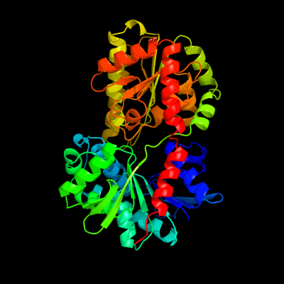
| 1 | d1uqta_
|
|
 |
100.0 |
100 |
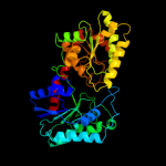
Fold:UDP-Glycosyltransferase/glycogen phosphorylase
Superfamily:UDP-Glycosyltransferase/glycogen phosphorylase
Family:Trehalose-6-phosphate synthase, OtsA |
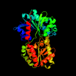
| 2 | c1uquB_
|
|
 |
100.0 |
100 |
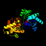
PDB header:synthase
Chain: B: PDB Molecule:alpha, alpha-trehalose-phosphate synthase;
PDBTitle: trehalose-6-phosphate from e. coli bound with udp-glucose.
|
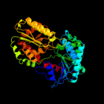
| 3 | c3s29C_
|
|
 |
100.0 |
13 |
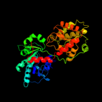
PDB header:transferase
Chain: C: PDB Molecule:sucrose synthase 1;
PDBTitle: the crystal structure of sucrose synthase-1 from arabidopsis thaliana2 and its functional implications.
|
| 4 | c3o3cD_
|
|
 |
100.0 |
13 |
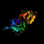
PDB header:transferase
Chain: D: PDB Molecule:glycogen [starch] synthase isoform 2;
PDBTitle: glycogen synthase basal state udp complex
|
| 5 | c3nb0A_
|
|
 |
100.0 |
13 |
PDB header:transferase
Chain: A: PDB Molecule:glycogen [starch] synthase isoform 2;
PDBTitle: glucose-6-phosphate activated form of yeast glycogen synthase
|
| 6 | c2r60A_
|
|
 |
100.0 |
13 |
PDB header:transferase
Chain: A: PDB Molecule:glycosyl transferase, group 1;
PDBTitle: structure of apo sucrose phosphate synthase (sps) of2 halothermothrix orenii
|
| 7 | c2x6rA_
|
|
 |
100.0 |
16 |
PDB header:isomerase
Chain: A: PDB Molecule:trehalose-synthase tret;
PDBTitle: crystal structure of trehalose synthase tret from p.2 horikoshi produced by soaking in trehalose
|
| 8 | c2xmpB_
|
|
 |
100.0 |
17 |
PDB header:sugar binding protein
Chain: B: PDB Molecule:trehalose-synthase tret;
PDBTitle: crystal structure of trehalose synthase tret mutant e326a2 from p.horishiki in complex with udp
|
| 9 | c3c4vB_
|
|
 |
100.0 |
16 |
PDB header:transferase
Chain: B: PDB Molecule:predicted glycosyltransferases;
PDBTitle: structure of the retaining glycosyltransferase msha:the2 first step in mycothiol biosynthesis. organism:3 corynebacterium glutamicum : complex with udp and 1l-ins-1-4 p.
|
| 10 | d2bisa1
|
|
 |
100.0 |
16 |
Fold:UDP-Glycosyltransferase/glycogen phosphorylase
Superfamily:UDP-Glycosyltransferase/glycogen phosphorylase
Family:Glycosyl transferases group 1 |
| 11 | c2qzsA_
|
|
 |
100.0 |
15 |
PDB header:transferase
Chain: A: PDB Molecule:glycogen synthase;
PDBTitle: crystal structure of wild-type e.coli gs in complex with adp2 and glucose(wtgsb)
|
| 12 | d1rzua_
|
|
 |
100.0 |
14 |
Fold:UDP-Glycosyltransferase/glycogen phosphorylase
Superfamily:UDP-Glycosyltransferase/glycogen phosphorylase
Family:Glycosyl transferases group 1 |
| 13 | c3okaA_
|
|
 |
100.0 |
17 |
PDB header:transferase
Chain: A: PDB Molecule:gdp-mannose-dependent alpha-(1-6)-phosphatidylinositol
PDBTitle: crystal structure of corynebacterium glutamicum pimb' in complex with2 gdp-man (triclinic crystal form)
|
| 14 | c3oy2A_
|
|
 |
100.0 |
15 |
PDB header:viral protein,transferase
Chain: A: PDB Molecule:glycosyltransferase b736l;
PDBTitle: crystal structure of a putative glycosyltransferase from paramecium2 bursaria chlorella virus ny2a
|
| 15 | c2x0dA_
|
|
 |
100.0 |
13 |
PDB header:transferase
Chain: A: PDB Molecule:wsaf;
PDBTitle: apo structure of wsaf
|
| 16 | c2gejA_
|
|
 |
100.0 |
17 |
PDB header:transferase
Chain: A: PDB Molecule:phosphatidylinositol mannosyltransferase (pima);
PDBTitle: crystal structure of phosphatidylinositol mannosyltransferase (pima)2 from mycobacterium smegmatis in complex with gdp-man
|
| 17 | c2q6vA_
|
|
 |
100.0 |
12 |
PDB header:transferase
Chain: A: PDB Molecule:glucuronosyltransferase gumk;
PDBTitle: crystal structure of gumk in complex with udp
|
| 18 | c2jjmH_
|
|
 |
100.0 |
15 |
PDB header:transferase
Chain: H: PDB Molecule:glycosyl transferase, group 1 family protein;
PDBTitle: crystal structure of a family gt4 glycosyltransferase from2 bacillus anthracis orf ba1558.
|
| 19 | d2iw1a1
|
|
 |
100.0 |
14 |
Fold:UDP-Glycosyltransferase/glycogen phosphorylase
Superfamily:UDP-Glycosyltransferase/glycogen phosphorylase
Family:Glycosyl transferases group 1 |
| 20 | c3rhzB_
|
|
 |
100.0 |
14 |
PDB header:transferase
Chain: B: PDB Molecule:nucleotide sugar synthetase-like protein;
PDBTitle: structure and functional analysis of a new subfamily of2 glycosyltransferases required for glycosylation of serine-rich3 streptococcal adhesions
|
| 21 | d1f6da_ |
|
not modelled |
100.0 |
14 |
Fold:UDP-Glycosyltransferase/glycogen phosphorylase
Superfamily:UDP-Glycosyltransferase/glycogen phosphorylase
Family:UDP-N-acetylglucosamine 2-epimerase |
| 22 | c3ot5D_ |
|
not modelled |
100.0 |
12 |
PDB header:isomerase
Chain: D: PDB Molecule:udp-n-acetylglucosamine 2-epimerase;
PDBTitle: 2.2 angstrom resolution crystal structure of putative udp-n-2 acetylglucosamine 2-epimerase from listeria monocytogenes
|
| 23 | c3dzcA_ |
|
not modelled |
100.0 |
13 |
PDB header:isomerase
Chain: A: PDB Molecule:udp-n-acetylglucosamine 2-epimerase;
PDBTitle: 2.35 angstrom resolution structure of wecb (vc0917), a udp-n-2 acetylglucosamine 2-epimerase from vibrio cholerae.
|
| 24 | d1v4va_ |
|
not modelled |
100.0 |
15 |
Fold:UDP-Glycosyltransferase/glycogen phosphorylase
Superfamily:UDP-Glycosyltransferase/glycogen phosphorylase
Family:UDP-N-acetylglucosamine 2-epimerase |
| 25 | c2xcuC_ |
|
not modelled |
99.9 |
12 |
PDB header:transferase
Chain: C: PDB Molecule:3-deoxy-d-manno-2-octulosonic acid transferase;
PDBTitle: membrane-embedded monofunctional glycosyltransferase waaa of aquifex2 aeolicus, comlex with cmp
|
| 26 | c2iv3B_ |
|
not modelled |
99.9 |
14 |
PDB header:transferase
Chain: B: PDB Molecule:glycosyltransferase;
PDBTitle: crystal structure of avigt4, a glycosyltransferase involved2 in avilamycin a biosynthesis
|
| 27 | d2bfwa1 |
|
not modelled |
99.9 |
16 |
Fold:UDP-Glycosyltransferase/glycogen phosphorylase
Superfamily:UDP-Glycosyltransferase/glycogen phosphorylase
Family:Glycosyl transferases group 1 |
| 28 | d1o6ca_ |
|
not modelled |
99.9 |
13 |
Fold:UDP-Glycosyltransferase/glycogen phosphorylase
Superfamily:UDP-Glycosyltransferase/glycogen phosphorylase
Family:UDP-N-acetylglucosamine 2-epimerase |
| 29 | d2f9fa1 |
|
not modelled |
99.9 |
16 |
Fold:UDP-Glycosyltransferase/glycogen phosphorylase
Superfamily:UDP-Glycosyltransferase/glycogen phosphorylase
Family:Glycosyl transferases group 1 |
| 30 | c2vsnB_ |
|
not modelled |
99.9 |
15 |
PDB header:transferase
Chain: B: PDB Molecule:xcogt;
PDBTitle: structure and topological arrangement of an o-glcnac2 transferase homolog: insight into molecular control of3 intracellular glycosylation
|
| 31 | c3ia7A_ |
|
not modelled |
99.9 |
12 |
PDB header:transferase
Chain: A: PDB Molecule:calg4;
PDBTitle: crystal structure of calg4, the calicheamicin glycosyltransferase
|
| 32 | c3pe3D_ |
|
not modelled |
99.9 |
14 |
PDB header:transferase
Chain: D: PDB Molecule:udp-n-acetylglucosamine--peptide n-
PDBTitle: structure of human o-glcnac transferase and its complex with a peptide2 substrate
|
| 33 | d1f0ka_ |
|
not modelled |
99.8 |
15 |
Fold:UDP-Glycosyltransferase/glycogen phosphorylase
Superfamily:UDP-Glycosyltransferase/glycogen phosphorylase
Family:Peptidoglycan biosynthesis glycosyltransferase MurG |
| 34 | c3iaaB_ |
|
not modelled |
99.8 |
15 |
PDB header:transferase
Chain: B: PDB Molecule:calg2;
PDBTitle: crystal structure of calg2, calicheamicin glycosyltransferase, tdp2 bound form
|
| 35 | c3qhpB_ |
|
not modelled |
99.8 |
16 |
PDB header:transferase
Chain: B: PDB Molecule:type 1 capsular polysaccharide biosynthesis protein j
PDBTitle: crystal structure of the catalytic domain of cholesterol-alpha-2 glucosyltransferase from helicobacter pylori
|
| 36 | c3othB_ |
|
not modelled |
99.7 |
12 |
PDB header:transferase/antibiotic
Chain: B: PDB Molecule:calg1;
PDBTitle: crystal structure of calg1, calicheamicin glycostyltransferase, tdp2 and calicheamicin alpha3i bound form
|
| 37 | c2iyaB_ |
|
not modelled |
99.7 |
13 |
PDB header:transferase
Chain: B: PDB Molecule:oleandomycin glycosyltransferase;
PDBTitle: the crystal structure of macrolide glycosyltransferases: a2 blueprint for antibiotic engineering
|
| 38 | c2iyfA_ |
|
not modelled |
99.7 |
14 |
PDB header:transferase
Chain: A: PDB Molecule:oleandomycin glycosyltransferase;
PDBTitle: the crystal structure of macrolide glycosyltransferases: a2 blueprint for antibiotic engineering
|
| 39 | c2p6pB_ |
|
not modelled |
99.7 |
11 |
PDB header:transferase
Chain: B: PDB Molecule:glycosyl transferase;
PDBTitle: x-ray crystal structure of c-c bond-forming dtdp-d-olivose-transferase2 urdgt2
|
| 40 | c3d0qB_ |
|
not modelled |
99.5 |
10 |
PDB header:transferase
Chain: B: PDB Molecule:protein calg3;
PDBTitle: crystal structure of calg3 from micromonospora echinospora determined2 in space group i222
|
| 41 | d1iira_ |
|
not modelled |
99.4 |
12 |
Fold:UDP-Glycosyltransferase/glycogen phosphorylase
Superfamily:UDP-Glycosyltransferase/glycogen phosphorylase
Family:Gtf glycosyltransferase |
| 42 | d1pn3a_ |
|
not modelled |
99.1 |
12 |
Fold:UDP-Glycosyltransferase/glycogen phosphorylase
Superfamily:UDP-Glycosyltransferase/glycogen phosphorylase
Family:Gtf glycosyltransferase |
| 43 | d1rrva_ |
|
not modelled |
98.9 |
11 |
Fold:UDP-Glycosyltransferase/glycogen phosphorylase
Superfamily:UDP-Glycosyltransferase/glycogen phosphorylase
Family:Gtf glycosyltransferase |
| 44 | c2c4mA_ |
|
not modelled |
98.7 |
14 |
PDB header:transferase
Chain: A: PDB Molecule:glycogen phosphorylase;
PDBTitle: starch phosphorylase: structural studies explain oxyanion-2 dependent kinetic stability and regulatory control.
|
| 45 | c3q3hA_ |
|
not modelled |
98.5 |
15 |
PDB header:transferase
Chain: A: PDB Molecule:hmw1c-like glycosyltransferase;
PDBTitle: crystal structure of the actinobacillus pleuropneumoniae hmw1c2 glycosyltransferase in complex with udp-glc
|
| 46 | d1l5wa_ |
|
not modelled |
98.4 |
15 |
Fold:UDP-Glycosyltransferase/glycogen phosphorylase
Superfamily:UDP-Glycosyltransferase/glycogen phosphorylase
Family:Oligosaccharide phosphorylase |
| 47 | d2gj4a1 |
|
not modelled |
98.3 |
18 |
Fold:UDP-Glycosyltransferase/glycogen phosphorylase
Superfamily:UDP-Glycosyltransferase/glycogen phosphorylase
Family:Oligosaccharide phosphorylase |
| 48 | c3ddsB_ |
|
not modelled |
98.3 |
16 |
PDB header:transferase
Chain: B: PDB Molecule:glycogen phosphorylase, liver form;
PDBTitle: crystal structure of glycogen phosphorylase complexed with an2 anthranilimide based inhibitor gsk261
|
| 49 | d1ygpa_ |
|
not modelled |
98.3 |
17 |
Fold:UDP-Glycosyltransferase/glycogen phosphorylase
Superfamily:UDP-Glycosyltransferase/glycogen phosphorylase
Family:Oligosaccharide phosphorylase |
| 50 | d2atia1 |
|
not modelled |
98.2 |
18 |
Fold:UDP-Glycosyltransferase/glycogen phosphorylase
Superfamily:UDP-Glycosyltransferase/glycogen phosphorylase
Family:Oligosaccharide phosphorylase |
| 51 | d2c1xa1 |
|
not modelled |
97.4 |
13 |
Fold:UDP-Glycosyltransferase/glycogen phosphorylase
Superfamily:UDP-Glycosyltransferase/glycogen phosphorylase
Family:UDPGT-like |
| 52 | d2acva1 |
|
not modelled |
96.7 |
11 |
Fold:UDP-Glycosyltransferase/glycogen phosphorylase
Superfamily:UDP-Glycosyltransferase/glycogen phosphorylase
Family:UDPGT-like |
| 53 | c3hbmA_ |
|
not modelled |
96.0 |
13 |
PDB header:hydrolase
Chain: A: PDB Molecule:udp-sugar hydrolase;
PDBTitle: crystal structure of pseg from campylobacter jejuni
|
| 54 | d2vcha1 |
|
not modelled |
95.7 |
13 |
Fold:UDP-Glycosyltransferase/glycogen phosphorylase
Superfamily:UDP-Glycosyltransferase/glycogen phosphorylase
Family:UDPGT-like |
| 55 | c2o6lA_ |
|
not modelled |
94.7 |
17 |
PDB header:transferase
Chain: A: PDB Molecule:udp-glucuronosyltransferase 2b7;
PDBTitle: crystal structure of the udp-glucuronic acid binding domain2 of the human drug metabolizing udp-glucuronosyltransferase3 2b7
|
| 56 | c3hbjA_ |
|
not modelled |
93.9 |
12 |
PDB header:transferase
Chain: A: PDB Molecule:flavonoid 3-o-glucosyltransferase;
PDBTitle: structure of ugt78g1 complexed with udp
|
| 57 | d2pq6a1 |
|
not modelled |
92.8 |
10 |
Fold:UDP-Glycosyltransferase/glycogen phosphorylase
Superfamily:UDP-Glycosyltransferase/glycogen phosphorylase
Family:UDPGT-like |
| 58 | c3l7mC_ |
|
not modelled |
87.4 |
14 |
PDB header:structural protein
Chain: C: PDB Molecule:teichoic acid biosynthesis protein f;
PDBTitle: structure of the wall teichoic acid polymerase tagf, h548a
|
| 59 | d1sc6a1 |
|
not modelled |
81.2 |
23 |
Fold:NAD(P)-binding Rossmann-fold domains
Superfamily:NAD(P)-binding Rossmann-fold domains
Family:Formate/glycerate dehydrogenases, NAD-domain |
| 60 | c2jzcA_ |
|
not modelled |
75.0 |
9 |
PDB header:transferase
Chain: A: PDB Molecule:udp-n-acetylglucosamine transferase subunit
PDBTitle: nmr solution structure of alg13: the sugar donor subunit of2 a yeast n-acetylglucosamine transferase. northeast3 structural genomics consortium target yg1
|
| 61 | d1pswa_ |
|
not modelled |
74.5 |
11 |
Fold:UDP-Glycosyltransferase/glycogen phosphorylase
Superfamily:UDP-Glycosyltransferase/glycogen phosphorylase
Family:ADP-heptose LPS heptosyltransferase II |
| 62 | d1dxya1 |
|
not modelled |
73.7 |
19 |
Fold:NAD(P)-binding Rossmann-fold domains
Superfamily:NAD(P)-binding Rossmann-fold domains
Family:Formate/glycerate dehydrogenases, NAD-domain |
| 63 | c1xdwA_ |
|
not modelled |
73.1 |
11 |
PDB header:oxidoreductase
Chain: A: PDB Molecule:nad+-dependent (r)-2-hydroxyglutarate
PDBTitle: nad+-dependent (r)-2-hydroxyglutarate dehydrogenase from2 acidaminococcus fermentans
|
| 64 | c1ybaC_ |
|
not modelled |
57.3 |
22 |
PDB header:oxidoreductase
Chain: C: PDB Molecule:d-3-phosphoglycerate dehydrogenase;
PDBTitle: the active form of phosphoglycerate dehydrogenase
|
| 65 | d1uana_ |
|
not modelled |
57.2 |
23 |
Fold:LmbE-like
Superfamily:LmbE-like
Family:LmbE-like |
| 66 | c3tovB_ |
|
not modelled |
53.2 |
10 |
PDB header:transferase
Chain: B: PDB Molecule:glycosyl transferase family 9;
PDBTitle: the crystal structure of the glycosyl transferase family 9 from2 veillonella parvula dsm 2008
|
| 67 | c1dxyA_ |
|
not modelled |
52.8 |
20 |
PDB header:oxidoreductase
Chain: A: PDB Molecule:d-2-hydroxyisocaproate dehydrogenase;
PDBTitle: structure of d-2-hydroxyisocaproate dehydrogenase
|
| 68 | c1qp8A_ |
|
not modelled |
44.0 |
16 |
PDB header:oxidoreductase
Chain: A: PDB Molecule:formate dehydrogenase;
PDBTitle: crystal structure of a putative formate dehydrogenase from2 pyrobaculum aerophilum
|
| 69 | c3crnA_ |
|
not modelled |
41.1 |
19 |
PDB header:signaling protein
Chain: A: PDB Molecule:response regulator receiver domain protein, chey-like;
PDBTitle: crystal structure of response regulator receiver domain protein (chey-2 like) from methanospirillum hungatei jf-1
|
| 70 | d1peya_ |
|
not modelled |
39.7 |
18 |
Fold:Flavodoxin-like
Superfamily:CheY-like
Family:CheY-related |
| 71 | c3dfiA_ |
|
not modelled |
38.1 |
15 |
PDB header:hydrolase
Chain: A: PDB Molecule:pseudoaglycone deacetylase dbv21;
PDBTitle: the crystal structure of antimicrobial reagent a40926 pseudoaglycone2 deacetylase dbv21
|
| 72 | c1wwkA_ |
|
not modelled |
31.2 |
19 |
PDB header:oxidoreductase
Chain: A: PDB Molecule:phosphoglycerate dehydrogenase;
PDBTitle: crystal structure of phosphoglycerate dehydrogenase from pyrococcus2 horikoshii ot3
|
| 73 | c3dhnA_ |
|
not modelled |
30.6 |
16 |
PDB header:isomerase, lyase
Chain: A: PDB Molecule:nad-dependent epimerase/dehydratase;
PDBTitle: crystal structure of the putative epimerase q89z24_bactn2 from bacteroides thetaiotaomicron. northeast structural3 genomics consortium target btr310.
|
| 74 | c2omeA_ |
|
not modelled |
28.2 |
15 |
PDB header:oxidoreductase
Chain: A: PDB Molecule:c-terminal-binding protein 2;
PDBTitle: crystal structure of human ctbp2 dehydrogenase complexed with nad(h)
|
| 75 | c3rfxB_ |
|
not modelled |
27.0 |
26 |
PDB header:oxidoreductase
Chain: B: PDB Molecule:uronate dehydrogenase;
PDBTitle: crystal structure of uronate dehydrogenase from agrobacterium2 tumefaciens, y136a mutant complexed with nad
|
| 76 | c3gvxA_ |
|
not modelled |
27.0 |
14 |
PDB header:oxidoreductase
Chain: A: PDB Molecule:glycerate dehydrogenase related protein;
PDBTitle: crystal structure of glycerate dehydrogenase related2 protein from thermoplasma acidophilum
|
| 77 | c3gg9C_ |
|
not modelled |
22.7 |
20 |
PDB header:oxidoreductase
Chain: C: PDB Molecule:d-3-phosphoglycerate dehydrogenase oxidoreductase protein;
PDBTitle: crystal structure of putative d-3-phosphoglycerate dehydrogenase2 oxidoreductase from ralstonia solanacearum
|
| 78 | c3bazA_ |
|
not modelled |
22.5 |
19 |
PDB header:oxidoreductase
Chain: A: PDB Molecule:hydroxyphenylpyruvate reductase;
PDBTitle: structure of hydroxyphenylpyruvate reductase from coleus blumei in2 complex with nadp+
|
| 79 | c1j4aA_ |
|
not modelled |
22.3 |
18 |
PDB header:oxidoreductase
Chain: A: PDB Molecule:d-lactate dehydrogenase;
PDBTitle: insights into domain closure, substrate specificity and2 catalysis of d-lactate dehydrogenase from lactobacillus3 bulgaricus
|
| 80 | d1diha1 |
|
not modelled |
21.7 |
15 |
Fold:NAD(P)-binding Rossmann-fold domains
Superfamily:NAD(P)-binding Rossmann-fold domains
Family:Glyceraldehyde-3-phosphate dehydrogenase-like, N-terminal domain |
| 81 | c3dlsA_ |
|
not modelled |
20.7 |
16 |
PDB header:transferase
Chain: A: PDB Molecule:pas domain-containing serine/threonine-protein kinase;
PDBTitle: crystal structure of human pas kinase bound to adp
|
| 82 | c2ogwB_ |
|
not modelled |
20.6 |
12 |
PDB header:transport protein
Chain: B: PDB Molecule:high-affinity zinc uptake system protein znua
PDBTitle: structure of abc type zinc transporter from e. coli
|
| 83 | c2cukC_ |
|
not modelled |
20.4 |
17 |
PDB header:oxidoreductase
Chain: C: PDB Molecule:glycerate dehydrogenase/glyoxylate reductase;
PDBTitle: crystal structure of tt0316 protein from thermus thermophilus hb8
|
| 84 | c2oemA_ |
|
not modelled |
20.2 |
14 |
PDB header:isomerase
Chain: A: PDB Molecule:2,3-diketo-5-methylthiopentyl-1-phosphate enolase;
PDBTitle: crystal structure of a rubisco-like protein from geobacillus2 kaustophilus liganded with mg2+ and 2,3-diketohexane 1-phosphate
|
| 85 | c1drwA_ |
|
not modelled |
20.0 |
11 |
PDB header:oxidoreductase
Chain: A: PDB Molecule:dihydrodipicolinate reductase;
PDBTitle: escherichia coli dhpr/nhdh complex
|
| 86 | c1gdhA_ |
|
not modelled |
19.6 |
15 |
PDB header:oxidoreductase(choh (d)-nad(p)+ (a))
Chain: A: PDB Molecule:d-glycerate dehydrogenase;
PDBTitle: crystal structure of a nad-dependent d-glycerate2 dehydrogenase at 2.4 angstroms resolution
|
| 87 | c2d69B_ |
|
not modelled |
19.4 |
23 |
PDB header:lyase
Chain: B: PDB Molecule:ribulose bisphosphate carboxylase;
PDBTitle: crystal structure of the complex of sulfate ion and octameric2 ribulose-1,5-bisphosphate carboxylase/oxygenase (rubisco) from3 pyrococcus horikoshii ot3 (form-2 crystal)
|
| 88 | c3ct7E_ |
|
not modelled |
19.2 |
9 |
PDB header:isomerase
Chain: E: PDB Molecule:d-allulose-6-phosphate 3-epimerase;
PDBTitle: crystal structure of d-allulose 6-phosphate 3-epimerase2 from escherichia coli k-12
|
| 89 | c2h1fB_ |
|
not modelled |
18.1 |
15 |
PDB header:transferase
Chain: B: PDB Molecule:lipopolysaccharide heptosyltransferase-1;
PDBTitle: e. coli heptosyltransferase waac with adp
|
| 90 | d1dz3a_ |
|
not modelled |
17.9 |
16 |
Fold:Flavodoxin-like
Superfamily:CheY-like
Family:CheY-related |
| 91 | c3n0rA_ |
|
not modelled |
17.8 |
23 |
PDB header:signaling protein
Chain: A: PDB Molecule:response regulator;
PDBTitle: structure of the phyr stress response regulator at 1.25 angstrom2 resolution
|
| 92 | c3h05A_ |
|
not modelled |
17.5 |
4 |
PDB header:structural genomics, unknown function
Chain: A: PDB Molecule:uncharacterized protein vpa0413;
PDBTitle: the crystal structure of a putative nicotinate-nucleotide2 adenylyltransferase from vibrio parahaemolyticus
|
| 93 | c2ps3A_ |
|
not modelled |
17.4 |
13 |
PDB header:metal transport
Chain: A: PDB Molecule:high-affinity zinc uptake system protein znua;
PDBTitle: structure and metal binding properties of znua, a2 periplasmic zinc transporter from escherichia coli
|
| 94 | c2zviB_ |
|
not modelled |
17.2 |
13 |
PDB header:isomerase
Chain: B: PDB Molecule:2,3-diketo-5-methylthiopentyl-1-phosphate
PDBTitle: crystal structure of 2,3-diketo-5-methylthiopentyl-1-2 phosphate enolase from bacillus subtilis
|
| 95 | c2g76A_ |
|
not modelled |
17.0 |
19 |
PDB header:oxidoreductase
Chain: A: PDB Molecule:d-3-phosphoglycerate dehydrogenase;
PDBTitle: crystal structure of human 3-phosphoglycerate dehydrogenase
|
| 96 | d1zq1a2 |
|
not modelled |
16.5 |
11 |
Fold:Glutaminase/Asparaginase
Superfamily:Glutaminase/Asparaginase
Family:Glutaminase/Asparaginase |
| 97 | c1rcxH_ |
|
not modelled |
16.3 |
15 |
PDB header:lyase (carbon-carbon)
Chain: H: PDB Molecule:ribulose bisphosphate carboxylase/oxygenase;
PDBTitle: non-activated spinach rubisco in complex with its substrate2 ribulose-1,5-bisphosphate
|
| 98 | c3ieeA_ |
|
not modelled |
16.1 |
15 |
PDB header:structural genomics, unknown function
Chain: A: PDB Molecule:putative exported protein;
PDBTitle: crystal structure of hypothetical protein bf3319 from bacteroides2 fragilis (yp_212931.1) from bacteroides fragilis nctc 9343 at 1.70 a3 resolution
|
| 99 | d2dlda1 |
|
not modelled |
16.1 |
17 |
Fold:NAD(P)-binding Rossmann-fold domains
Superfamily:NAD(P)-binding Rossmann-fold domains
Family:Formate/glycerate dehydrogenases, NAD-domain |