Phyre Home Retrieve Phyre Job Id
Emaill.a.kelley@imperial.ac.uk
DescriptionP32715
DateThu Jan 5 11:50:37 GMT 2012
Unique Job ID30aed27db6b14bb7

Summary 

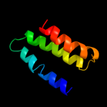
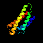
Top model
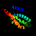
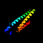
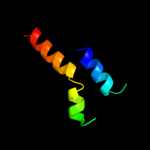
Image coloured by rainbow N → C terminus
Model (left) based on template d2r6gf1
Top template information
Fold:MalF N-terminal region-like
Superfamily:MalF N-terminal region-like
Family:MalF N-terminal region-like
Confidence and coverage
Confidence: 32.8% Coverage: 9%
63 residues ( 9% of your sequence) have been modelled with 32.8% confidence by the single highest scoring template.
You may wish to submit your sequence to Phyrealarm. This will automatically scan your sequence every week for new potential templates as they appear in the Phyre2 library.
Please note: You must be registered and logged in to use Phyrealarm.
3D viewing
Interactive 3D view in Jmol

Sequence analysis 

Secondary structure and disorder prediction 

   1........10.........20.........30.........40.........50.........60
Sequence  MSALNSLPLPVVRLLAFFHEELSERRPGRVPQTVQLWVGCLLVILISMTFEIPFVALSLA
Secondary structure 


SS confidence 


Disorder  ????????
??


?
?


Disorder confidence 


   .........70.........80.........90.........100.........110.........120
Sequence  VLFYGIQSNAFYTKFVAILFVVATVLEIGSLFLIYKWSYGEPLIRLIIAGPILMGCMFLM
Secondary structure 


SS confidence 


Disorder 


????


Disorder confidence 


   .........130.........140.........150.........160.........170.........180
Sequence  RTHRLGLVFFAVAIVAIYGQTFPAMLDYPEVVVRLTLWCIVVGLYPTLLMTLIGVLWFPS
Secondary structure 


SS confidence 


Disorder 


????


Disorder confidence 


   .........190.........200.........210.........220.........230.........240
Sequence  RAISQMHQALNDRLDDAISHLTDSLAPLPETRIEREALALQKLNVFCLADDANWRTQNAW
Secondary structure 


SS confidence 


Disorder 


?

??????


?????????


Disorder confidence 


   .........250.........260.........270.........280.........290.........300
Sequence  WQSCVATVTYIYSTLNRYDPTSFADSQAIIEFRQKLASEINKLQHAVAEGQCWQSDWRIS
Secondary structure 


SS confidence 


Disorder 


?
????????????


???????


Disorder confidence 


   .........310.........320.........330.........340.........350.........360
Sequence  ESEAMAARECNLENICQTLLQLGQMDPNTPPTPAAKPPSMAADAFTNPDYMRYAVKTLLA
Secondary structure 


SS confidence 


Disorder 


?


??????????????????????


Disorder confidence 


   .........370.........380.........390.........400.........410.........420
Sequence  CLICYTFYSGVDWEGIHTCMLTCVIVANPNVGSSYQKMVLRFGGAFCGAILALLFTLLVM
Secondary structure 


SS confidence 


Disorder 


???


Disorder confidence 


   .........430.........440.........450.........460.........470.........480
Sequence  PWLDNIVELLFVLAPIFLLGAWIATSSERSSYIGTQMVVTFALATLENVFGPVYDLVEIR
Secondary structure 


SS confidence 


Disorder 


????


??????


Disorder confidence 


   .........490.........500.........510.........520.........530.........540
Sequence  DRALGIIIGTVVSAVIYTFVWPESEARTLPQKLAGTLGMLSKVMRIPRQQEVTALRTYLQ
Secondary structure 


SS confidence 


Disorder 


????????


Disorder confidence 


   .........550.........560.........570.........580.........590.........600
Sequence  IRIGLHAAFNACEEMCQRVALERQLDSEERALLIERSQTVIRQGRDLLHAWDATWNSAQA
Secondary structure 


SS confidence 


Disorder 


??????????


??
Disorder confidence 


   .........610.........620.........630.........640.........650.........660
Sequence  LDNALQPDRAGQFADALEKYAAGLATALSRSPQITLEETPASQAILPTLLKQEQHVCQLF
Secondary structure 


SS confidence 


Disorder  ???????


????
?????????


Disorder confidence 


   .........670.........680...
Sequence  ARLPDWTAPALTPATEQAQGATQ
Secondary structure 


SS confidence 


Disorder 


?


????????????
Disorder confidence 


Confidence Key
High(9)                    Low (0)
?Disordered
Alpha helix
Beta strand

Domain analysis 

Hover over an aligned region to see model and summary info

Please note, only up to the top 20 hits are modelled to reduce computer load

RankAligned region

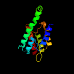
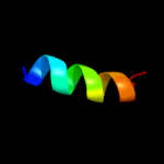
PDB 2r6g chain F domain 1

3D model

Region: 404 - 466
Aligned: 63
Modelled: 63
Confidence: 32.8%
Identity: 10%
Fold: MalF N-terminal region-like
Superfamily: MalF N-terminal region-like
Family: MalF N-terminal region-like

Phyre2

PDB 1oy8 chain A

3D model

Region: 345 - 502
Aligned: 143
Modelled: 143
Confidence: 21.7%
Identity: 13%
PDB header:membrane protein
Chain: A: PDB Molecule:acriflavine resistance protein b;
PDBTitle: structural basis of multiple drug binding capacity of the acrb2 multidrug efflux pump

Phyre2

PDB 2w2e chain A

3D model

Region: 323 - 524
Aligned: 197
Modelled: 202
Confidence: 21.0%
Identity: 11%
PDB header:membrane protein
Chain: A: PDB Molecule:aquaporin;
PDBTitle: 1.15 angstrom crystal structure of p.pastoris aquaporin,2 aqy1, in a closed conformation at ph 3.5

Phyre2

PDB 1kpl chain A

3D model

Region: 357 - 519
Aligned: 123
Modelled: 131
Confidence: 20.0%
Identity: 12%
Fold: Clc chloride channel
Superfamily: Clc chloride channel
Family: Clc chloride channel

Phyre2

PDB 3nd0 chain A

3D model

Region: 357 - 513
Aligned: 117
Modelled: 125
Confidence: 19.4%
Identity: 15%
PDB header:transport protein
Chain: A: PDB Molecule:sll0855 protein;
PDBTitle: x-ray crystal structure of a slow cyanobacterial cl-/h+ antiporter

Phyre2

PDB 2ht2 chain B

3D model

Region: 358 - 515
Aligned: 118
Modelled: 139
Confidence: 16.1%
Identity: 16%
PDB header:membrane protein
Chain: B: PDB Molecule:h(+)/cl(-) exchange transporter clca;
PDBTitle: structure of the escherichia coli clc chloride channel2 y445h mutant and fab complex

Phyre2

PDB 1st6 chain A domain 4

3D model

Region: 182 - 289
Aligned: 104
Modelled: 108
Confidence: 13.8%
Identity: 13%
Fold: Four-helical up-and-down bundle
Superfamily: alpha-catenin/vinculin-like
Family: alpha-catenin/vinculin

Phyre2

PDB 1ots chain A

3D model

Region: 351 - 519
Aligned: 129
Modelled: 150
Confidence: 13.0%
Identity: 17%
Fold: Clc chloride channel
Superfamily: Clc chloride channel
Family: Clc chloride channel

Phyre2

PDB 1szi chain A

3D model

Region: 548 - 651
Aligned: 102
Modelled: 104
Confidence: 12.9%
Identity: 12%
Fold: Four-helical up-and-down bundle
Superfamily: Mannose-6-phosphate receptor binding protein 1 (Tip47), C-terminal domain
Family: Mannose-6-phosphate receptor binding protein 1 (Tip47), C-terminal domain

Phyre2

PDB 1no1 chain C

3D model

Region: 17 - 30
Aligned: 14
Modelled: 14
Confidence: 11.3%
Identity: 36%
PDB header:replication
Chain: C: PDB Molecule:replisome organizer;
PDBTitle: structure of truncated variant of b.subtilis spp1 phage g39p helicase2 loader/inhibitor protein

Phyre2

PDB 1no1 chain A

3D model

Region: 17 - 30
Aligned: 14
Modelled: 14
Confidence: 11.3%
Identity: 36%
Fold: Replisome organizer (g39p helicase loader/inhibitor protein)
Superfamily: Replisome organizer (g39p helicase loader/inhibitor protein)
Family: Replisome organizer (g39p helicase loader/inhibitor protein)

Phyre2

PDB 2ksf chain A

3D model

Region: 403 - 499
Aligned: 96
Modelled: 97
Confidence: 11.2%
Identity: 8%
PDB header:transferase
Chain: A: PDB Molecule:sensor protein kdpd;
PDBTitle: backbone structure of the membrane domain of e. coli2 histidine kinase receptor kdpd, center for structures of3 membrane proteins (csmp) target 4312c

Phyre2

PDB 1bhb chain A

3D model

Region: 11 - 51
Aligned: 41
Modelled: 41
Confidence: 9.9%
Identity: 12%
PDB header:photoreceptor
Chain: A: PDB Molecule:bacteriorhodopsin;
PDBTitle: three-dimensional structure of (1-71) bacterioopsin2 solubilized in methanol-chloroform and sds micelles3 determined by 15n-1h heteronuclear nmr spectroscopy

Phyre2

PDB 1b9u chain A

3D model

Region: 485 - 500
Aligned: 16
Modelled: 16
Confidence: 9.5%
Identity: 19%
PDB header:hydrolase
Chain: A: PDB Molecule:protein (atp synthase);
PDBTitle: membrane domain of the subunit b of the e.coli atp synthase

Phyre2

PDB 3su8 chain X

3D model

Region: 5 - 36
Aligned: 31
Modelled: 32
Confidence: 8.5%
Identity: 13%
PDB header:apoptosis/signaling protein
Chain: X: PDB Molecule:plexin-b1;
PDBTitle: crystal structure of a truncated intracellular domain of plexin-b1 in2 complex with rac1

Phyre2

PDB 2d57 chain A

3D model

Region: 354 - 502
Aligned: 147
Modelled: 149
Confidence: 8.4%
Identity: 18%
PDB header:transport protein
Chain: A: PDB Molecule:aquaporin-4;
PDBTitle: double layered 2d crystal structure of aquaporin-4 (aqp4m23) at 3.2 a2 resolution by electron crystallography

Phyre2

PDB 3g9d chain B

3D model

Region: 398 - 412
Aligned: 15
Modelled: 15
Confidence: 8.3%
Identity: 27%
PDB header:hydrolase
Chain: B: PDB Molecule:dinitrogenase reductase activacting
PDBTitle: crystal structure glycohydrolase

Phyre2

PDB 1st6 chain A domain 3

3D model

Region: 183 - 290
Aligned: 105
Modelled: 108
Confidence: 8.3%
Identity: 10%
Fold: Four-helical up-and-down bundle
Superfamily: alpha-catenin/vinculin-like
Family: alpha-catenin/vinculin

Phyre2

PDB 2b5f chain D

3D model

Region: 354 - 500
Aligned: 145
Modelled: 147
Confidence: 8.3%
Identity: 15%
PDB header:transport protein,membrane protein
Chain: D: PDB Molecule:aquaporin;
PDBTitle: crystal structure of the spinach aquaporin sopip2;1 in an2 open conformation to 3.9 resolution

Phyre2

PDB 2woc chain A

3D model

Region: 398 - 412
Aligned: 15
Modelled: 15
Confidence: 8.0%
Identity: 33%
PDB header:hydrolase
Chain: A: PDB Molecule:adp-ribosyl-[dinitrogen reductase] glycohydrolase;
PDBTitle: crystal structure of the dinitrogenase reductase-activating2 glycohydrolase (drag) from rhodospirillum rubrum

Phyre2
1

d2r6gf1
2

c1oy8A_
3

c2w2eA_
4

d1kpla_
5

c3nd0A_
6

c2ht2B_
7

d1st6a4
8

d1otsa_
9

d1szia_
10

c1no1C_
11

d1no1a_
12

c2ksfA_
13

c1bhbA_
14

c1b9uA_
15

c3su8X_
16

c2d57A_
17

c3g9dB_
18

d1st6a3
19

c2b5fD_
20

c2wocA_
21


22


23


24


25


26


27


28


29


30


31


32


33


34


35


36


37


38


39


40


41


42


43


44


45


46


47


48


49


50


Detailed template information 

#
Template Alignment Coverage3D Model Confidence
% i.d. Template Information
1d2r6gf1


32.8 10 Fold:MalF N-terminal region-like
Superfamily:MalF N-terminal region-like
Family:MalF N-terminal region-like
2c1oy8A_


21.7 13 PDB header:membrane protein
Chain: A: PDB Molecule:acriflavine resistance protein b;
PDBTitle: structural basis of multiple drug binding capacity of the acrb2 multidrug efflux pump
3c2w2eA_


21.0 11 PDB header:membrane protein
Chain: A: PDB Molecule:aquaporin;
PDBTitle: 1.15 angstrom crystal structure of p.pastoris aquaporin,2 aqy1, in a closed conformation at ph 3.5
4d1kpla_


20.0 12 Fold:Clc chloride channel
Superfamily:Clc chloride channel
Family:Clc chloride channel
5c3nd0A_


19.4 15 PDB header:transport protein
Chain: A: PDB Molecule:sll0855 protein;
PDBTitle: x-ray crystal structure of a slow cyanobacterial cl-/h+ antiporter
6c2ht2B_


16.1 16 PDB header:membrane protein
Chain: B: PDB Molecule:h(+)/cl(-) exchange transporter clca;
PDBTitle: structure of the escherichia coli clc chloride channel2 y445h mutant and fab complex
7d1st6a4


13.8 13 Fold:Four-helical up-and-down bundle
Superfamily:alpha-catenin/vinculin-like
Family:alpha-catenin/vinculin
8d1otsa_


13.0 17 Fold:Clc chloride channel
Superfamily:Clc chloride channel
Family:Clc chloride channel
9d1szia_


12.9 12 Fold:Four-helical up-and-down bundle
Superfamily:Mannose-6-phosphate receptor binding protein 1 (Tip47), C-terminal domain
Family:Mannose-6-phosphate receptor binding protein 1 (Tip47), C-terminal domain
10c1no1C_


11.3 36 PDB header:replication
Chain: C: PDB Molecule:replisome organizer;
PDBTitle: structure of truncated variant of b.subtilis spp1 phage g39p helicase2 loader/inhibitor protein
11d1no1a_


11.3 36 Fold:Replisome organizer (g39p helicase loader/inhibitor protein)
Superfamily:Replisome organizer (g39p helicase loader/inhibitor protein)
Family:Replisome organizer (g39p helicase loader/inhibitor protein)
12c2ksfA_


11.2 8 PDB header:transferase
Chain: A: PDB Molecule:sensor protein kdpd;
PDBTitle: backbone structure of the membrane domain of e. coli2 histidine kinase receptor kdpd, center for structures of3 membrane proteins (csmp) target 4312c
13c1bhbA_


9.9 12 PDB header:photoreceptor
Chain: A: PDB Molecule:bacteriorhodopsin;
PDBTitle: three-dimensional structure of (1-71) bacterioopsin2 solubilized in methanol-chloroform and sds micelles3 determined by 15n-1h heteronuclear nmr spectroscopy
14c1b9uA_


9.5 19 PDB header:hydrolase
Chain: A: PDB Molecule:protein (atp synthase);
PDBTitle: membrane domain of the subunit b of the e.coli atp synthase
15c3su8X_


8.5 13 PDB header:apoptosis/signaling protein
Chain: X: PDB Molecule:plexin-b1;
PDBTitle: crystal structure of a truncated intracellular domain of plexin-b1 in2 complex with rac1
16c2d57A_


8.4 18 PDB header:transport protein
Chain: A: PDB Molecule:aquaporin-4;
PDBTitle: double layered 2d crystal structure of aquaporin-4 (aqp4m23) at 3.2 a2 resolution by electron crystallography
17c3g9dB_


8.3 27 PDB header:hydrolase
Chain: B: PDB Molecule:dinitrogenase reductase activacting
PDBTitle: crystal structure glycohydrolase
18d1st6a3


8.3 10 Fold:Four-helical up-and-down bundle
Superfamily:alpha-catenin/vinculin-like
Family:alpha-catenin/vinculin
19c2b5fD_


8.3 15 PDB header:transport protein,membrane protein
Chain: D: PDB Molecule:aquaporin;
PDBTitle: crystal structure of the spinach aquaporin sopip2;1 in an2 open conformation to 3.9 resolution
20c2wocA_


8.0 33 PDB header:hydrolase
Chain: A: PDB Molecule:adp-ribosyl-[dinitrogen reductase] glycohydrolase;
PDBTitle: crystal structure of the dinitrogenase reductase-activating2 glycohydrolase (drag) from rhodospirillum rubrum
21d2nwwa1


not modelled 7.9 20 Fold:Proton glutamate symport protein
Superfamily:Proton glutamate symport protein
Family:Proton glutamate symport protein
22c2f2bA_


not modelled 7.8 13 PDB header:membrane protein
Chain: A: PDB Molecule:aquaporin aqpm;
PDBTitle: crystal structure of integral membrane protein aquaporin aqpm at 1.68a2 resolution
23c2xq2A_


not modelled 7.7 19 PDB header:transport protein
Chain: A: PDB Molecule:sodium/glucose cotransporter;
PDBTitle: structure of the k294a mutant of vsglt
24c3hfwA_


not modelled 7.5 8 PDB header:hydrolase
Chain: A: PDB Molecule:protein adp-ribosylarginine hydrolase;
PDBTitle: crystal structure of human adp-ribosylhydrolase 1 (harh1)
25d1st6a5


not modelled 7.4 7 Fold:Four-helical up-and-down bundle
Superfamily:alpha-catenin/vinculin-like
Family:alpha-catenin/vinculin
26c3gd8A_


not modelled 7.3 19 PDB header:membrane protein
Chain: A: PDB Molecule:aquaporin-4;
PDBTitle: crystal structure of human aquaporin 4 at 1.8 and its mechanism of2 conductance
27d2e74d2


not modelled 7.3 33 Fold:Single transmembrane helix
Superfamily:ISP transmembrane anchor
Family:ISP transmembrane anchor
28c2yvxD_


not modelled 7.3 10 PDB header:transport protein
Chain: D: PDB Molecule:mg2+ transporter mgte;
PDBTitle: crystal structure of magnesium transporter mgte
29d1rc2a_


not modelled 7.2 15 Fold:Aquaporin-like
Superfamily:Aquaporin-like
Family:Aquaporin-like
30c3dh4A_


not modelled 7.0 13 PDB header:transport protein
Chain: A: PDB Molecule:sodium/glucose cotransporter;
PDBTitle: crystal structure of sodium/sugar symporter with bound galactose from2 vibrio parahaemolyticus
31c2qtyB_


not modelled 6.8 40 PDB header:hydrolase
Chain: B: PDB Molecule:poly(adp-ribose) glycohydrolase arh3;
PDBTitle: crystal structure of mouse adp-ribosylhydrolase 3 (marh3)
32c2yzwA_


not modelled 6.7 29 PDB header:hydrolase
Chain: A: PDB Molecule:adp-ribosylglycohydrolase;
PDBTitle: adp-ribosylglycohydrolase-related protein complex
33c2hjdA_


not modelled 6.7 16 PDB header:signaling protein
Chain: A: PDB Molecule:quorum-sensing antiactivator;
PDBTitle: crystal structure of a second quorum sensing antiactivator tram2 from2 a. tumefaciens strain a6
34d1fx8a_


not modelled 6.7 11 Fold:Aquaporin-like
Superfamily:Aquaporin-like
Family:Aquaporin-like
35c1ldaA_


not modelled 6.7 11 PDB header:transport protein
Chain: A: PDB Molecule:glycerol uptake facilitator protein;
PDBTitle: crystal structure of the e. coli glycerol facilitator (glpf) without2 substrate glycerol
36c3k3gA_


not modelled 6.5 14 PDB header:transport protein
Chain: A: PDB Molecule:urea transporter;
PDBTitle: crystal structure of the urea transporter from desulfovibrio vulgaris2 bound to 1,3-dimethylurea
37c3orgB_


not modelled 6.5 15 PDB header:transport protein
Chain: B: PDB Molecule:cmclc;
PDBTitle: crystal structure of a eukaryotic clc transporter
38c3ig3A_


not modelled 6.0 29 PDB header:signaling protein, membrane protein
Chain: A: PDB Molecule:plxna3 protein;
PDBTitle: crystal strucure of mouse plexin a3 intracellular domain
39c2l16A_


not modelled 5.9 14 PDB header:protein transport
Chain: A: PDB Molecule:sec-independent protein translocase protein tatad;
PDBTitle: solution structure of bacillus subtilits tatad protein in dpc micelles
40c3hm6X_


not modelled 5.8 13 PDB header:signaling protein
Chain: X: PDB Molecule:plexin-b1;
PDBTitle: crystal structure of the cytoplasmic domain of human plexin b1
41d1t5ja_


not modelled 5.8 31 Fold:ADP-ribosylglycohydrolase
Superfamily:ADP-ribosylglycohydrolase
Family:ADP-ribosylglycohydrolase
42d1rfya_


not modelled 5.6 15 Fold:Long alpha-hairpin
Superfamily:Transcriptional repressor TraM
Family:Transcriptional repressor TraM
43d1rzhh2


not modelled 5.3 38 Fold:Single transmembrane helix
Superfamily:Photosystem II reaction centre subunit H, transmembrane region
Family:Photosystem II reaction centre subunit H, transmembrane region
44d1ymga1


not modelled 5.2 14 Fold:Aquaporin-like
Superfamily:Aquaporin-like
Family:Aquaporin-like
45c1ymgA_


not modelled 5.2 14 PDB header:membrane protein
Chain: A: PDB Molecule:lens fiber major intrinsic protein;
PDBTitle: the channel architecture of aquaporin o at 2.2 angstrom resolution
46d1v54l_


not modelled 5.2 13 Fold:Single transmembrane helix
Superfamily:Mitochondrial cytochrome c oxidase subunit VIIc (aka VIIIa)
Family:Mitochondrial cytochrome c oxidase subunit VIIc (aka VIIIa)
47c2wscL_


not modelled 5.2 18 PDB header:photosynthesis
Chain: L: PDB Molecule:photosystem i reaction center subunit xi,
PDBTitle: improved model of plant photosystem i
48c2fhdA_


not modelled 5.0 35 PDB header:cell cycle
Chain: A: PDB Molecule:dna repair protein rhp9/crb2;
PDBTitle: crystal structure of crb2 tandem tudor domains
49c2o01L_


not modelled 5.0 18 PDB header:photosynthesis
Chain: L: PDB Molecule:photosystem i reaction center subunit xi,
PDBTitle: the structure of a plant photosystem i supercomplex at 3.42 angstrom resolution
50d2axti1


not modelled 5.0 8 Fold:Single transmembrane helix
Superfamily:Photosystem II reaction center protein I, PsbI
Family:PsbI-like

Binding site prediction 

Due to computational demand, binding site predictions are not run for batch jobs

If you want to predict binding sites, please manually submit your model of choice to 3DLigandSite


Phyre is for academic use only

Please cite: Protein structure prediction on the web: a case study using the Phyre server
Kelley LA and Sternberg MJE. Nature Protocols 4, 363 - 371 (2009) [pdf] [Import into BibTeX]
 
If you use the binding site predictions from 3DLigandSite, please also cite:
3DLigandSite: predicting ligand-binding sites using similar structures.
Wass MN, Kelley LA and Sternberg MJ Nucleic Acids Research 38, W469-73 (2010) [PubMed]
 
© Structural Bioinformatics Group
Imperial College London
Lawrence Kelley, Benjamin Jefferys
Disclaimer
Terms and Conditions
Component software
Template detection: HHpred 1.51
Secondary structure prediction: Psi-pred 2.5
Disorder prediction: Disopred 2.4
Transmembrane prediction: Memsat_SVM
Multi-template modelling and ab initio: Poing 1.0