1 c3rfzB_
100.0
28
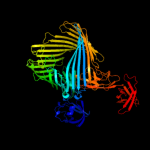
PDB header: cell adhesion/transport/chaperoneChain: B: PDB Molecule: outer membrane usher protein, type 1 fimbrial synthesis;PDBTitle: crystal structure of the fimd usher bound to its cognate fimc:fimh2 substrate
2 c2vqiA_
100.0
41
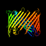
PDB header: transportChain: A: PDB Molecule: outer membrane usher protein papc;PDBTitle: structure of the p pilus usher (papc) translocation pore
3 c3ohnA_
100.0
29
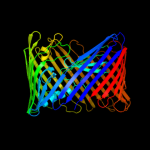
PDB header: membrane proteinChain: A: PDB Molecule: outer membrane usher protein fimd;PDBTitle: crystal structure of the fimd translocation domain
4 d3bwud1
100.0
24
Fold: FimD N-terminal domain-likeSuperfamily: FimD N-terminal domain-likeFamily: Usher N-domain5 d1zdva1
100.0
26
Fold: FimD N-terminal domain-likeSuperfamily: FimD N-terminal domain-likeFamily: Usher N-domain6 c3fcgB_
99.9
34
PDB header: membrane protein, protein transportChain: B: PDB Molecule: f1 capsule-anchoring protein;PDBTitle: crystal structure analysis of the middle domain of the2 caf1a usher
7 d1zdxa1
99.9
22
Fold: FimD N-terminal domain-likeSuperfamily: FimD N-terminal domain-likeFamily: Usher N-domain8 c3l48B_
99.7
38
PDB header: transport proteinChain: B: PDB Molecule: outer membrane usher protein papc;PDBTitle: crystal structure of the c-terminal domain of the papc usher
9 c2xetB_
99.7
25
PDB header: transport proteinChain: B: PDB Molecule: f1 capsule-anchoring protein;PDBTitle: conserved hydrophobic clusters on the surface of the caf1a usher2 c-terminal domain are important for f1 antigen assembly
10 d1uwya1
90.8
10
Fold: Prealbumin-likeSuperfamily: Carboxypeptidase regulatory domain-likeFamily: Carboxypeptidase regulatory domain11 c3mn8A_
88.4
12
PDB header: hydrolaseChain: A: PDB Molecule: lp15968p;PDBTitle: structure of drosophila melanogaster carboxypeptidase d isoform 1b2 short
12 d1h8la1
88.3
14
Fold: Prealbumin-likeSuperfamily: Carboxypeptidase regulatory domain-likeFamily: Carboxypeptidase regulatory domain13 c2nsmA_
88.3
7
PDB header: hydrolaseChain: A: PDB Molecule: carboxypeptidase n catalytic chain;PDBTitle: crystal structure of the human carboxypeptidase n (kininase i)2 catalytic domain
14 c3pdgA_
87.3
20
PDB header: unknown functionChain: A: PDB Molecule: fibronectin(iii)-like module;PDBTitle: structures of clostridium thermocellum cbha fibronectin(iii)-like2 modules
15 c1uwyA_
87.3
14
PDB header: hydrolaseChain: A: PDB Molecule: carboxypeptidase m;PDBTitle: crystal structure of human carboxypeptidase m
16 c3pe9B_
86.9
14
PDB header: unknown functionChain: B: PDB Molecule: fibronectin(iii)-like module;PDBTitle: structures of clostridium thermocellum cbha fibronectin(iii)-like2 modules
17 c1h8lA_
82.9
14
PDB header: carboxypeptidaseChain: A: PDB Molecule: carboxypeptidase gp180 residues 503-882;PDBTitle: duck carboxypeptidase d domain ii in complex with gemsa
18 d1w0na_
82.5
18
Fold: Galactose-binding domain-likeSuperfamily: Galactose-binding domain-likeFamily: Family 36 carbohydrate binding module, CBM3619 c3pe9D_
76.7
11
PDB header: unknown functionChain: D: PDB Molecule: fibronectin(iii)-like module;PDBTitle: structures of clostridium thermocellum cbha fibronectin(iii)-like2 modules
20 c1d2pA_
75.8
12
PDB header: structural proteinChain: A: PDB Molecule: collagen adhesin;PDBTitle: crystal structure of two b repeat units (b1b2) of the2 collagen binding protein (cna) of staphylococcus aureus
21 c1u00A_
not modelled
75.6
22
PDB header: chaperoneChain: A: PDB Molecule: chaperone protein hsca;PDBTitle: hsca substrate binding domain complexed with the iscu2 recognition peptide elppvkihc
22 c3dpqE_
not modelled
72.4
22
PDB header: chaperone, peptide binding proteinChain: E: PDB Molecule: chaperone protein dnak;PDBTitle: crystal structure of the substrate binding domain of e.2 coli dnak in complex with a long pyrrhocoricin-derived3 inhibitor peptide (form b)
23 c2op6A_
not modelled
71.9
20
PDB header: peptide binding proteinChain: A: PDB Molecule: heat shock 70 kda protein d;PDBTitle: peptide-binding domain of heat shock 70 kda protein d precursor from2 c.elegans
24 c3n8eA_
not modelled
70.8
24
PDB header: chaperoneChain: A: PDB Molecule: stress-70 protein, mitochondrial;PDBTitle: substrate binding domain of the human heat shock 70kda protein 92 (mortalin)
25 c3d33B_
not modelled
70.5
7
PDB header: unknown functionChain: B: PDB Molecule: domain of unknown function with an immunoglobulin-likePDBTitle: crystal structure of a duf3244 family protein with an immunoglobulin-2 like beta-sandwich fold (bvu_0276) from bacteroides vulgatus atcc3 8482 at 1.70 a resolution
26 c1bprA_
not modelled
67.2
20
PDB header: molecular chaperoneChain: A: PDB Molecule: dnak;PDBTitle: nmr structure of the substrate binding domain of dnak,2 minimized average structure
27 c3pe9A_
not modelled
66.8
11
PDB header: unknown functionChain: A: PDB Molecule: fibronectin(iii)-like module;PDBTitle: structures of clostridium thermocellum cbha fibronectin(iii)-like2 modules
28 c3pe9C_
not modelled
66.8
11
PDB header: unknown functionChain: C: PDB Molecule: fibronectin(iii)-like module;PDBTitle: structures of clostridium thermocellum cbha fibronectin(iii)-like2 modules
29 c2r32A_
not modelled
58.6
25
PDB header: immune systemChain: A: PDB Molecule: gcn4-pii/tumor necrosis factor ligandPDBTitle: crystal structure of human gitrl variant
30 d1ci3m2
not modelled
58.2
16
Fold: Barrel-sandwich hybridSuperfamily: Rudiment single hybrid motifFamily: Cytochrome f, small domain31 d1u00a2
not modelled
57.6
22
Fold: Heat shock protein 70kD (HSP70), peptide-binding domainSuperfamily: Heat shock protein 70kD (HSP70), peptide-binding domainFamily: Heat shock protein 70kD (HSP70), peptide-binding domain32 c3dqgC_
not modelled
57.3
24
PDB header: chaperoneChain: C: PDB Molecule: heat shock 70 kda protein f;PDBTitle: peptide-binding domain of heat shock 70 kda protein f, mitochondrial2 precursor, from caenorhabditis elegans.
33 c3e8vA_
not modelled
56.1
15
PDB header: structural genomics, unknown functionChain: A: PDB Molecule: possible transglutaminase-family protein;PDBTitle: crystal structure of a possible transglutaminase-family2 protein proteolytic fragment from bacteroides fragilis
34 d1yuwa1
not modelled
55.1
22
Fold: Heat shock protein 70kD (HSP70), peptide-binding domainSuperfamily: Heat shock protein 70kD (HSP70), peptide-binding domainFamily: Heat shock protein 70kD (HSP70), peptide-binding domain35 d1v8ha1
not modelled
53.3
17
Fold: Immunoglobulin-like beta-sandwichSuperfamily: E set domainsFamily: SoxZ-like36 d2ag4a1
not modelled
53.2
9
Fold: Ganglioside M2 (gm2) activatorSuperfamily: Ganglioside M2 (gm2) activatorFamily: Ganglioside M2 (gm2) activator37 d1dkza2
not modelled
51.3
22
Fold: Heat shock protein 70kD (HSP70), peptide-binding domainSuperfamily: Heat shock protein 70kD (HSP70), peptide-binding domainFamily: Heat shock protein 70kD (HSP70), peptide-binding domain38 c3k1dA_
not modelled
47.1
16
PDB header: transferaseChain: A: PDB Molecule: 1,4-alpha-glucan-branching enzyme;PDBTitle: crystal structure of glycogen branching enzyme synonym: 1,4-alpha-d-2 glucan:1,4-alpha-d-glucan 6-glucosyl-transferase from mycobacterium3 tuberculosis h37rv
39 d1e2wa2
not modelled
46.2
18
Fold: Barrel-sandwich hybridSuperfamily: Rudiment single hybrid motifFamily: Cytochrome f, small domain40 c3c12A_
not modelled
39.1
15
PDB header: biosynthetic proteinChain: A: PDB Molecule: flagellar protein;PDBTitle: crystal structure of flgd from xanthomonas campestris:2 insights into the hook capping essential for flagellar3 assembly
41 c2oxgE_
not modelled
31.5
15
PDB header: transport proteinChain: E: PDB Molecule: soxz protein;PDBTitle: the soxyz complex of paracoccus pantotrophus
42 d2vzsa4
not modelled
29.3
22
Fold: Galactose-binding domain-likeSuperfamily: Galactose-binding domain-likeFamily: beta-Galactosidase/glucuronidase, N-terminal domain43 d1cxla1
not modelled
27.9
10
Fold: Immunoglobulin-like beta-sandwichSuperfamily: E set domainsFamily: E-set domains of sugar-utilizing enzymes44 c2x5pA_
not modelled
27.4
18
PDB header: protein bindingChain: A: PDB Molecule: fibronectin binding protein;PDBTitle: crystal structure of the streptococcus pyogenes fibronectin binding2 protein fbab-b
45 c1e2vB_
not modelled
24.8
17
PDB header: electron transport proteinsChain: B: PDB Molecule: cytochrome f;PDBTitle: n153q mutant of cytochrome f from chlamydomonas reinhardtii
46 d2je8a4
not modelled
22.5
11
Fold: Galactose-binding domain-likeSuperfamily: Galactose-binding domain-likeFamily: beta-Galactosidase/glucuronidase, N-terminal domain47 c2jxmB_
not modelled
21.9
15
PDB header: electron transportChain: B: PDB Molecule: cytochrome f;PDBTitle: ensemble of twenty structures of the prochlorothrix2 hollandica plastocyanin- cytochrome f complex
48 c1q90A_
not modelled
21.8
19
PDB header: photosynthesisChain: A: PDB Molecule: apocytochrome f;PDBTitle: structure of the cytochrome b6f (plastohydroquinone : plastocyanin2 oxidoreductase) from chlamydomonas reinhardtii
49 c3rghA_
not modelled
21.1
19
PDB header: cell adhesionChain: A: PDB Molecule: filamin-a;PDBTitle: structure of filamin a immunoglobulin-like repeat 10 from homo sapiens
50 c3bryB_
not modelled
21.0
17
PDB header: transport proteinChain: B: PDB Molecule: tbux;PDBTitle: crystal structure of the ralstonia pickettii toluene2 transporter tbux
51 d1wlha1
not modelled
20.5
13
Fold: Immunoglobulin-like beta-sandwichSuperfamily: E set domainsFamily: Filamin repeat (rod domain)52 d2dj4a1
not modelled
20.3
25
Fold: Immunoglobulin-like beta-sandwichSuperfamily: E set domainsFamily: Filamin repeat (rod domain)53 d2bp3a1
not modelled
19.8
31
Fold: Immunoglobulin-like beta-sandwichSuperfamily: E set domainsFamily: Filamin repeat (rod domain)54 c2xk0A_
not modelled
19.8
23
PDB header: transcriptionChain: A: PDB Molecule: polycomb protein pcl;PDBTitle: solution structure of the tudor domain from drosophila2 polycomblike (pcl)
55 c3iswA_
not modelled
19.3
31
PDB header: structural proteinChain: A: PDB Molecule: filamin-a;PDBTitle: crystal structure of filamin-a immunoglobulin-like repeat 21 bound to2 an n-terminal peptide of cftr
56 c2ww8A_
not modelled
18.5
12
PDB header: cell adhesionChain: A: PDB Molecule: cell wall surface anchor family protein;PDBTitle: structure of the pilus adhesin (rrga) from streptococcus2 pneumoniae
57 c2pz4A_
not modelled
18.4
14
PDB header: cell adhesionChain: A: PDB Molecule: protein gbs052;PDBTitle: crystal structure of spab (gbs52), the minor pilin in gram-positive2 pathogen streptococcus agalactiae
58 c2eqjA_
not modelled
18.2
33
PDB header: transcriptionChain: A: PDB Molecule: metal-response element-binding transcriptionPDBTitle: solution structure of the tudor domain of metal-response2 element-binding transcription factor 2
59 c3b9eA_
not modelled
18.1
7
PDB header: hydrolaseChain: A: PDB Molecule: chitinase a;PDBTitle: crystal structure of inactive mutant e315m chitinase a from2 vibrio harveyi
60 d2a9da1
not modelled
17.8
16
Fold: Immunoglobulin-like beta-sandwichSuperfamily: E set domainsFamily: Molybdenum-containing oxidoreductases-like dimerisation domain61 d2di8a1
not modelled
17.3
16
Fold: Immunoglobulin-like beta-sandwichSuperfamily: E set domainsFamily: Filamin repeat (rod domain)62 d1edqa1
not modelled
17.2
17
Fold: Immunoglobulin-like beta-sandwichSuperfamily: E set domainsFamily: E-set domains of sugar-utilizing enzymes63 d2dmca1
not modelled
16.8
17
Fold: Immunoglobulin-like beta-sandwichSuperfamily: E set domainsFamily: Filamin repeat (rod domain)64 c2k78A_
not modelled
16.6
12
PDB header: heme-binding proteinChain: A: PDB Molecule: iron-regulated surface determinant protein c;PDBTitle: solution structure of the isdc neat domain bound to zinc2 protoporphyrin
65 d1pfsa_
not modelled
16.1
24
Fold: OB-foldSuperfamily: Nucleic acid-binding proteinsFamily: Phage ssDNA-binding proteins66 d2o6pa1
not modelled
16.0
12
Fold: Immunoglobulin-like beta-sandwichSuperfamily: NEAT domain-likeFamily: NEAT domain67 c2o6pA_
not modelled
15.3
12
PDB header: transport proteinChain: A: PDB Molecule: iron-regulated surface determinant protein c;PDBTitle: crystal structure of the heme-isdc complex
68 d2gr7a1
not modelled
14.6
22
Fold: Pili subunitsSuperfamily: Pili subunitsFamily: YadA C-terminal domain-like69 c2gr7C_
not modelled
14.6
22
PDB header: membrane proteinChain: C: PDB Molecule: adhesin;PDBTitle: hia 992-1098
70 c2brqB_
not modelled
14.6
31
PDB header: structural proteinChain: B: PDB Molecule: filamin a;PDBTitle: crystal structure of the filamin a repeat 21 complexed with2 the integrin beta7 cytoplasmic tail peptide
71 d2dica1
not modelled
14.1
19
Fold: Immunoglobulin-like beta-sandwichSuperfamily: E set domainsFamily: Filamin repeat (rod domain)72 d2w0pa1
not modelled
14.0
31
Fold: Immunoglobulin-like beta-sandwichSuperfamily: E set domainsFamily: Filamin repeat (rod domain)73 d2vmha1
not modelled
13.8
10
Fold: Galactose-binding domain-likeSuperfamily: Galactose-binding domain-likeFamily: NPCBM-like74 d1qfha1
not modelled
13.7
13
Fold: Immunoglobulin-like beta-sandwichSuperfamily: E set domainsFamily: Filamin repeat (rod domain)75 c2ds4A_
not modelled
13.6
19
PDB header: protein bindingChain: A: PDB Molecule: tripartite motif protein 45;PDBTitle: solution structure of the filamin domain from human2 tripartite motif protein 45
76 c3brzA_
not modelled
13.5
15
PDB header: transport proteinChain: A: PDB Molecule: todx;PDBTitle: crystal structure of the pseudomonas putida toluene2 transporter todx
77 d1psoe_
not modelled
13.4
10
Fold: Acid proteasesSuperfamily: Acid proteasesFamily: Pepsin-like78 c3lemA_
not modelled
13.3
22
PDB header: hydrolaseChain: A: PDB Molecule: fructosyltransferase;PDBTitle: crystal structure of fructosyltransferase (d191a) from a. japonicus in2 complex with nystose
79 c2jf1A_
not modelled
13.1
31
PDB header: cell adhesionChain: A: PDB Molecule: filamin-a;PDBTitle: crystal structure of the filamin a repeat 21 complexed with2 the integrin beta2 cytoplasmic tail peptide
80 d2d7oa1
not modelled
13.0
21
Fold: Immunoglobulin-like beta-sandwichSuperfamily: E set domainsFamily: Filamin repeat (rod domain)81 d2e9ia1
not modelled
13.0
10
Fold: Immunoglobulin-like beta-sandwichSuperfamily: E set domainsFamily: Filamin repeat (rod domain)82 d1qfha2
not modelled
13.0
25
Fold: Immunoglobulin-like beta-sandwichSuperfamily: E set domainsFamily: Filamin repeat (rod domain)83 c3nm7C_
not modelled
12.8
16
PDB header: nucleic acid binding proteinChain: C: PDB Molecule: uncharacterized protein;PDBTitle: crystal structure of borrelia burgdorferi pur-alpha
84 d2dmba1
not modelled
12.2
19
Fold: Immunoglobulin-like beta-sandwichSuperfamily: E set domainsFamily: Filamin repeat (rod domain)85 d1yq2a3
not modelled
12.1
14
Fold: Galactose-binding domain-likeSuperfamily: Galactose-binding domain-likeFamily: beta-Galactosidase/glucuronidase, N-terminal domain86 d2diaa1
not modelled
12.0
25
Fold: Immunoglobulin-like beta-sandwichSuperfamily: E set domainsFamily: Filamin repeat (rod domain)87 d1v05a_
not modelled
11.9
25
Fold: Immunoglobulin-like beta-sandwichSuperfamily: E set domainsFamily: Filamin repeat (rod domain)88 c2e5pA_
not modelled
11.9
22
PDB header: transcriptionChain: A: PDB Molecule: phd finger protein 1;PDBTitle: solution structure of the tudor domain of phd finger2 protein 1 (phf1 protein)
89 c2k7pA_
not modelled
11.5
25
PDB header: structural proteinChain: A: PDB Molecule: filamin-a;PDBTitle: filamin a ig-like domains 16-17
90 d2diba1
not modelled
11.3
13
Fold: Immunoglobulin-like beta-sandwichSuperfamily: E set domainsFamily: Filamin repeat (rod domain)91 d2je6i3
not modelled
11.3
25
Fold: Eukaryotic type KH-domain (KH-domain type I)Superfamily: Eukaryotic type KH-domain (KH-domain type I)Family: Eukaryotic type KH-domain (KH-domain type I)92 c2brqA_
not modelled
11.2
31
PDB header: structural proteinChain: A: PDB Molecule: filamin a;PDBTitle: crystal structure of the filamin a repeat 21 complexed with2 the integrin beta7 cytoplasmic tail peptide
93 c2w0pB_
not modelled
11.2
31
PDB header: cell adhesionChain: B: PDB Molecule: filamin-a;PDBTitle: crystal structure of the filamin a repeat 21 complexed with2 the migfilin peptide
94 c3iswB_
not modelled
11.2
31
PDB header: structural proteinChain: B: PDB Molecule: filamin-a;PDBTitle: crystal structure of filamin-a immunoglobulin-like repeat 21 bound to2 an n-terminal peptide of cftr
95 c1ksrA_
not modelled
11.2
13
PDB header: actin binding proteinChain: A: PDB Molecule: gelation factor;PDBTitle: the repeating segments of the f-actin cross-linking2 gelation factor (abp-120) have an immunoglobulin fold, nmr,3 20 structures
96 c2e9jA_
not modelled
11.2
19
PDB header: structural proteinChain: A: PDB Molecule: filamin-b;PDBTitle: solution structure of the 14th filamin domain from human2 filamin-b
97 d2d7ma1
not modelled
10.9
19
Fold: Immunoglobulin-like beta-sandwichSuperfamily: E set domainsFamily: Filamin repeat (rod domain)98 c3cnkB_
not modelled
10.9
25
PDB header: structural proteinChain: B: PDB Molecule: filamin-a;PDBTitle: crystal structure of the dimerization domain of human2 filamin a
99 c2vnvC_
not modelled
10.7
11
PDB header: sugar-binding proteinChain: C: PDB Molecule: bcla;PDBTitle: crystal structure of bcla lectin from burkholderia2 cenocepacia in complex with alpha-methyl-mannoside at 1.73 angstrom resolution