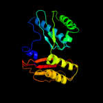
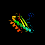
1 c2qbfC_
100.0
100
PDB header: ribosomeChain: C: PDB Molecule: 30s ribosomal protein s3;PDBTitle: crystal structure of the bacterial ribosome from escherichia2 coli in complex with ribosome recycling factor (rrf). this3 file contains the 30s subunit of the second 70s ribosome.4 the entire crystal structure contains two 70s ribosomes and5 is described in remark 400.
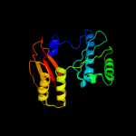
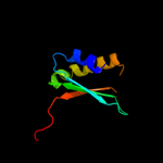
2 c1pnxC_
100.0
52
PDB header: ribosomeChain: C: PDB Molecule: 30s ribosomal protein s3;PDBTitle: crystal structure of the wild type ribosome from e. coli,2 30s subunit of 70s ribosome. this file, 1pnx, contains3 only molecules of the 30s ribosomal subunit. the 50s4 subunit is in the pdb file 1pny.
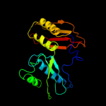
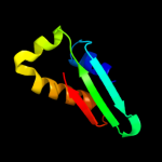
3 c2gy9C_
100.0
100
PDB header: ribosomeChain: C: PDB Molecule: 30s ribosomal subunit protein s3;PDBTitle: structure of the 30s subunit of a pre-translocational e.2 coli ribosome obtained by fitting atomic models for rna and3 protein components into cryo-em map emd-1056
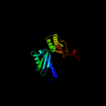
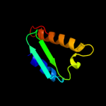
4 c1hnwC_
100.0
52
PDB header: ribosomeChain: C: PDB Molecule: 30s ribosomal protein s3;PDBTitle: structure of the thermus thermophilus 30s ribosomal subunit2 in complex with tetracycline
5 c3bbnC_
100.0
42
PDB header: ribosomeChain: C: PDB Molecule: ribosomal protein s3;PDBTitle: homology model for the spinach chloroplast 30s subunit2 fitted to 9.4a cryo-em map of the 70s chlororibosome.
6 c2xznC_
100.0
21
PDB header: ribosomeChain: C: PDB Molecule: kh domain containing protein;PDBTitle: crystal structure of the eukaryotic 40s ribosomal2 subunit in complex with initiation factor 1. this file3 contains the 40s subunit and initiation factor for4 molecule 2
7 c2zkqc_
100.0
25
PDB header: ribosomal protein/rnaChain: C: PDB Molecule: rna expansion segment es4;PDBTitle: structure of a mammalian ribosomal 40s subunit within an2 80s complex obtained by docking homology models of the rna3 and proteins into an 8.7 a cryo-em map
8 c1s1hC_
100.0
25
PDB header: ribosomeChain: C: PDB Molecule: 40s ribosomal protein s3;PDBTitle: structure of the ribosomal 80s-eef2-sordarin complex from2 yeast obtained by docking atomic models for rna and protein3 components into a 11.7 a cryo-em map. this file, 1s1h,4 contains 40s subunit. the 60s ribosomal subunit is in file5 1s1i.
9 c3izbB_
100.0
26
PDB header: ribosomeChain: B: PDB Molecule: 40s ribosomal protein rps3 (s3p);PDBTitle: localization of the small subunit ribosomal proteins into a 6.1 a2 cryo-em map of saccharomyces cerevisiae translating 80s ribosome
10 d2uubc2
100.0
62
Fold: Ribosomal protein S3 C-terminal domainSuperfamily: Ribosomal protein S3 C-terminal domainFamily: Ribosomal protein S3 C-terminal domain11 d2qalc2
100.0
100
Fold: Ribosomal protein S3 C-terminal domainSuperfamily: Ribosomal protein S3 C-terminal domainFamily: Ribosomal protein S3 C-terminal domain12 d2qalc1
100.0
100
Fold: Alpha-lytic protease prodomain-likeSuperfamily: Prokaryotic type KH domain (KH-domain type II)Family: Prokaryotic type KH domain (KH-domain type II)13 d2uubc1
100.0
43
Fold: Alpha-lytic protease prodomain-likeSuperfamily: Prokaryotic type KH domain (KH-domain type II)Family: Prokaryotic type KH domain (KH-domain type II)14 d1wh9a_
99.4
22
Fold: Alpha-lytic protease prodomain-likeSuperfamily: Prokaryotic type KH domain (KH-domain type II)Family: Prokaryotic type KH domain (KH-domain type II)15 c3gkuB_
94.2
18
PDB header: rna binding proteinChain: B: PDB Molecule: probable rna-binding protein;PDBTitle: crystal structure of a probable rna-binding protein from clostridium2 symbiosum atcc 14940
16 c2pt7G_
92.7
15
PDB header: hydrolase/protein bindingChain: G: PDB Molecule: hypothetical protein;PDBTitle: crystal structure of cag virb11 (hp0525) and an inhibitory protein2 (hp1451)
17 d1hh2p3
58.2
26
Fold: Alpha-lytic protease prodomain-likeSuperfamily: Prokaryotic type KH domain (KH-domain type II)Family: Prokaryotic type KH domain (KH-domain type II)18 d2asba3
53.9
21
Fold: Alpha-lytic protease prodomain-likeSuperfamily: Prokaryotic type KH domain (KH-domain type II)Family: Prokaryotic type KH domain (KH-domain type II)19 d1tuaa1
32.8
18
Fold: Eukaryotic type KH-domain (KH-domain type I)Superfamily: Eukaryotic type KH-domain (KH-domain type I)Family: Eukaryotic type KH-domain (KH-domain type I)20 c2hh3A_
27.3
32
PDB header: rna binding proteinChain: A: PDB Molecule: kh-type splicing regulatory protein;PDBTitle: solution structure of the third kh domain of ksrp
21 c2hh2A_
not modelled
26.2
33
PDB header: rna binding proteinChain: A: PDB Molecule: kh-type splicing regulatory protein;PDBTitle: solution structure of the fourth kh domain of ksrp
22 c2cy1A_
not modelled
26.1
22
PDB header: transcriptionChain: A: PDB Molecule: nusa protein homolog;PDBTitle: crystal structure of ape1850
23 d2axya1
not modelled
26.1
43
Fold: Eukaryotic type KH-domain (KH-domain type I)Superfamily: Eukaryotic type KH-domain (KH-domain type I)Family: Eukaryotic type KH-domain (KH-domain type I)24 c2asbA_
not modelled
22.7
21
PDB header: transcription/rnaChain: A: PDB Molecule: transcription elongation protein nusa;PDBTitle: structure of a mycobacterium tuberculosis nusa-rna complex
25 d1dt4a_
not modelled
22.6
15
Fold: Eukaryotic type KH-domain (KH-domain type I)Superfamily: Eukaryotic type KH-domain (KH-domain type I)Family: Eukaryotic type KH-domain (KH-domain type I)26 d1j4wa1
not modelled
21.4
39
Fold: Eukaryotic type KH-domain (KH-domain type I)Superfamily: Eukaryotic type KH-domain (KH-domain type I)Family: Eukaryotic type KH-domain (KH-domain type I)27 c2anrA_
not modelled
19.7
33
PDB header: rna-binding protein/rnaChain: A: PDB Molecule: neuro-oncological ventral antigen 1;PDBTitle: crystal structure (ii) of nova-1 kh1/kh2 domain tandem with 25nt rna2 hairpin
28 d1j4wa2
not modelled
19.3
26
Fold: Eukaryotic type KH-domain (KH-domain type I)Superfamily: Eukaryotic type KH-domain (KH-domain type I)Family: Eukaryotic type KH-domain (KH-domain type I)29 d1dtja_
not modelled
18.4
15
Fold: Eukaryotic type KH-domain (KH-domain type I)Superfamily: Eukaryotic type KH-domain (KH-domain type I)Family: Eukaryotic type KH-domain (KH-domain type I)30 d1khma_
not modelled
17.8
30
Fold: Eukaryotic type KH-domain (KH-domain type I)Superfamily: Eukaryotic type KH-domain (KH-domain type I)Family: Eukaryotic type KH-domain (KH-domain type I)31 d1ec6a_
not modelled
17.6
15
Fold: Eukaryotic type KH-domain (KH-domain type I)Superfamily: Eukaryotic type KH-domain (KH-domain type I)Family: Eukaryotic type KH-domain (KH-domain type I)32 d1wf3a2
not modelled
17.4
17
Fold: Alpha-lytic protease prodomain-likeSuperfamily: Prokaryotic type KH domain (KH-domain type II)Family: Prokaryotic type KH domain (KH-domain type II)33 d1we8a_
not modelled
16.2
19
Fold: Eukaryotic type KH-domain (KH-domain type I)Superfamily: Eukaryotic type KH-domain (KH-domain type I)Family: Eukaryotic type KH-domain (KH-domain type I)34 c1j4wA_
not modelled
15.5
41
PDB header: transcription/dnaChain: A: PDB Molecule: fuse binding protein;PDBTitle: complex of the kh3 and kh4 domains of fbp with a2 single_stranded 29mer dna oligonucleotide from the fuse3 element of the c-myc oncogene
35 c1ztgD_
not modelled
15.3
44
PDB header: dna, rna binding protein/dnaChain: D: PDB Molecule: poly(rc)-binding protein 1;PDBTitle: human alpha polyc binding protein kh1
36 d1wvna1
not modelled
15.2
26
Fold: Eukaryotic type KH-domain (KH-domain type I)Superfamily: Eukaryotic type KH-domain (KH-domain type I)Family: Eukaryotic type KH-domain (KH-domain type I)37 d1viga_
not modelled
15.0
16
Fold: Eukaryotic type KH-domain (KH-domain type I)Superfamily: Eukaryotic type KH-domain (KH-domain type I)Family: Eukaryotic type KH-domain (KH-domain type I)38 d1zzka1
not modelled
14.0
33
Fold: Eukaryotic type KH-domain (KH-domain type I)Superfamily: Eukaryotic type KH-domain (KH-domain type I)Family: Eukaryotic type KH-domain (KH-domain type I)39 d1x4na1
not modelled
13.9
19
Fold: Eukaryotic type KH-domain (KH-domain type I)Superfamily: Eukaryotic type KH-domain (KH-domain type I)Family: Eukaryotic type KH-domain (KH-domain type I)40 c1tuaA_
not modelled
13.7
31
PDB header: structural genomics, unknown functionChain: A: PDB Molecule: hypothetical protein ape0754;PDBTitle: 1.5 a crystal structure of a protein of unknown function2 ape0754 from aeropyrum pernix
41 c2jvfA_
not modelled
13.7
19
PDB header: de novo proteinChain: A: PDB Molecule: de novo protein m7;PDBTitle: solution structure of m7, a computationally-designed2 artificial protein
42 d2ctla1
not modelled
13.2
25
Fold: Eukaryotic type KH-domain (KH-domain type I)Superfamily: Eukaryotic type KH-domain (KH-domain type I)Family: Eukaryotic type KH-domain (KH-domain type I)43 d2ba0a3
not modelled
12.7
39
Fold: Eukaryotic type KH-domain (KH-domain type I)Superfamily: Eukaryotic type KH-domain (KH-domain type I)Family: Eukaryotic type KH-domain (KH-domain type I)44 d1x4ma1
not modelled
12.3
33
Fold: Eukaryotic type KH-domain (KH-domain type I)Superfamily: Eukaryotic type KH-domain (KH-domain type I)Family: Eukaryotic type KH-domain (KH-domain type I)45 c2dgrA_
not modelled
12.1
26
PDB header: rna binding proteinChain: A: PDB Molecule: ring finger and kh domain-containing protein 1;PDBTitle: solution structure of the second kh domain in ring finger2 and kh domain containing protein 1
46 c1k0rB_
not modelled
11.8
21
PDB header: transcriptionChain: B: PDB Molecule: nusa;PDBTitle: crystal structure of mycobacterium tuberculosis nusa
47 d2ctja1
not modelled
11.6
22
Fold: Eukaryotic type KH-domain (KH-domain type I)Superfamily: Eukaryotic type KH-domain (KH-domain type I)Family: Eukaryotic type KH-domain (KH-domain type I)48 c3af5A_
not modelled
11.2
19
PDB header: hydrolaseChain: A: PDB Molecule: putative uncharacterized protein ph1404;PDBTitle: the crystal structure of an archaeal cpsf subunit, ph1404 from2 pyrococcus horikoshii
49 c3krmB_
not modelled
11.1
33
PDB header: rna binding proteinChain: B: PDB Molecule: insulin-like growth factor 2 mrna-binding proteinPDBTitle: imp1 kh34
50 c1hh2P_
not modelled
11.1
26
PDB header: transcription regulationChain: P: PDB Molecule: n utilization substance protein a;PDBTitle: crystal structure of nusa from thermotoga maritima
51 d2ctea1
not modelled
10.6
33
Fold: Eukaryotic type KH-domain (KH-domain type I)Superfamily: Eukaryotic type KH-domain (KH-domain type I)Family: Eukaryotic type KH-domain (KH-domain type I)52 d2cmea1
not modelled
10.6
50
Fold: SARS ORF9b-likeSuperfamily: 'SARS ORF9b-likeFamily: SARS ORF9b-like53 c2fenA_
not modelled
10.6
43
PDB header: isomeraseChain: A: PDB Molecule: 3-carboxy-cis,cis-muconate lactonizing enzyme;PDBTitle: 3-carboxy-cis,cis-muconate lactonizing enzyme from agrobacterium2 radiobacter s2
54 c2qndA_
not modelled
10.3
24
PDB header: rna binding proteinChain: A: PDB Molecule: fmr1 protein;PDBTitle: crystal structure of the kh1-kh2 domains from human fragile x mental2 retardation protein
55 d2ctka1
not modelled
10.1
27
Fold: Eukaryotic type KH-domain (KH-domain type I)Superfamily: Eukaryotic type KH-domain (KH-domain type I)Family: Eukaryotic type KH-domain (KH-domain type I)56 c2jzxA_
not modelled
9.8
46
PDB header: rna binding proteinChain: A: PDB Molecule: poly(rc)-binding protein 2;PDBTitle: pcbp2 kh1-kh2 domains
57 c2jvzA_
not modelled
9.3
36
PDB header: splicingChain: A: PDB Molecule: far upstream element-binding protein 2;PDBTitle: solution nmr structure of the second and third kh domains2 of ksrp
58 c1l2fA_
not modelled
9.3
26
PDB header: transcriptionChain: A: PDB Molecule: n utilization substance protein a;PDBTitle: crystal structure of nusa from thermotoga maritima: a2 structure-based role of the n-terminal domain
59 c2w2rA_
not modelled
9.1
15
PDB header: viral proteinChain: A: PDB Molecule: matrix protein;PDBTitle: structure of the vesicular stomatitis virus matrix protein
60 d1hsta_
not modelled
8.7
14
Fold: DNA/RNA-binding 3-helical bundleSuperfamily: "Winged helix" DNA-binding domainFamily: Linker histone H1/H561 d2ctma1
not modelled
8.6
19
Fold: Eukaryotic type KH-domain (KH-domain type I)Superfamily: Eukaryotic type KH-domain (KH-domain type I)Family: Eukaryotic type KH-domain (KH-domain type I)62 d1egaa2
not modelled
8.2
25
Fold: Alpha-lytic protease prodomain-likeSuperfamily: Prokaryotic type KH domain (KH-domain type II)Family: Prokaryotic type KH domain (KH-domain type II)63 d1myna_
not modelled
8.1
42
Fold: Knottins (small inhibitors, toxins, lectins)Superfamily: Scorpion toxin-likeFamily: Insect defensins64 c1egaB_
not modelled
7.6
18
PDB header: hydrolaseChain: B: PDB Molecule: protein (gtp-binding protein era);PDBTitle: crystal structure of a widely conserved gtpase era
65 d2gysa1
not modelled
7.5
40
Fold: Immunoglobulin-like beta-sandwichSuperfamily: Fibronectin type IIIFamily: Fibronectin type III66 d1c3ca_
not modelled
7.4
43
Fold: L-aspartase-likeSuperfamily: L-aspartase-likeFamily: L-aspartase/fumarase67 c1qysA_
not modelled
7.3
13
PDB header: de novo proteinChain: A: PDB Molecule: top7;PDBTitle: crystal structure of top7: a computationally designed2 protein with a novel fold
68 c2pfmA_
not modelled
7.2
43
PDB header: lyaseChain: A: PDB Molecule: adenylosuccinate lyase;PDBTitle: crystal structure of adenylosuccinate lyase (purb) from bacillus2 anthracis
69 d2ctfa1
not modelled
6.9
26
Fold: Eukaryotic type KH-domain (KH-domain type I)Superfamily: Eukaryotic type KH-domain (KH-domain type I)Family: Eukaryotic type KH-domain (KH-domain type I)70 c3gtdB_
not modelled
6.7
57
PDB header: lyaseChain: B: PDB Molecule: fumarate hydratase class ii;PDBTitle: 2.4 angstrom crystal structure of fumarate hydratase from rickettsia2 prowazekii
71 d1tj7a_
not modelled
6.7
50
Fold: L-aspartase-likeSuperfamily: L-aspartase-likeFamily: L-aspartase/fumarase72 d1lg7a_
not modelled
6.6
12
Fold: VSV matrix proteinSuperfamily: VSV matrix proteinFamily: VSV matrix protein73 d1tjva_
not modelled
6.1
50
Fold: L-aspartase-likeSuperfamily: L-aspartase-likeFamily: L-aspartase/fumarase74 d2fmra_
not modelled
6.0
38
Fold: Eukaryotic type KH-domain (KH-domain type I)Superfamily: Eukaryotic type KH-domain (KH-domain type I)Family: Eukaryotic type KH-domain (KH-domain type I)75 c3ocfB_
not modelled
6.0
57
PDB header: lyaseChain: B: PDB Molecule: fumarate lyase:delta crystallin;PDBTitle: crystal structure of fumarate lyase:delta crystallin from brucella2 melitensis in native form
76 c3ej9D_
not modelled
5.9
44
PDB header: hydrolaseChain: D: PDB Molecule: beta-subunit of trans-3-chloroacrylic acid dehalogenase;PDBTitle: structural and mechanistic analysis of trans-3-chloroacrylic acid2 dehalogenase activity
77 d1f1oa_
not modelled
5.4
43
Fold: L-aspartase-likeSuperfamily: L-aspartase-likeFamily: L-aspartase/fumarase78 d1eu3a2
not modelled
5.3
19
Fold: beta-Grasp (ubiquitin-like)Superfamily: Superantigen toxins, C-terminal domainFamily: Superantigen toxins, C-terminal domain79 c3no9C_
not modelled
5.3
43
PDB header: lyaseChain: C: PDB Molecule: fumarate hydratase class ii;PDBTitle: crystal structure of apo fumarate hydratase from mycobacterium2 tuberculosis
80 d1dofa_
not modelled
5.2
14
Fold: L-aspartase-likeSuperfamily: L-aspartase-likeFamily: L-aspartase/fumarase81 c3e04C_
not modelled
5.1
57
PDB header: lyaseChain: C: PDB Molecule: fumarate hydratase;PDBTitle: crystal structure of human fumarate hydratase
82 d1yfma_
not modelled
5.0
57
Fold: L-aspartase-likeSuperfamily: L-aspartase-likeFamily: L-aspartase/fumarase