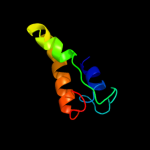
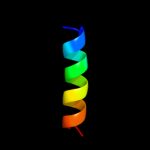
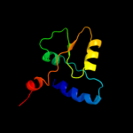
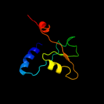
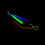
1 d2cx6a1
32.0
19
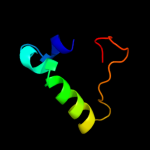
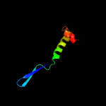
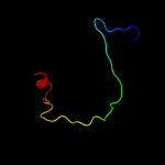
Fold: Barstar-likeSuperfamily: Barstar-relatedFamily: Barstar-related2 d3saka_
26.9
24
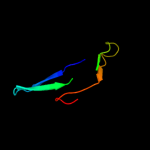
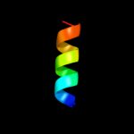
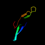
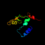
Fold: p53 tetramerization domainSuperfamily: p53 tetramerization domainFamily: p53 tetramerization domain3 c3bvhE_
25.2
15
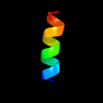
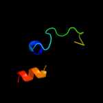
PDB header: blood clottingChain: E: PDB Molecule: fibrinogen beta chain;PDBTitle: crystal structure of recombinant gammad364a fibrinogen fragment d with2 the peptide ligand gly-pro-arg-pro-amide
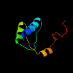
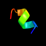
4 d2o3aa1
24.3
17
Fold: alpha/beta knotSuperfamily: alpha/beta knotFamily: AF0751-like5 c3peiA_
22.8
20
PDB header: hydrolaseChain: A: PDB Molecule: cytosol aminopeptidase;PDBTitle: crystal structure of cytosol aminopeptidase from francisella2 tularensis
6 c3nw0B_
22.0
14
PDB header: metal binding proteinChain: B: PDB Molecule: melanoma-associated antigen g1;PDBTitle: crystal structure of mageg1 and nse1 complex
7 c3zy1A_
20.4
43
PDB header: transcriptionChain: A: PDB Molecule: tumor protein 63;PDBTitle: crystal structure of the human p63 tetramerization domain
8 c4a9zD_
20.0
43
PDB header: transcriptionChain: D: PDB Molecule: tumor protein 63;PDBTitle: crystal structure of human p63 tetramerization domain
9 d1gyta2
19.9
20
Fold: Phosphorylase/hydrolase-likeSuperfamily: Zn-dependent exopeptidasesFamily: Leucine aminopeptidase, C-terminal domain10 d1zc6a2
19.8
17
Fold: Ribonuclease H-like motifSuperfamily: Actin-like ATPase domainFamily: BadF/BadG/BcrA/BcrD-like11 c2hpcH_
19.5
15
PDB header: blood clottingChain: H: PDB Molecule: fibrinogen beta chain;PDBTitle: crystal structure of fragment d from human fibrinogen complexed with2 gly-pro-arg-pro-amide.
12 c3qu3A_
18.8
33
PDB header: dna binding proteinChain: A: PDB Molecule: interferon regulatory factor 7;PDBTitle: crystal structure of irf-7 dbd apo form
13 c1lwuH_
18.0
10
PDB header: blood clottingChain: H: PDB Molecule: fibrinogen beta chain;PDBTitle: crystal structure of fragment d from lamprey fibrinogen complexed with2 the peptide gly-his-arg-pro-amide
14 c3d35A_
17.1
16
PDB header: transferaseChain: A: PDB Molecule: regulator of ty1 transposition protein 109;PDBTitle: crystal structure of rtt109-ac-coa complex
15 c3h8gC_
17.0
16
PDB header: hydrolaseChain: C: PDB Molecule: cytosol aminopeptidase;PDBTitle: bestatin complex structure of leucine aminopeptidase from pseudomonas2 putida
16 d2fh1a2
16.3
56
Fold: Gelsolin-likeSuperfamily: Actin depolymerizing proteinsFamily: Gelsolin-like17 c2c4rL_
15.9
50
PDB header: hydrolaseChain: L: PDB Molecule: ribonuclease e;PDBTitle: catalytic domain of e. coli rnase e
18 c3rayA_
15.5
13
PDB header: transcriptionChain: A: PDB Molecule: pr domain-containing protein 11;PDBTitle: crystal structure of methyltransferase domain of human pr domain-2 containing protein 11
19 c3jruB_
15.0
19
PDB header: hydrolaseChain: B: PDB Molecule: probable cytosol aminopeptidase;PDBTitle: crystal structure of leucyl aminopeptidase (pepa) from xoo0834,2 xanthomonas oryzae pv. oryzae kacc10331
20 c3kr5E_
14.5
20
PDB header: hydrolaseChain: E: PDB Molecule: m17 leucyl aminopeptidase;PDBTitle: structure of a protease 4
21 d1v8ca2
not modelled
14.1
35
Fold: TBP-likeSuperfamily: MoaD-related protein, C-terminal domainFamily: MoaD-related protein, C-terminal domain22 c2gutA_
not modelled
13.4
29
PDB header: transcriptionChain: A: PDB Molecule: arc/mediator, positive cofactor 2 glutamine/q-PDBTitle: solution structure of the trans-activation domain of the2 human co-activator arc105
23 d1lama1
not modelled
13.3
16
Fold: Phosphorylase/hydrolase-likeSuperfamily: Zn-dependent exopeptidasesFamily: Leucine aminopeptidase, C-terminal domain24 c2vcpE_
not modelled
13.1
27
PDB header: structural proteinChain: E: PDB Molecule: neural wiskott-aldrich syndrome protein;PDBTitle: crystal structure of n-wasp vc domain in complex with2 skeletal actin
25 d1d0na5
not modelled
13.0
45
Fold: Gelsolin-likeSuperfamily: Actin depolymerizing proteinsFamily: Gelsolin-like26 d1e7ua2
not modelled
12.9
8
Fold: C2 domain-likeSuperfamily: C2 domain (Calcium/lipid-binding domain, CaLB)Family: PLC-like (P variant)27 c3ghgK_
not modelled
12.6
15
PDB header: blood clottingChain: K: PDB Molecule: fibrinogen beta chain;PDBTitle: crystal structure of human fibrinogen
28 c1gytG_
not modelled
12.4
20
PDB header: hydrolaseChain: G: PDB Molecule: cytosol aminopeptidase;PDBTitle: e. coli aminopeptidase a (pepa)
29 d1npha2
not modelled
12.2
56
Fold: Gelsolin-likeSuperfamily: Actin depolymerizing proteinsFamily: Gelsolin-like30 d2a90a2
not modelled
11.1
43
Fold: WWE domainSuperfamily: WWE domainFamily: WWE domain31 c3k13A_
not modelled
11.1
24
PDB header: transferaseChain: A: PDB Molecule: 5-methyltetrahydrofolate-homocysteine methyltransferase;PDBTitle: structure of the pterin-binding domain metr of 5-2 methyltetrahydrofolate-homocysteine methyltransferase from3 bacteroides thetaiotaomicron
32 d1lwub1
not modelled
11.0
10
Fold: Fibrinogen C-terminal domain-likeSuperfamily: Fibrinogen C-terminal domain-likeFamily: Fibrinogen C-terminal domain-like33 c3nztA_
not modelled
10.9
27
PDB header: ligaseChain: A: PDB Molecule: glutamate--cysteine ligase;PDBTitle: 2.0 angstrom crystal structure of glutamate--cysteine ligase (gsha)2 ftom francisella tularensis in complex with amp
34 d2tsra_
not modelled
10.8
20
Fold: Thymidylate synthase/dCMP hydroxymethylaseSuperfamily: Thymidylate synthase/dCMP hydroxymethylaseFamily: Thymidylate synthase/dCMP hydroxymethylase35 c1ei3E_
not modelled
10.8
15
PDB header: PDB COMPND: 36 c2dymE_
not modelled
10.6
17
PDB header: protein turnover/protein turnoverChain: E: PDB Molecule: autophagy protein 5;PDBTitle: the crystal structure of saccharomyces cerevisiae atg5-2 atg16(1-46) complex
37 c3fpjA_
not modelled
10.5
19
PDB header: biosynthetic protein, transferaseChain: A: PDB Molecule: putative uncharacterized protein;PDBTitle: crystal structure of e81q mutant of mtnas in complex with s-2 adenosylmethionine
38 d2czla1
not modelled
10.5
12
Fold: Periplasmic binding protein-like IISuperfamily: Periplasmic binding protein-like IIFamily: Phosphate binding protein-like39 c3cz7A_
not modelled
10.3
16
PDB header: replicationChain: A: PDB Molecule: regulator of ty1 transposition protein 109;PDBTitle: molecular basis for the autoregulation of the protein acetyl2 transferase rtt109
40 c2zfnA_
not modelled
9.9
16
PDB header: transferaseChain: A: PDB Molecule: regulator of ty1 transposition protein 109;PDBTitle: self-acetylation mediated histone h3 lysine 56 acetylation by rtt109
41 c3mwbA_
not modelled
9.7
14
PDB header: lyaseChain: A: PDB Molecule: prephenate dehydratase;PDBTitle: the crystal structure of prephenate dehydratase in complex with l-phe2 from arthrobacter aurescens to 2.0a
42 d1kzfa_
not modelled
9.6
17
Fold: Acyl-CoA N-acyltransferases (Nat)Superfamily: Acyl-CoA N-acyltransferases (Nat)Family: Autoinducer synthetase43 c3luyA_
not modelled
9.4
20
PDB header: isomeraseChain: A: PDB Molecule: probable chorismate mutase;PDBTitle: putative chorismate mutase from bifidobacterium adolescentis
44 d1wh6a_
not modelled
9.4
22
Fold: lambda repressor-like DNA-binding domainsSuperfamily: lambda repressor-like DNA-binding domainsFamily: CUT domain45 d2it9a1
not modelled
9.3
21
Fold: ssDNA-binding transcriptional regulator domainSuperfamily: ssDNA-binding transcriptional regulator domainFamily: PMN2A0962/syc2379c-like46 c3ix6B_
not modelled
9.1
26
PDB header: transferaseChain: B: PDB Molecule: thymidylate synthase;PDBTitle: crystal structure of thymidylate synthase thya from brucella2 melitensis
47 c2hc9A_
not modelled
8.7
6
PDB header: hydrolaseChain: A: PDB Molecule: leucine aminopeptidase 1;PDBTitle: structure of caenorhabditis elegans leucine aminopeptidase-zinc2 complex (lap1)
48 c2a3zC_
not modelled
8.6
31
PDB header: structural proteinChain: C: PDB Molecule: wiskott-aldrich syndrome protein;PDBTitle: ternary complex of the wh2 domain of wasp with actin-dnase i
49 c2kerA_
not modelled
8.6
38
PDB header: hydrolase inhibitorChain: A: PDB Molecule: alpha-amylase inhibitor z-2685;PDBTitle: alpha-amylase inhibitor parvulustat (z-2685) from2 streptomyces parvulus
50 d1tf5a1
not modelled
8.4
17
Fold: Pre-protein crosslinking domain of SecASuperfamily: Pre-protein crosslinking domain of SecAFamily: Pre-protein crosslinking domain of SecA51 d2o0ma1
not modelled
8.3
9
Fold: NagB/RpiA/CoA transferase-likeSuperfamily: NagB/RpiA/CoA transferase-likeFamily: SorC sugar-binding domain-like52 c2o0mA_
not modelled
8.3
9
PDB header: transcriptionChain: A: PDB Molecule: transcriptional regulator, sorc family;PDBTitle: the crystal structure of the putative sorc family transcriptional2 regulator from enterococcus faecalis
53 d1nkta1
not modelled
8.2
18
Fold: Pre-protein crosslinking domain of SecASuperfamily: Pre-protein crosslinking domain of SecAFamily: Pre-protein crosslinking domain of SecA54 d1ok0a_
not modelled
8.2
50
Fold: alpha-Amylase inhibitor tendamistatSuperfamily: alpha-Amylase inhibitor tendamistatFamily: alpha-Amylase inhibitor tendamistat55 d1fzda_
not modelled
8.0
10
Fold: Fibrinogen C-terminal domain-likeSuperfamily: Fibrinogen C-terminal domain-likeFamily: Fibrinogen C-terminal domain-like56 d1uw4a_
not modelled
8.0
4
Fold: Ferredoxin-likeSuperfamily: RNA-binding domain, RBDFamily: Smg-4/UPF357 d1v66a_
not modelled
7.7
36
Fold: LEM/SAP HeH motifSuperfamily: SAP domainFamily: SAP domain58 d1m1jb1
not modelled
7.5
15
Fold: Fibrinogen C-terminal domain-likeSuperfamily: Fibrinogen C-terminal domain-likeFamily: Fibrinogen C-terminal domain-like59 c2e1nA_
not modelled
7.5
23
PDB header: circadian clock proteinChain: A: PDB Molecule: pex;PDBTitle: crystal structure of the cyanobacterium circadian clock modifier pex
60 d2a90a1
not modelled
7.5
7
Fold: WWE domainSuperfamily: WWE domainFamily: WWE domain61 d2oz4a2
not modelled
7.4
13
Fold: Immunoglobulin-like beta-sandwichSuperfamily: ImmunoglobulinFamily: I set domains62 d1yvoa1
not modelled
7.1
11
Fold: Acyl-CoA N-acyltransferases (Nat)Superfamily: Acyl-CoA N-acyltransferases (Nat)Family: N-acetyl transferase, NAT63 d1o6la_
not modelled
7.0
15
Fold: Protein kinase-like (PK-like)Superfamily: Protein kinase-like (PK-like)Family: Protein kinases, catalytic subunit64 d1oe4a_
not modelled
6.8
30
Fold: Uracil-DNA glycosylase-likeSuperfamily: Uracil-DNA glycosylase-likeFamily: Single-strand selective monofunctional uracil-DNA glycosylase SMUG165 c3dwdB_
not modelled
6.8
20
PDB header: transport proteinChain: B: PDB Molecule: adp-ribosylation factor gtpase-activating protein 1;PDBTitle: crystal structure of the arfgap domain of human arfgap1
66 d1t62a_
not modelled
6.7
29
Fold: PUA domain-likeSuperfamily: PUA domain-likeFamily: Hypothetical protein EF313367 d2qqsa2
not modelled
6.7
24
Fold: SH3-like barrelSuperfamily: Tudor/PWWP/MBTFamily: Tudor domain68 c3jqxA_
not modelled
6.7
9
PDB header: cell adhesionChain: A: PDB Molecule: colh protein;PDBTitle: crystal structure of clostridium histolyticum colh collagenase2 collagen binding domain 3 at 2.2 angstrom resolution in the presence3 of calcium and cadmium
69 c3a9lB_
not modelled
6.6
14
PDB header: hydrolaseChain: B: PDB Molecule: poly-gamma-glutamate hydrolase;PDBTitle: structure of bacteriophage poly-gamma-glutamate hydrolase
70 c3km3B_
not modelled
6.6
19
PDB header: hydrolaseChain: B: PDB Molecule: deoxycytidine triphosphate deaminase;PDBTitle: crystal structure of eoxycytidine triphosphate deaminase from2 anaplasma phagocytophilum at 2.1a resolution
71 d1n6za_
not modelled
6.5
40
Fold: Hypothetical protein Yml108wSuperfamily: Hypothetical protein Yml108wFamily: Hypothetical protein Yml108w72 d1ufwa_
not modelled
6.5
17
Fold: Ferredoxin-likeSuperfamily: RNA-binding domain, RBDFamily: Canonical RBD73 c1vbiA_
not modelled
6.4
30
PDB header: oxidoreductaseChain: A: PDB Molecule: type 2 malate/lactate dehydrogenase;PDBTitle: crystal structure of type 2 malate/lactate dehydrogenase from thermus2 thermophilus hb8
74 d1tqza1
not modelled
6.4
29
Fold: PH domain-like barrelSuperfamily: PH domain-likeFamily: Necap1 N-terminal domain-like75 c1jjoE_
not modelled
6.3
9
PDB header: signaling proteinChain: E: PDB Molecule: neuroserpin;PDBTitle: crystal structure of mouse neuroserpin (cleaved form)
76 c2yy8B_
not modelled
6.1
17
PDB header: transferaseChain: B: PDB Molecule: upf0106 protein ph0461;PDBTitle: crystal structure of archaeal trna-methylase for position2 56 (atrm56) from pyrococcus horikoshii, complexed with s-3 adenosyl-l-methionine
77 c3m3nW_
not modelled
6.1
31
PDB header: structural proteinChain: W: PDB Molecule: neural wiskott-aldrich syndrome protein;PDBTitle: structure of a longitudinal actin dimer assembled by tandem w domains
78 c3f02C_
not modelled
6.0
14
PDB header: hydrolase inhibitorChain: C: PDB Molecule: neuroserpin;PDBTitle: cleaved human neuroserpin
79 d1v8ha1
not modelled
6.0
22
Fold: Immunoglobulin-like beta-sandwichSuperfamily: E set domainsFamily: SoxZ-like80 d1x4fa1
not modelled
6.0
8
Fold: Ferredoxin-likeSuperfamily: RNA-binding domain, RBDFamily: Canonical RBD81 d1ua4a_
not modelled
5.9
15
Fold: Ribokinase-likeSuperfamily: Ribokinase-likeFamily: ADP-specific Phosphofructokinase/Glucokinase82 c3lyrA_
not modelled
5.9
25
PDB header: transcription activatorChain: A: PDB Molecule: transcription factor coe1;PDBTitle: human early b-cell factor 1 (ebf1) dna-binding domain
83 c3kgbA_
not modelled
5.9
15
PDB header: transferaseChain: A: PDB Molecule: thymidylate synthase 1/2;PDBTitle: crystal structure of thymidylate synthase 1/2 from encephalitozoon2 cuniculi at 2.2 a resolution
84 c2jx2A_
not modelled
5.8
14
PDB header: transcriptionChain: A: PDB Molecule: negative elongation factor e;PDBTitle: solution conformation of rna-bound nelf-e rrm
85 d1l2la_
not modelled
5.8
18
Fold: Ribokinase-likeSuperfamily: Ribokinase-likeFamily: ADP-specific Phosphofructokinase/Glucokinase86 c1deqO_
not modelled
5.8
15
PDB header: PDB COMPND: 87 c3s9xA_
not modelled
5.7
21
PDB header: structural genomics, unknown functionChain: A: PDB Molecule: asch domain;PDBTitle: high resolution crystal structure of asch domain from lactobacillus2 crispatus jv v101
88 c2qsrA_
not modelled
5.7
15
PDB header: transcriptionChain: A: PDB Molecule: transcription-repair coupling factor;PDBTitle: crystal structure of c-terminal domain of transcription-repair2 coupling factor
89 d1umya_
not modelled
5.6
27
Fold: TIM beta/alpha-barrelSuperfamily: Homocysteine S-methyltransferaseFamily: Homocysteine S-methyltransferase90 d2irfg_
not modelled
5.5
30
Fold: DNA/RNA-binding 3-helical bundleSuperfamily: "Winged helix" DNA-binding domainFamily: Interferon regulatory factor91 c1hleB_
not modelled
5.5
15
PDB header: hydrolase inhibitor(serine proteinase)Chain: B: PDB Molecule: horse leukocyte elastase inhibitor;PDBTitle: crystal structure of cleaved equine leucocyte elastase2 inhibitor determined at 1.95 angstroms resolution
92 c2bn5A_
not modelled
5.5
50
PDB header: nuclear proteinChain: A: PDB Molecule: psi;PDBTitle: p-element somatic inhibitor protein complex with u1-70k2 proline-rich peptide
93 d1duvg1
not modelled
5.5
22
Fold: ATC-likeSuperfamily: Aspartate/ornithine carbamoyltransferaseFamily: Aspartate/ornithine carbamoyltransferase94 d2o4aa1
not modelled
5.4
9
Fold: lambda repressor-like DNA-binding domainsSuperfamily: lambda repressor-like DNA-binding domainsFamily: CUT domain95 d1px5a1
not modelled
5.4
18
Fold: PAP/OAS1 substrate-binding domainSuperfamily: PAP/OAS1 substrate-binding domainFamily: 2'-5'-oligoadenylate synthetase 1, OAS1, second domain96 d1tswa_
not modelled
5.4
23
Fold: Thymidylate synthase/dCMP hydroxymethylaseSuperfamily: Thymidylate synthase/dCMP hydroxymethylaseFamily: Thymidylate synthase/dCMP hydroxymethylase97 d2b4jc1
not modelled
5.3
9
Fold: N-cbl likeSuperfamily: HIV integrase-binding domainFamily: HIV integrase-binding domain98 c1wtjB_
not modelled
5.3
15
PDB header: oxidoreductaseChain: B: PDB Molecule: ureidoglycolate dehydrogenase;PDBTitle: crystal structure of delta1-piperideine-2-carboxylate2 reductase from pseudomonas syringae pvar.tomato
99 c2h4qB_
not modelled
5.3
20
PDB header: hydrolase inhibitorChain: B: PDB Molecule: heterochromatin-associated protein ment;PDBTitle: crystal structure of a m-loop deletion variant of ment in2 the cleaved conformation


