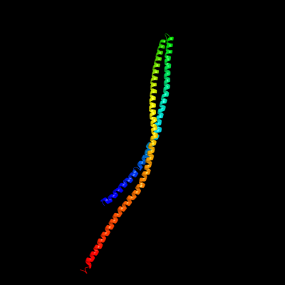
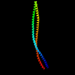
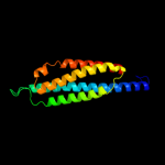
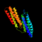
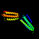
1 c1qu7A_
99.9
84
PDB header: signaling proteinChain: A: PDB Molecule: methyl-accepting chemotaxis protein i;PDBTitle: four helical-bundle structure of the cytoplasmic domain of a serine2 chemotaxis receptor
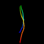
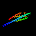
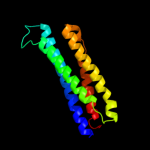
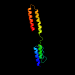
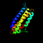
2 c2ch7A_
99.7
21
PDB header: chemotaxisChain: A: PDB Molecule: methyl-accepting chemotaxis protein;PDBTitle: crystal structure of the cytoplasmic domain of a bacterial2 chemoreceptor from thermotoga maritima
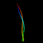
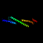
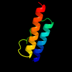
3 c3g67A_
99.5
21
PDB header: signaling proteinChain: A: PDB Molecule: methyl-accepting chemotaxis protein;PDBTitle: crystal structure of a soluble chemoreceptor from thermotoga2 maritima
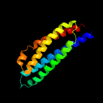
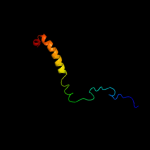
4 c2d4uA_
98.3
34
PDB header: signaling proteinChain: A: PDB Molecule: methyl-accepting chemotaxis protein i;PDBTitle: crystal structure of the ligand binding domain of the bacterial serine2 chemoreceptor tsr
5 c3lnrA_
98.1
12
PDB header: signaling proteinChain: A: PDB Molecule: aerotaxis transducer aer2;PDBTitle: crystal structure of poly-hamp domains from the p. aeruginosa soluble2 receptor aer2
6 d2asra_
98.0
99
Fold: Four-helical up-and-down bundleSuperfamily: Aspartate receptor, ligand-binding domainFamily: Aspartate receptor, ligand-binding domain7 d2liga_
98.0
69
Fold: Four-helical up-and-down bundleSuperfamily: Aspartate receptor, ligand-binding domainFamily: Aspartate receptor, ligand-binding domain8 d1vlta_
97.5
70
Fold: Four-helical up-and-down bundleSuperfamily: Aspartate receptor, ligand-binding domainFamily: Aspartate receptor, ligand-binding domain9 d2asxa1
97.0
27
Fold: HAMP domain-likeSuperfamily: HAMP domain-likeFamily: HAMP domain10 c2rm8A_
96.7
19
PDB header: signaling proteinChain: A: PDB Molecule: sensory rhodopsin ii transducer;PDBTitle: the solution structure of phototactic transducer protein2 htrii linker region from natronomonas pharaonis
11 c1sj8A_
94.9
9
PDB header: structural proteinChain: A: PDB Molecule: talin 1;PDBTitle: crystal structure of talin residues 482-789
12 c3zrwB_
94.3
19
PDB header: signaling proteinChain: B: PDB Molecule: af1503 protein, osmolarity sensor protein envz;PDBTitle: the structure of the dimeric hamp-dhp fusion a291v mutant
13 c2wpqA_
93.0
12
PDB header: membrane proteinChain: A: PDB Molecule: trimeric autotransporter adhesin fragment;PDBTitle: salmonella enterica sada 479-519 fused to gcn4 adaptors (2 sadak3, in-register fusion)
14 c3ojaB_
80.6
12
PDB header: protein bindingChain: B: PDB Molecule: anopheles plasmodium-responsive leucine-rich repeat proteinPDBTitle: crystal structure of lrim1/apl1c complex
15 c3dyjA_
72.5
9
PDB header: structural proteinChain: A: PDB Molecule: talin-1;PDBTitle: crystal structure a talin rod fragment
16 c2kbbA_
64.8
12
PDB header: structural proteinChain: A: PDB Molecule: talin-1;PDBTitle: nmr structure of the talin rod domain, 1655-1822
17 c1deqO_
61.7
16
PDB header: PDB COMPND: 18 c2qihA_
56.6
12
PDB header: cell adhesionChain: A: PDB Molecule: protein uspa1;PDBTitle: crystal structure of 527-665 fragment of uspa1 protein from2 moraxella catarrhalis
19 c3hd7A_
53.6
26
PDB header: exocytosisChain: A: PDB Molecule: vesicle-associated membrane protein 2;PDBTitle: helical extension of the neuronal snare complex into the membrane,2 spacegroup c 1 2 1
20 c1ei3E_
52.3
16
PDB header: PDB COMPND: 21 c1urqA_
not modelled
52.1
21
PDB header: transport proteinChain: A: PDB Molecule: m-tomosyn isoform;PDBTitle: crystal structure of neuronal q-snares in complex with2 r-snare motif of tomosyn
22 c3b5nF_
not modelled
48.2
12
PDB header: membrane proteinChain: F: PDB Molecule: protein sso1;PDBTitle: structure of the yeast plasma membrane snare complex
23 c3ipdB_
not modelled
46.6
13
PDB header: exocytosisChain: B: PDB Molecule: syntaxin-1a;PDBTitle: helical extension of the neuronal snare complex into the2 membrane, spacegroup i 21 21 21
24 c1sfcJ_
not modelled
44.8
13
PDB header: transport proteinChain: J: PDB Molecule: protein (syntaxin 1a);PDBTitle: neuronal synaptic fusion complex
25 c1n7sB_
not modelled
42.6
15
PDB header: transport proteinChain: B: PDB Molecule: syntaxin 1a;PDBTitle: high resolution structure of a truncated neuronal snare2 complex
26 c1ei3C_
not modelled
41.6
6
PDB header: PDB COMPND: 27 c1deqF_
not modelled
40.2
5
PDB header: PDB COMPND: 28 c2ieqC_
not modelled
37.7
14
PDB header: viral proteinChain: C: PDB Molecule: spike glycoprotein;PDBTitle: core structure of s2 from the human coronavirus nl63 spike2 glycoprotein
29 c1n7sA_
not modelled
37.1
25
PDB header: transport proteinChain: A: PDB Molecule: vesicle-associated membrane protein 2;PDBTitle: high resolution structure of a truncated neuronal snare2 complex
30 c3ghgK_
not modelled
36.9
15
PDB header: blood clottingChain: K: PDB Molecule: fibrinogen beta chain;PDBTitle: crystal structure of human fibrinogen
31 c2npsA_
not modelled
35.4
25
PDB header: transport proteinChain: A: PDB Molecule: vesicle-associated membrane protein 4;PDBTitle: crystal structure of the early endosomal snare complex
32 c2vs0B_
not modelled
33.5
13
PDB header: cell invasionChain: B: PDB Molecule: virulence factor esxa;PDBTitle: structural analysis of homodimeric staphylococcal aureus2 virulence factor esxa
33 c2kseA_
not modelled
33.5
13
PDB header: transferaseChain: A: PDB Molecule: sensor protein qsec;PDBTitle: backbone structure of the membrane domain of e. coli2 histidine kinase receptor qsec, center for structures of3 membrane proteins (csmp) target 4311c
34 c2npsB_
not modelled
33.5
16
PDB header: transport proteinChain: B: PDB Molecule: syntaxin 13;PDBTitle: crystal structure of the early endosomal snare complex
35 c3b5nE_
not modelled
32.5
15
PDB header: membrane proteinChain: E: PDB Molecule: synaptobrevin homolog 1;PDBTitle: structure of the yeast plasma membrane snare complex
36 c1sfcI_
not modelled
30.4
26
PDB header: transport proteinChain: I: PDB Molecule: protein (synaptobrevin 2);PDBTitle: neuronal synaptic fusion complex
37 c2efrB_
not modelled
27.5
13
PDB header: contractile proteinChain: B: PDB Molecule: general control protein gcn4 and tropomyosin 1 alpha chain;PDBTitle: crystal structure of the c-terminal tropomyosin fragment with n- and2 c-terminal extensions of the leucine zipper at 1.8 angstroms3 resolution
38 c1i49A_
not modelled
25.0
13
PDB header: signaling proteinChain: A: PDB Molecule: arfaptin 2;PDBTitle: crystal structure analysis of arfaptin
39 c1gl2A_
not modelled
22.4
13
PDB header: membrane proteinChain: A: PDB Molecule: endobrevin;PDBTitle: crystal structure of an endosomal snare core complex
40 c1m1jA_
not modelled
21.4
6
PDB header: blood clottingChain: A: PDB Molecule: fibrinogen alpha subunit;PDBTitle: crystal structure of native chicken fibrinogen with two different2 bound ligands
41 c3gvmA_
not modelled
18.4
11
PDB header: viral proteinChain: A: PDB Molecule: putative uncharacterized protein sag1039;PDBTitle: structure of the homodimeric wxg-100 family protein from streptococcus2 agalactiae
42 c2bezC_
not modelled
17.3
16
PDB header: viral proteinChain: C: PDB Molecule: e2 glycoprotein;PDBTitle: structure of a proteolitically resistant core from the2 severe acute respiratory syndrome coronavirus s2 fusion3 protein
43 c1l4aD_
not modelled
17.1
7
PDB header: endocytosis/exocytosisChain: D: PDB Molecule: s-snap25 fusion protein;PDBTitle: x-ray structure of the neuronal complexin/snare complex2 from the squid loligo pealei
44 c3arcl_
not modelled
16.8
35
PDB header: electron transport, photosynthesisChain: L: PDB Molecule: photosystem ii reaction center protein l;PDBTitle: crystal structure of oxygen-evolving photosystem ii at 1.9 angstrom2 resolution
45 c2npsD_
not modelled
16.2
5
PDB header: transport proteinChain: D: PDB Molecule: syntaxin-6;PDBTitle: crystal structure of the early endosomal snare complex
46 c1sfcD_
not modelled
15.7
10
PDB header: transport proteinChain: D: PDB Molecule: protein (snap-25b);PDBTitle: neuronal synaptic fusion complex
47 c3cwgA_
not modelled
15.4
9
PDB header: transcriptionChain: A: PDB Molecule: signal transducer and activator of transcriptionPDBTitle: unphosphorylated mouse stat3 core fragment
48 c1nafA_
not modelled
15.4
15
PDB header: signaling protein, membrane proteinChain: A: PDB Molecule: adp-ribosylation factor binding protein gga1;PDBTitle: crystal structure of the human gga1 gat domain
49 c1kmiZ_
not modelled
15.3
7
PDB header: signaling proteinChain: Z: PDB Molecule: chemotaxis protein chez;PDBTitle: crystal structure of an e.coli chemotaxis protein, chez
50 d1ez3a_
not modelled
14.8
12
Fold: STAT-likeSuperfamily: t-snare proteinsFamily: t-snare proteins51 c2d3eD_
not modelled
14.5
8
PDB header: contractile proteinChain: D: PDB Molecule: general control protein gcn4 and tropomyosin 1PDBTitle: crystal structure of the c-terminal fragment of rabbit2 skeletal alpha-tropomyosin
52 c2d4yA_
not modelled
14.1
9
PDB header: structural proteinChain: A: PDB Molecule: flagellar hook-associated protein 1;PDBTitle: crystal structure of a 49k fragment of hap1 (flgk)
53 c3prrL_
not modelled
13.7
35
PDB header: photosynthesisChain: L: PDB Molecule: photosystem ii reaction center protein l;PDBTitle: crystal structure of cyanobacterial photosystem ii in complex with2 terbutryn (part 2 of 2). this file contains second monomer of psii3 dimer
54 c3kziL_
not modelled
13.7
35
PDB header: electron transportChain: L: PDB Molecule: photosystem ii reaction center protein l;PDBTitle: crystal structure of monomeric form of cyanobacterial photosystem ii
55 c3prqL_
not modelled
13.7
35
PDB header: photosynthesisChain: L: PDB Molecule: photosystem ii reaction center protein l;PDBTitle: crystal structure of cyanobacterial photosystem ii in complex with2 terbutryn (part 1 of 2). this file contains first monomer of psii3 dimer
56 c1s5lL_
not modelled
13.7
35
PDB header: photosynthesisChain: L: PDB Molecule: photosystem ii reaction center l protein;PDBTitle: architecture of the photosynthetic oxygen evolving center
57 c3bz1L_
not modelled
13.7
35
PDB header: electron transportChain: L: PDB Molecule: photosystem ii reaction center protein l;PDBTitle: crystal structure of cyanobacterial photosystem ii (part 12 of 2). this file contains first monomer of psii dimer
58 d2axtl1
not modelled
13.7
35
Fold: Single transmembrane helixSuperfamily: Photosystem II reaction center protein L, PsbLFamily: PsbL-like59 c3bz2L_
not modelled
13.7
35
PDB header: electron transportChain: L: PDB Molecule: photosystem ii reaction center protein l;PDBTitle: crystal structure of cyanobacterial photosystem ii (part 22 of 2). this file contains second monomer of psii dimer
60 c3a0hL_
not modelled
13.7
35
PDB header: electron transportChain: L: PDB Molecule: photosystem ii reaction center protein l;PDBTitle: crystal structure of i-substituted photosystem ii complex
61 c3a0bl_
not modelled
13.7
35
PDB header: electron transportChain: L: PDB Molecule: photosystem ii reaction center protein l;PDBTitle: crystal structure of br-substituted photosystem ii complex
62 c3a0bL_
not modelled
13.7
35
PDB header: electron transportChain: L: PDB Molecule: photosystem ii reaction center protein l;PDBTitle: crystal structure of br-substituted photosystem ii complex
63 c1s5ll_
not modelled
13.7
35
PDB header: photosynthesisChain: L: PDB Molecule: photosystem ii reaction center l protein;PDBTitle: architecture of the photosynthetic oxygen evolving center
64 c3arcL_
not modelled
13.7
35
PDB header: electron transport, photosynthesisChain: L: PDB Molecule: photosystem ii reaction center protein l;PDBTitle: crystal structure of oxygen-evolving photosystem ii at 1.9 angstrom2 resolution
65 c3a0hl_
not modelled
13.7
35
PDB header: electron transportChain: L: PDB Molecule: photosystem ii reaction center protein l;PDBTitle: crystal structure of i-substituted photosystem ii complex
66 c2axtL_
not modelled
13.7
35
PDB header: electron transportChain: L: PDB Molecule: photosystem ii reaction center l protein;PDBTitle: crystal structure of photosystem ii from thermosynechococcus elongatus
67 c2axtl_
not modelled
13.7
35
PDB header: electron transportChain: L: PDB Molecule: photosystem ii reaction center l protein;PDBTitle: crystal structure of photosystem ii from thermosynechococcus elongatus
68 c1l7cA_
not modelled
13.4
13
PDB header: cell adhesionChain: A: PDB Molecule: alpha e-catenin;PDBTitle: alpha-catenin fragment, residues 385-651
69 c1s94A_
not modelled
13.0
11
PDB header: endocytosis/exocytosisChain: A: PDB Molecule: s-syntaxin;PDBTitle: crystal structure of the habc domain of neuronal syntaxin from the2 squid loligo pealei
70 d1s94a_
not modelled
13.0
11
Fold: STAT-likeSuperfamily: t-snare proteinsFamily: t-snare proteins71 c1zvaA_
not modelled
12.9
10
PDB header: viral proteinChain: A: PDB Molecule: e2 glycoprotein;PDBTitle: a structure-based mechanism of sars virus membrane fusion
72 c3ok8A_
not modelled
12.4
8
PDB header: protein bindingChain: A: PDB Molecule: brain-specific angiogenesis inhibitor 1-associated proteinPDBTitle: i-bar of pinkbar
73 d1i4da_
not modelled
12.2
13
Fold: BAR/IMD domain-likeSuperfamily: BAR/IMD domain-likeFamily: Arfaptin, Rac-binding fragment74 d1eq1a_
not modelled
11.9
9
Fold: Apolipophorin-IIISuperfamily: Apolipophorin-IIIFamily: Apolipophorin-III75 d1r0da_
not modelled
10.7
11
Fold: I/LWEQ domainSuperfamily: I/LWEQ domainFamily: I/LWEQ domain76 c3dtpA_
not modelled
10.6
18
PDB header: contractile proteinChain: A: PDB Molecule: myosin 2 heavy chain chimera of smooth andPDBTitle: tarantula heavy meromyosin obtained by flexible docking to2 tarantula muscle thick filament cryo-em 3d-map
77 c1eboE_
not modelled
10.2
18
PDB header: viral proteinChain: E: PDB Molecule: ebola virus envelope protein chimera consistingPDBTitle: crystal structure of the ebola virus membrane-fusion2 subunit, gp2, from the envelope glycoprotein ectodomain
78 c3b5nL_
not modelled
10.2
11
PDB header: membrane proteinChain: L: PDB Molecule: protein transport protein sec9;PDBTitle: structure of the yeast plasma membrane snare complex
79 c1gl2D_
not modelled
10.2
25
PDB header: membrane proteinChain: D: PDB Molecule: syntaxin 8;PDBTitle: crystal structure of an endosomal snare core complex
80 c3c98B_
not modelled
10.1
10
PDB header: endocytosis/exocytosisChain: B: PDB Molecule: syntaxin-1a;PDBTitle: revised structure of the munc18a-syntaxin1 complex
81 c1l4aC_
not modelled
10.1
7
PDB header: endocytosis/exocytosisChain: C: PDB Molecule: s-snap25 fusion protein;PDBTitle: x-ray structure of the neuronal complexin/snare complex2 from the squid loligo pealei
82 c3gxvD_
not modelled
10.0
15
PDB header: hydrolase/replicationChain: D: PDB Molecule: replicative dna helicase;PDBTitle: three-dimensional structure of n-terminal domain of dnab2 helicase from helicobacter pylori and its interactions with3 primase
83 c2dnxA_
not modelled
9.8
9
PDB header: transport proteinChain: A: PDB Molecule: syntaxin-12;PDBTitle: solution structure of rsgi ruh-063, an n-terminal domain of2 syntaxin 12 from human cdna
84 c3gxvC_
not modelled
9.6
15
PDB header: hydrolase/replicationChain: C: PDB Molecule: replicative dna helicase;PDBTitle: three-dimensional structure of n-terminal domain of dnab2 helicase from helicobacter pylori and its interactions with3 primase
85 d1oxza_
not modelled
9.5
15
Fold: Spectrin repeat-likeSuperfamily: GAT-like domainFamily: GAT domain86 c1oxzA_
not modelled
9.5
15
PDB header: membrane proteinChain: A: PDB Molecule: adp-ribosylation factor binding protein gga1;PDBTitle: crystal structure of the human gga1 gat domain
87 c1wyyB_
not modelled
9.2
16
PDB header: viral proteinChain: B: PDB Molecule: e2 glycoprotein;PDBTitle: post-fusion hairpin conformation of the sars coronavirus spike2 glycoprotein
88 d1wr6a1
not modelled
9.1
9
Fold: Spectrin repeat-likeSuperfamily: GAT-like domainFamily: GAT domain89 c2l9uA_
not modelled
9.1
13
PDB header: membrane proteinChain: A: PDB Molecule: receptor tyrosine-protein kinase erbb-3;PDBTitle: spatial structure of dimeric erbb3 transmembrane domain
90 c1junB_
not modelled
9.0
25
PDB header: transcription regulationChain: B: PDB Molecule: c-jun homodimer;PDBTitle: nmr study of c-jun homodimer
91 d1t01a1
not modelled
8.9
12
Fold: Four-helical up-and-down bundleSuperfamily: alpha-catenin/vinculin-likeFamily: alpha-catenin/vinculin92 c1n73C_
not modelled
8.9
15
PDB header: blood clottingChain: C: PDB Molecule: fibrin gamma chain;PDBTitle: fibrin d-dimer, lamprey complexed with the peptide ligand: gly-his-2 arg-pro-amide
93 c1zv8I_
not modelled
8.9
7
PDB header: viral proteinChain: I: PDB Molecule: e2 glycoprotein;PDBTitle: a structure-based mechanism of sars virus membrane fusion
94 d1lvfa_
not modelled
8.8
15
Fold: STAT-likeSuperfamily: t-snare proteinsFamily: t-snare proteins95 c1ciiA_
not modelled
8.6
12
PDB header: transmembrane proteinChain: A: PDB Molecule: colicin ia;PDBTitle: colicin ia
96 c3hnwB_
not modelled
8.5
12
PDB header: structural genomics, unknown functionChain: B: PDB Molecule: uncharacterized protein;PDBTitle: crystal structure of a basic coiled-coil protein of unknown function2 from eubacterium eligens atcc 27750
97 c2qrxA_
not modelled
8.4
9
PDB header: dna binding proteinChain: A: PDB Molecule: gm27569p;PDBTitle: crystal structure of drosophila melanogaster translin2 protein
98 c2l16A_
not modelled
8.2
14
PDB header: protein transportChain: A: PDB Molecule: sec-independent protein translocase protein tatad;PDBTitle: solution structure of bacillus subtilits tatad protein in dpc micelles
99 c1y4cA_
not modelled
8.1
10
PDB header: de novo proteinChain: A: PDB Molecule: maltose binding protein fused with designedPDBTitle: designed helical protein fusion mbp