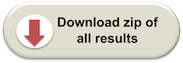
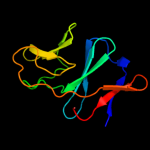
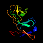
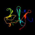
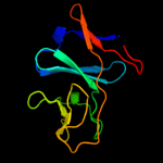
| 1 | d1fqta_
|
|
 |
100.0 |
39 |
Fold:ISP domain
Superfamily:ISP domain
Family:Rieske iron-sulfur protein (ISP) |
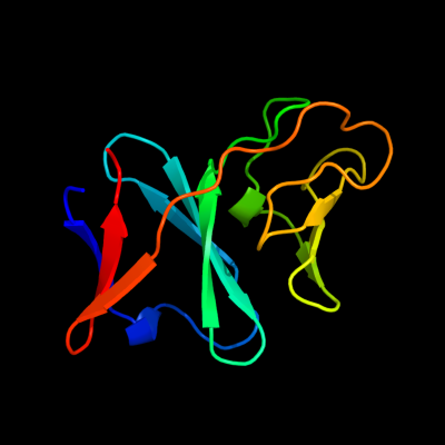
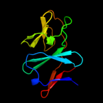
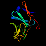
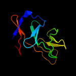
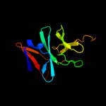
| 2 | c2qpzA_
|
|
 |
100.0 |
45 |
PDB header:metal binding protein
Chain: A: PDB Molecule:naphthalene 1,2-dioxygenase system ferredoxin
PDBTitle: naphthalene 1,2-dioxygenase rieske ferredoxin
|
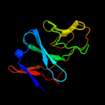
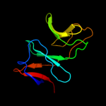
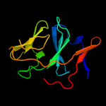
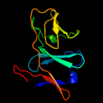
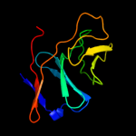
| 3 | c2i7fB_
|
|
 |
100.0 |
40 |
PDB header:oxidoreductase
Chain: B: PDB Molecule:ferredoxin component of dioxygenase;
PDBTitle: sphingomonas yanoikuyae b1 ferredoxin
|
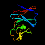
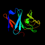
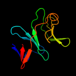
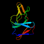
| 4 | c2de7E_
|
|
 |
100.0 |
34 |
PDB header:oxidoreductase
Chain: E: PDB Molecule:ferredoxin component of carbazole;
PDBTitle: the substrate-bound complex between oxygenase and2 ferredoxin in carbazole 1,9a-dioxygenase
|
| 5 | c3gceA_
|
|
 |
100.0 |
27 |
PDB header:oxidoreductase
Chain: A: PDB Molecule:ferredoxin component of carbazole 1,9a-
PDBTitle: ferredoxin of carbazole 1,9a-dioxygenase from nocardioides2 aromaticivorans ic177
|
| 6 | d3c0da1
|
|
 |
100.0 |
25 |
Fold:ISP domain
Superfamily:ISP domain
Family:NirD-like |
| 7 | c3dqyA_
|
|
 |
100.0 |
39 |
PDB header:oxidoreductase
Chain: A: PDB Molecule:toluene 1,2-dioxygenase system ferredoxin
PDBTitle: crystal structure of toluene 2,3-dioxygenase ferredoxin
|
| 8 | d2de6a1
|
|
 |
99.9 |
24 |
Fold:ISP domain
Superfamily:ISP domain
Family:Ring hydroxylating alpha subunit ISP domain |
| 9 | d1z01a1
|
|
 |
99.9 |
31 |
Fold:ISP domain
Superfamily:ISP domain
Family:Ring hydroxylating alpha subunit ISP domain |
| 10 | d1vm9a_
|
|
 |
99.9 |
27 |
Fold:ISP domain
Superfamily:ISP domain
Family:Rieske iron-sulfur protein (ISP) |
| 11 | c3d89A_
|
|
 |
99.9 |
22 |
PDB header:electron transport
Chain: A: PDB Molecule:rieske domain-containing protein;
PDBTitle: crystal structure of a soluble rieske ferredoxin from mus musculus
|
| 12 | d2jzaa1
|
|
 |
99.9 |
25 |
Fold:ISP domain
Superfamily:ISP domain
Family:NirD-like |
| 13 | c2de7B_
|
|
 |
99.9 |
25 |
PDB header:oxidoreductase
Chain: B: PDB Molecule:terminal oxygenase component of carbazole;
PDBTitle: the substrate-bound complex between oxygenase and2 ferredoxin in carbazole 1,9a-dioxygenase
|
| 14 | c3gkqB_
|
|
 |
99.9 |
28 |
PDB header:oxidoreductase
Chain: B: PDB Molecule:terminal oxygenase component of carbazole 1,9a-
PDBTitle: terminal oxygenase of carbazole 1,9a-dioxygenase from2 novosphingobium sp. ka1
|
| 15 | d2jo6a1
|
|
 |
99.9 |
17 |
Fold:ISP domain
Superfamily:ISP domain
Family:NirD-like |
| 16 | c3gcfC_
|
|
 |
99.9 |
28 |
PDB header:oxidoreductase
Chain: C: PDB Molecule:terminal oxygenase component of carbazole 1,9a-
PDBTitle: terminal oxygenase of carbazole 1,9a-dioxygenase from2 nocardioides aromaticivorans ic177
|
| 17 | c2zylA_
|
|
 |
99.9 |
19 |
PDB header:oxidoreductase
Chain: A: PDB Molecule:possible oxidoreductase;
PDBTitle: crystal structure of 3-ketosteroid-9-alpha-hydroxylase2 (ksha) from m. tuberculosis
|
| 18 | c1z01D_
|
|
 |
99.9 |
32 |
PDB header:oxidoreductase
Chain: D: PDB Molecule:2-oxo-1,2-dihydroquinoline 8-monooxygenase,
PDBTitle: 2-oxoquinoline 8-monooxygenase component: active site2 modulation by rieske-[2fe-2s] center oxidation/reduction
|
| 19 | c3n0qA_
|
|
 |
99.9 |
22 |
PDB header:oxidoreductase
Chain: A: PDB Molecule:putative aromatic-ring hydroxylating dioxygenase;
PDBTitle: crystal structure of a putative aromatic-ring hydroxylating2 dioxygenase (tm1040_3219) from silicibacter sp. tm1040 at 1.80 a3 resolution
|
| 20 | c3gteB_
|
|
 |
99.9 |
23 |
PDB header:electron transport, oxidoreductase
Chain: B: PDB Molecule:ddmc;
PDBTitle: crystal structure of dicamba monooxygenase with non-heme2 iron
|
| 21 | d1ulia1 |
|
not modelled |
99.9 |
16 |
Fold:ISP domain
Superfamily:ISP domain
Family:Ring hydroxylating alpha subunit ISP domain |
| 22 | d1wqla1 |
|
not modelled |
99.9 |
14 |
Fold:ISP domain
Superfamily:ISP domain
Family:Ring hydroxylating alpha subunit ISP domain |
| 23 | d2bmoa1 |
|
not modelled |
99.9 |
14 |
Fold:ISP domain
Superfamily:ISP domain
Family:Ring hydroxylating alpha subunit ISP domain |
| 24 | c1uljA_ |
|
not modelled |
99.9 |
16 |
PDB header:oxidoreductase
Chain: A: PDB Molecule:biphenyl dioxygenase large subunit;
PDBTitle: biphenyl dioxygenase (bpha1a2) in complex with the substrate
|
| 25 | d2b1xa1 |
|
not modelled |
99.9 |
23 |
Fold:ISP domain
Superfamily:ISP domain
Family:Ring hydroxylating alpha subunit ISP domain |
| 26 | c1wqlA_ |
|
not modelled |
99.9 |
15 |
PDB header:oxidoreductase
Chain: A: PDB Molecule:iron-sulfur protein large subunit of cumene dioxygenase;
PDBTitle: cumene dioxygenase (cuma1a2) from pseudomonas fluorescens ip01
|
| 27 | d1o7na1 |
|
not modelled |
99.9 |
18 |
Fold:ISP domain
Superfamily:ISP domain
Family:Ring hydroxylating alpha subunit ISP domain |
| 28 | d1g8kb_ |
|
not modelled |
99.9 |
22 |
Fold:ISP domain
Superfamily:ISP domain
Family:Rieske iron-sulfur protein (ISP) |
| 29 | c2gbxE_ |
|
not modelled |
99.9 |
19 |
PDB header:oxidoreductase
Chain: E: PDB Molecule:biphenyl 2,3-dioxygenase alpha subunit;
PDBTitle: crystal structure of biphenyl 2,3-dioxygenase from sphingomonas2 yanoikuyae b1 bound to biphenyl
|
| 30 | c2hmnA_ |
|
not modelled |
99.9 |
17 |
PDB header:oxidoreductase
Chain: A: PDB Molecule:naphthalene 1,2-dioxygenase alpha subunit;
PDBTitle: crystal structure of the naphthalene 1,2-dioxygenase f352v2 mutant bound to anthracene.
|
| 31 | c2b1xE_ |
|
not modelled |
99.9 |
19 |
PDB header:oxidoreductase
Chain: E: PDB Molecule:naphthalene dioxygenase large subunit;
PDBTitle: crystal structure of naphthalene 1,2-dioxygenase from rhodococcus sp.
|
| 32 | d3cx5e1 |
|
not modelled |
99.9 |
16 |
Fold:ISP domain
Superfamily:ISP domain
Family:Rieske iron-sulfur protein (ISP) |
| 33 | d1rfsa_ |
|
not modelled |
99.8 |
17 |
Fold:ISP domain
Superfamily:ISP domain
Family:Rieske iron-sulfur protein (ISP) |
| 34 | d1q90c_ |
|
not modelled |
99.8 |
19 |
Fold:ISP domain
Superfamily:ISP domain
Family:Rieske iron-sulfur protein (ISP) |
| 35 | d1riea_ |
|
not modelled |
99.8 |
19 |
Fold:ISP domain
Superfamily:ISP domain
Family:Rieske iron-sulfur protein (ISP) |
| 36 | d1nyka_ |
|
not modelled |
99.8 |
19 |
Fold:ISP domain
Superfamily:ISP domain
Family:Rieske iron-sulfur protein (ISP) |
| 37 | d2e74d1 |
|
not modelled |
99.8 |
20 |
Fold:ISP domain
Superfamily:ISP domain
Family:Rieske iron-sulfur protein (ISP) |
| 38 | c2e76D_ |
|
not modelled |
99.7 |
19 |
PDB header:photosynthesis
Chain: D: PDB Molecule:cytochrome b6-f complex iron-sulfur subunit;
PDBTitle: crystal structure of the cytochrome b6f complex with tridecyl-2 stigmatellin (tds) from m.laminosus
|
| 39 | c1p84E_ |
|
not modelled |
99.7 |
18 |
PDB header:oxidoreductase
Chain: E: PDB Molecule:ubiquinol-cytochrome c reductase iron-sulfur
PDBTitle: hdbt inhibited yeast cytochrome bc1 complex
|
| 40 | c2nvgA_ |
|
not modelled |
99.7 |
23 |
PDB header:oxidoreductase
Chain: A: PDB Molecule:ubiquinol-cytochrome c reductase iron-sulfur subunit;
PDBTitle: soluble domain of rieske iron sulfur protein.
|
| 41 | c2fyuE_ |
|
not modelled |
99.7 |
18 |
PDB header:oxidoreductase
Chain: E: PDB Molecule:ubiquinol-cytochrome c reductase iron-sulfur subunit,
PDBTitle: crystal structure of bovine heart mitochondrial bc1 with jg1442 inhibitor
|
| 42 | d1jm1a_ |
|
not modelled |
99.6 |
28 |
Fold:ISP domain
Superfamily:ISP domain
Family:Rieske iron-sulfur protein (ISP) |
| 43 | c2fynO_ |
|
not modelled |
99.6 |
20 |
PDB header:oxidoreductase
Chain: O: PDB Molecule:ubiquinol-cytochrome c reductase iron-sulfur
PDBTitle: crystal structure analysis of the double mutant rhodobacter2 sphaeroides bc1 complex
|
| 44 | d2hf1a1 |
|
not modelled |
69.9 |
14 |
Fold:Trm112p-like
Superfamily:Trm112p-like
Family:Trm112p-like |
| 45 | d2jnya1 |
|
not modelled |
69.8 |
14 |
Fold:Trm112p-like
Superfamily:Trm112p-like
Family:Trm112p-like |
| 46 | c2jr6A_ |
|
not modelled |
62.9 |
17 |
PDB header:structural genomics, unknown function
Chain: A: PDB Molecule:upf0434 protein nma0874;
PDBTitle: solution structure of upf0434 protein nma0874. northeast structural2 genomics target mr32
|
| 47 | d2pk7a1 |
|
not modelled |
62.9 |
9 |
Fold:Trm112p-like
Superfamily:Trm112p-like
Family:Trm112p-like |
| 48 | c2js4A_ |
|
not modelled |
60.6 |
17 |
PDB header:structural genomics, unknown function
Chain: A: PDB Molecule:upf0434 protein bb2007;
PDBTitle: solution nmr structure of bordetella bronchiseptica protein2 bb2007. northeast structural genomics consortium target3 bor54
|
| 49 | c2kpiA_ |
|
not modelled |
56.1 |
11 |
PDB header:structural genomics, unknown function
Chain: A: PDB Molecule:uncharacterized protein sco3027;
PDBTitle: solution nmr structure of streptomyces coelicolor sco30272 modeled with zn+2 bound, northeast structural genomics3 consortium target rr58
|
| 50 | c2w3nA_ |
|
not modelled |
36.3 |
29 |
PDB header:lyase
Chain: A: PDB Molecule:carbonic anhydrase 2;
PDBTitle: structure and inhibition of the co2-sensing carbonic2 anhydrase can2 from the pathogenic fungus cryptococcus3 neoformans
|
| 51 | c3lasA_ |
|
not modelled |
32.8 |
25 |
PDB header:lyase
Chain: A: PDB Molecule:putative carbonic anhydrase;
PDBTitle: crystal structure of carbonic anhydrase from streptococcus mutans to2 1.4 angstrom resolution
|
| 52 | d1ddza2 |
|
not modelled |
30.5 |
14 |
Fold:Resolvase-like
Superfamily:beta-carbonic anhydrase, cab
Family:beta-carbonic anhydrase, cab |
| 53 | c1ylkA_ |
|
not modelled |
28.9 |
31 |
PDB header:unknown function
Chain: A: PDB Molecule:hypothetical protein rv1284/mt1322;
PDBTitle: crystal structure of rv1284 from mycobacterium tuberculosis in complex2 with thiocyanate
|
| 54 | c1ddzA_ |
|
not modelled |
28.9 |
18 |
PDB header:lyase
Chain: A: PDB Molecule:carbonic anhydrase;
PDBTitle: x-ray structure of a beta-carbonic anhydrase from the red2 alga, porphyridium purpureum r-1
|
| 55 | c3ucoB_ |
|
not modelled |
28.8 |
25 |
PDB header:lyase/lyase inhibitor
Chain: B: PDB Molecule:carbonic anhydrase;
PDBTitle: coccomyxa beta-carbonic anhydrase in complex with iodide
|
| 56 | c3eyxB_ |
|
not modelled |
28.7 |
9 |
PDB header:lyase
Chain: B: PDB Molecule:carbonic anhydrase;
PDBTitle: crystal structure of carbonic anhydrase nce103 from2 saccharomyces cerevisiae
|
| 57 | c2a8cE_ |
|
not modelled |
27.5 |
15 |
PDB header:lyase
Chain: E: PDB Molecule:carbonic anhydrase 2;
PDBTitle: haemophilus influenzae beta-carbonic anhydrase
|
| 58 | d1ekja_ |
|
not modelled |
26.5 |
8 |
Fold:Resolvase-like
Superfamily:beta-carbonic anhydrase, cab
Family:beta-carbonic anhydrase, cab |
| 59 | d1ddza1 |
|
not modelled |
26.4 |
18 |
Fold:Resolvase-like
Superfamily:beta-carbonic anhydrase, cab
Family:beta-carbonic anhydrase, cab |
| 60 | c2a5vB_ |
|
not modelled |
24.9 |
23 |
PDB header:lyase
Chain: B: PDB Molecule:carbonic anhydrase (carbonate dehydratase) (carbonic
PDBTitle: crystal structure of m. tuberculosis beta carbonic anhydrase, rv3588c,2 tetrameric form
|
| 61 | d1i6pa_ |
|
not modelled |
24.8 |
14 |
Fold:Resolvase-like
Superfamily:beta-carbonic anhydrase, cab
Family:beta-carbonic anhydrase, cab |
| 62 | d1g5ca_ |
|
not modelled |
23.2 |
21 |
Fold:Resolvase-like
Superfamily:beta-carbonic anhydrase, cab
Family:beta-carbonic anhydrase, cab |
| 63 | c3eoiB_ |
|
not modelled |
12.6 |
9 |
PDB header:structural genomics, unknown function
Chain: B: PDB Molecule:pilm;
PDBTitle: crystal structure of putative protein pilm from escherichia coli b7a
|
| 64 | c2k5rA_ |
|
not modelled |
12.6 |
8 |
PDB header:structural genomics, unknown function
Chain: A: PDB Molecule:uncharacterized protein xf2673;
PDBTitle: solution nmr structure of xf2673 from xylella fastidiosa.2 northeast structural genomics consortium target xfr39
|
| 65 | c2xk0A_ |
|
not modelled |
12.0 |
17 |
PDB header:transcription
Chain: A: PDB Molecule:polycomb protein pcl;
PDBTitle: solution structure of the tudor domain from drosophila2 polycomblike (pcl)
|
| 66 | d2j7ja2 |
|
not modelled |
11.0 |
33 |
Fold:beta-beta-alpha zinc fingers
Superfamily:beta-beta-alpha zinc fingers
Family:Classic zinc finger, C2H2 |
| 67 | d1ubdc1 |
|
not modelled |
10.6 |
33 |
Fold:beta-beta-alpha zinc fingers
Superfamily:beta-beta-alpha zinc fingers
Family:Classic zinc finger, C2H2 |
| 68 | d1r2za3 |
|
not modelled |
10.2 |
18 |
Fold:Glucocorticoid receptor-like (DNA-binding domain)
Superfamily:Glucocorticoid receptor-like (DNA-binding domain)
Family:C-terminal, Zn-finger domain of MutM-like DNA repair proteins |
| 69 | c3hg9B_ |
|
not modelled |
10.1 |
5 |
PDB header:structural genomics, unknown function
Chain: B: PDB Molecule:pilm;
PDBTitle: crystal structure of putative pilm protein from pseudomonas aeruginosa2 2192
|
| 70 | d1l1ta3 |
|
not modelled |
8.8 |
18 |
Fold:Glucocorticoid receptor-like (DNA-binding domain)
Superfamily:Glucocorticoid receptor-like (DNA-binding domain)
Family:C-terminal, Zn-finger domain of MutM-like DNA repair proteins |
| 71 | d1tdza3 |
|
not modelled |
8.8 |
27 |
Fold:Glucocorticoid receptor-like (DNA-binding domain)
Superfamily:Glucocorticoid receptor-like (DNA-binding domain)
Family:C-terminal, Zn-finger domain of MutM-like DNA repair proteins |
| 72 | d2c42a2 |
|
not modelled |
8.1 |
8 |
Fold:Thiamin diphosphate-binding fold (THDP-binding)
Superfamily:Thiamin diphosphate-binding fold (THDP-binding)
Family:PFOR PP module |
| 73 | d1h7va_ |
|
not modelled |
7.9 |
15 |
Fold:Rubredoxin-like
Superfamily:Rubredoxin-like
Family:Rubredoxin |
| 74 | d1e44b_ |
|
not modelled |
7.6 |
38 |
Fold:Ribonuclease domain of colicin E3
Superfamily:Ribonuclease domain of colicin E3
Family:Ribonuclease domain of colicin E3 |
| 75 | c2w8xB_ |
|
not modelled |
7.6 |
15 |
PDB header:membrane protein
Chain: B: PDB Molecule:ion-channel modulator raklp;
PDBTitle: structure of the tick ion-channel modulator ra-klp
|
| 76 | d2aqaa1 |
|
not modelled |
7.5 |
17 |
Fold:Rubredoxin-like
Superfamily:Nop10-like SnoRNP
Family:Nucleolar RNA-binding protein Nop10-like |
| 77 | c2opfA_ |
|
not modelled |
7.5 |
18 |
PDB header:hydrolase/dna
Chain: A: PDB Molecule:endonuclease viii;
PDBTitle: crystal structure of the dna repair enzyme endonuclease-viii (nei)2 from e. coli (r252a) in complex with ap-site containing dna substrate
|
| 78 | c3izbP_ |
|
not modelled |
7.3 |
40 |
PDB header:ribosome
Chain: P: PDB Molecule:40s ribosomal protein rps11 (s17p);
PDBTitle: localization of the small subunit ribosomal proteins into a 6.1 a2 cryo-em map of saccharomyces cerevisiae translating 80s ribosome
|
| 79 | d1dgwa_ |
|
not modelled |
7.2 |
14 |
Fold:Double-stranded beta-helix
Superfamily:RmlC-like cupins
Family:Germin/Seed storage 7S protein |
| 80 | c2kn9A_ |
|
not modelled |
7.2 |
15 |
PDB header:electron transport
Chain: A: PDB Molecule:rubredoxin;
PDBTitle: solution structure of zinc-substituted rubredoxin b (rv3250c) from2 mycobacterium tuberculosis. seattle structural genomics center for3 infectious disease target mytud.01635.a
|
| 81 | c2j6aA_ |
|
not modelled |
7.1 |
17 |
PDB header:transferase
Chain: A: PDB Molecule:protein trm112;
PDBTitle: crystal structure of s. cerevisiae ynr046w, a zinc finger2 protein from the erf1 methyltransferase complex.
|
| 82 | c2eqjA_ |
|
not modelled |
6.9 |
17 |
PDB header:transcription
Chain: A: PDB Molecule:metal-response element-binding transcription
PDBTitle: solution structure of the tudor domain of metal-response2 element-binding transcription factor 2
|
| 83 | d1ee8a3 |
|
not modelled |
6.6 |
18 |
Fold:Glucocorticoid receptor-like (DNA-binding domain)
Superfamily:Glucocorticoid receptor-like (DNA-binding domain)
Family:C-terminal, Zn-finger domain of MutM-like DNA repair proteins |
| 84 | c3e59A_ |
|
not modelled |
6.5 |
11 |
PDB header:transferase
Chain: A: PDB Molecule:pyoverdine biosynthesis protein pvca;
PDBTitle: crystal structure of the pvca (pa2254) protein from pseudomonas2 aeruginosa
|
| 85 | d1ibia1 |
|
not modelled |
6.5 |
23 |
Fold:Glucocorticoid receptor-like (DNA-binding domain)
Superfamily:Glucocorticoid receptor-like (DNA-binding domain)
Family:LIM domain |
| 86 | d1k3xa3 |
|
not modelled |
6.4 |
18 |
Fold:Glucocorticoid receptor-like (DNA-binding domain)
Superfamily:Glucocorticoid receptor-like (DNA-binding domain)
Family:C-terminal, Zn-finger domain of MutM-like DNA repair proteins |
| 87 | c2e5pA_ |
|
not modelled |
6.3 |
17 |
PDB header:transcription
Chain: A: PDB Molecule:phd finger protein 1;
PDBTitle: solution structure of the tudor domain of phd finger2 protein 1 (phf1 protein)
|
| 88 | d1k82a3 |
|
not modelled |
6.2 |
18 |
Fold:Glucocorticoid receptor-like (DNA-binding domain)
Superfamily:Glucocorticoid receptor-like (DNA-binding domain)
Family:C-terminal, Zn-finger domain of MutM-like DNA repair proteins |
| 89 | d1dl6a_ |
|
not modelled |
6.2 |
25 |
Fold:Rubredoxin-like
Superfamily:Zinc beta-ribbon
Family:Transcriptional factor domain |
| 90 | c3iz6P_ |
|
not modelled |
6.2 |
40 |
PDB header:ribosome
Chain: P: PDB Molecule:40s ribosomal protein s11 (s17p);
PDBTitle: localization of the small subunit ribosomal proteins into a 5.5 a2 cryo-em map of triticum aestivum translating 80s ribosome
|
| 91 | d2apob1 |
|
not modelled |
6.1 |
23 |
Fold:Rubredoxin-like
Superfamily:Nop10-like SnoRNP
Family:Nucleolar RNA-binding protein Nop10-like |
| 92 | d2dara1 |
|
not modelled |
5.8 |
29 |
Fold:Glucocorticoid receptor-like (DNA-binding domain)
Superfamily:Glucocorticoid receptor-like (DNA-binding domain)
Family:LIM domain |
| 93 | d1hq0a_ |
|
not modelled |
5.8 |
19 |
Fold:CNF1/YfiH-like putative cysteine hydrolases
Superfamily:CNF1/YfiH-like putative cysteine hydrolases
Family:Type 1 cytotoxic necrotizing factor, catalytic domain |
| 94 | c2xznQ_ |
|
not modelled |
5.6 |
40 |
PDB header:ribosome
Chain: Q: PDB Molecule:ribosomal protein s17 containing protein;
PDBTitle: crystal structure of the eukaryotic 40s ribosomal2 subunit in complex with initiation factor 1. this file3 contains the 40s subunit and initiation factor for4 molecule 2
|
| 95 | d1uika1 |
|
not modelled |
5.5 |
23 |
Fold:Double-stranded beta-helix
Superfamily:RmlC-like cupins
Family:Germin/Seed storage 7S protein |
| 96 | c3dkqB_ |
|
not modelled |
5.5 |
14 |
PDB header:oxidoreductase
Chain: B: PDB Molecule:pkhd-type hydroxylase sbal_3634;
PDBTitle: crystal structure of putative oxygenase (yp_001051978.1) from2 shewanella baltica os155 at 2.26 a resolution
|
| 97 | d2ey4e1 |
|
not modelled |
5.3 |
23 |
Fold:Rubredoxin-like
Superfamily:Nop10-like SnoRNP
Family:Nucleolar RNA-binding protein Nop10-like |
| 98 | c2e5qA_ |
|
not modelled |
5.2 |
12 |
PDB header:transcription
Chain: A: PDB Molecule:phd finger protein 19;
PDBTitle: solution structure of the tudor domain of phd finger2 protein 19, isoform b [homo sapiens]
|