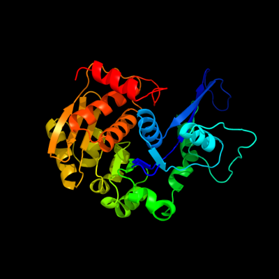
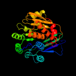
1 c3kzhA_
100.0
31
PDB header: transferaseChain: A: PDB Molecule: probable sugar kinase;PDBTitle: crystal structure of a putative sugar kinase from2 clostridium perfringens
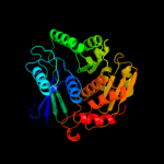
2 d1rkda_
100.0
21
Fold: Ribokinase-likeSuperfamily: Ribokinase-likeFamily: Ribokinase-like3 c2nwhA_
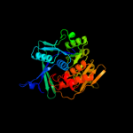
100.0
27
PDB header: signaling protein,transferaseChain: A: PDB Molecule: carbohydrate kinase;PDBTitle: carbohydrate kinase from agrobacterium tumefaciens
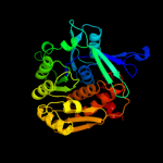
4 c2c49A_
100.0
18
PDB header: transferaseChain: A: PDB Molecule: sugar kinase mj0406;PDBTitle: crystal structure of methanocaldococcus jannaschii2 nucleoside kinase - an archaeal member of the ribokinase3 family
5 c3pl2D_
100.0
18
PDB header: transferaseChain: D: PDB Molecule: sugar kinase, ribokinase family;PDBTitle: crystal structure of a 5-keto-2-deoxygluconokinase (ncgl0155, cgl0158)2 from corynebacterium glutamicum atcc 13032 kitasato at 1.89 a3 resolution
6 c2rbcA_
100.0
15
PDB header: transferaseChain: A: PDB Molecule: sugar kinase;PDBTitle: crystal structure of a putative ribokinase from agrobacterium2 tumefaciens
7 c2pkkA_
100.0
18
PDB header: transferaseChain: A: PDB Molecule: adenosine kinase;PDBTitle: crystal structure of m tuberculosis adenosine kinase complexed with 2-2 fluro adenosine
8 d2fv7a1
100.0
19
Fold: Ribokinase-likeSuperfamily: Ribokinase-likeFamily: Ribokinase-like9 c3in1A_
100.0
22
PDB header: transferaseChain: A: PDB Molecule: uncharacterized sugar kinase ydjh;PDBTitle: crystal structure of a putative ribokinase in complex with2 adp from e.coli
10 c2qcvA_
100.0
18
PDB header: transferaseChain: A: PDB Molecule: putative 5-dehydro-2-deoxygluconokinase;PDBTitle: crystal structure of a putative 5-dehydro-2-deoxygluconokinase (iolc)2 from bacillus halodurans c-125 at 1.90 a resolution
11 c3b1qD_
100.0
19
PDB header: transferaseChain: D: PDB Molecule: ribokinase, putative;PDBTitle: structure of burkholderia thailandensis nucleoside kinase (bthnk) in2 complex with inosine
12 d1vm7a_
100.0
20
Fold: Ribokinase-likeSuperfamily: Ribokinase-likeFamily: Ribokinase-like13 c3cqdB_
100.0
19
PDB header: transferaseChain: B: PDB Molecule: 6-phosphofructokinase isozyme 2;PDBTitle: structure of the tetrameric inhibited form of2 phosphofructokinase-2 from escherichia coli
14 c3go6B_
100.0
19
PDB header: transferaseChain: B: PDB Molecule: ribokinase rbsk;PDBTitle: crystal structure of m. tuberculosis ribokinase (rv2436) in2 complex with ribose and amp-pnp
15 c3iq0B_
100.0
16
PDB header: transferaseChain: B: PDB Molecule: putative ribokinase ii;PDBTitle: crystal structure of a putative ribokinase ii in complex2 with atp and mg+2 from e.coli
16 c2jg1C_
100.0
17
PDB header: transferaseChain: C: PDB Molecule: tagatose-6-phosphate kinase;PDBTitle: structure of staphylococcus aureus d-tagatose-6-phosphate2 kinase with cofactor and substrate
17 d2f02a1
100.0
19
Fold: Ribokinase-likeSuperfamily: Ribokinase-likeFamily: Ribokinase-like18 d1v19a_
100.0
19
Fold: Ribokinase-likeSuperfamily: Ribokinase-likeFamily: Ribokinase-like19 c2varB_
100.0
15
PDB header: transferaseChain: B: PDB Molecule: fructokinase;PDBTitle: crystal structure of sulfolobus solfataricus 2-keto-3-2 deoxygluconate kinase complexed with 2-keto-3-3 deoxygluconate
20 c3looC_
100.0
15
PDB header: transferaseChain: C: PDB Molecule: anopheles gambiae adenosine kinase;PDBTitle: crystal structure of anopheles gambiae adenosine kinase in complex2 with p1,p4-di(adenosine-5) tetraphosphate
21 c3i3yB_
not modelled
100.0
17
PDB header: transferaseChain: B: PDB Molecule: carbohydrate kinase;PDBTitle: crystal structure of ribokinase in complex with d-ribose from2 klebsiella pneumoniae
22 d2abqa1
not modelled
100.0
16
Fold: Ribokinase-likeSuperfamily: Ribokinase-likeFamily: Ribokinase-like23 d1bx4a_
not modelled
100.0
15
Fold: Ribokinase-likeSuperfamily: Ribokinase-likeFamily: Ribokinase-like24 c2jg5B_
not modelled
100.0
18
PDB header: transferaseChain: B: PDB Molecule: fructose 1-phosphate kinase;PDBTitle: crystal structure of a putative phosphofructokinase from2 staphylococcus aureus
25 c2xtbA_
not modelled
100.0
15
PDB header: transferaseChain: A: PDB Molecule: adenosine kinase;PDBTitle: crystal structure of trypanosoma brucei rhodesiense2 adenosine kinase complexed with activator
26 d2dcna1
not modelled
100.0
15
Fold: Ribokinase-likeSuperfamily: Ribokinase-likeFamily: Ribokinase-like27 c3b3lC_
not modelled
100.0
15
PDB header: transferaseChain: C: PDB Molecule: ketohexokinase;PDBTitle: crystal structures of alternatively-spliced isoforms of human2 ketohexokinase
28 d2afba1
not modelled
100.0
16
Fold: Ribokinase-likeSuperfamily: Ribokinase-likeFamily: Ribokinase-like29 c3ktnA_
not modelled
100.0
15
PDB header: transferaseChain: A: PDB Molecule: carbohydrate kinase, pfkb family;PDBTitle: crystal structure of a putative 2-keto-3-deoxygluconate2 kinase from enterococcus faecalis
30 d2ajra1
not modelled
100.0
18
Fold: Ribokinase-likeSuperfamily: Ribokinase-likeFamily: Ribokinase-like31 d2absa1
not modelled
100.0
18
Fold: Ribokinase-likeSuperfamily: Ribokinase-likeFamily: Ribokinase-like32 c2absA_
not modelled
100.0
18
PDB header: signaling protein,transferaseChain: A: PDB Molecule: adenosine kinase;PDBTitle: crystal structure of t. gondii adenosine kinase complexed2 with amp-pcp
33 c3bf5A_
not modelled
100.0
15
PDB header: transferaseChain: A: PDB Molecule: ribokinase related protein;PDBTitle: crystal structure of putative ribokinase (10640157) from thermoplasma2 acidophilum at 1.91 a resolution
34 c3gbuD_
not modelled
100.0
18
PDB header: transferaseChain: D: PDB Molecule: uncharacterized sugar kinase ph1459;PDBTitle: crystal structure of an uncharacterized sugar kinase ph1459 from2 pyrococcus horikoshii in complex with atp
35 c3kd6B_
not modelled
100.0
18
PDB header: transferaseChain: B: PDB Molecule: carbohydrate kinase, pfkb family;PDBTitle: crystal structure of nucleoside kinase from chlorobium tepidum in2 complex with amp
36 d1tyya_
not modelled
100.0
19
Fold: Ribokinase-likeSuperfamily: Ribokinase-likeFamily: Ribokinase-like37 c3lhxA_
not modelled
100.0
18
PDB header: transferaseChain: A: PDB Molecule: ketodeoxygluconokinase;PDBTitle: crystal structure of a ketodeoxygluconokinase (kdgk) from2 shigella flexneri
38 c2qhpA_
not modelled
100.0
18
PDB header: transferaseChain: A: PDB Molecule: fructokinase;PDBTitle: crystal structure of fructokinase (np_810670.1) from bacteroides2 thetaiotaomicron vpi-5482 at 1.80 a resolution
39 c3julA_
not modelled
100.0
18
PDB header: transferaseChain: A: PDB Molecule: lin2199 protein;PDBTitle: crystal structure of listeria innocua d-tagatose-6-phosphate2 kinase bound with substrate
40 c3lkiA_
not modelled
100.0
17
PDB header: transferaseChain: A: PDB Molecule: fructokinase;PDBTitle: crystal structure of fructokinase with bound atp from2 xylella fastidiosa
41 c1tz6B_
not modelled
100.0
19
PDB header: transferaseChain: B: PDB Molecule: putative sugar kinase;PDBTitle: crystal structure of aminoimidazole riboside kinase from2 salmonella enterica complexed with aminoimidazole riboside3 and atp analog
42 c3hj6B_
not modelled
100.0
20
PDB header: transferaseChain: B: PDB Molecule: fructokinase;PDBTitle: structure of halothermothrix orenii fructokinase (frk)
43 d1vk4a_
not modelled
100.0
17
Fold: Ribokinase-likeSuperfamily: Ribokinase-likeFamily: Ribokinase-like44 c2ddmA_
not modelled
99.9
17
PDB header: transferaseChain: A: PDB Molecule: pyridoxine kinase;PDBTitle: crystal structure of pyridoxal kinase from the escherichia2 coli pdxk gene at 2.1 a resolution
45 c3mbjA_
not modelled
99.8
15
PDB header: transferaseChain: A: PDB Molecule: putative phosphomethylpyrimidine kinase;PDBTitle: crystal structure of a putative phosphomethylpyrimidine kinase2 (bt_4458) from bacteroides thetaiotaomicron vpi-5482 at 2.10 a3 resolution (rhombohedral form)
46 c2i5bC_
not modelled
99.7
21
PDB header: transferaseChain: C: PDB Molecule: phosphomethylpyrimidine kinase;PDBTitle: the crystal structure of an adp complex of bacillus2 subtilis pyridoxal kinase provides evidence for the3 parralel emergence of enzyme activity during evolution
47 d1vi9a_
not modelled
99.7
15
Fold: Ribokinase-likeSuperfamily: Ribokinase-likeFamily: PfkB-like kinase48 c3ibqA_
not modelled
99.7
18
PDB header: transferaseChain: A: PDB Molecule: pyridoxal kinase;PDBTitle: crystal structure of pyridoxal kinase from lactobacillus2 plantarum in complex with atp
49 d1ub0a_
not modelled
99.7
25
Fold: Ribokinase-likeSuperfamily: Ribokinase-likeFamily: Thiamin biosynthesis kinases50 d1lhpa_
not modelled
99.6
21
Fold: Ribokinase-likeSuperfamily: Ribokinase-likeFamily: PfkB-like kinase51 c3rm5B_
not modelled
99.5
16
PDB header: transferaseChain: B: PDB Molecule: hydroxymethylpyrimidine/phosphomethylpyrimidine kinasePDBTitle: structure of trifunctional thi20 from yeast
52 d1jxha_
not modelled
99.5
22
Fold: Ribokinase-likeSuperfamily: Ribokinase-likeFamily: Thiamin biosynthesis kinases53 c3dzvB_
not modelled
99.2
17
PDB header: transferaseChain: B: PDB Molecule: 4-methyl-5-(beta-hydroxyethyl)thiazole kinase;PDBTitle: crystal structure of 4-methyl-5-(beta-hydroxyethyl)thiazole2 kinase (np_816404.1) from enterococcus faecalis v583 at3 2.57 a resolution
54 d1v8aa_
not modelled
99.0
20
Fold: Ribokinase-likeSuperfamily: Ribokinase-likeFamily: Thiamin biosynthesis kinases55 d2ax3a1
not modelled
99.0
13
Fold: Ribokinase-likeSuperfamily: Ribokinase-likeFamily: YjeF C-terminal domain-like56 d1kyha_
not modelled
98.9
11
Fold: Ribokinase-likeSuperfamily: Ribokinase-likeFamily: YjeF C-terminal domain-like57 c2ax3A_
not modelled
98.7
13
PDB header: transferaseChain: A: PDB Molecule: hypothetical protein tm0922;PDBTitle: crystal structure of a putative carbohydrate kinase (tm0922) from2 thermotoga maritima msb8 at 2.25 a resolution
58 c2r3bA_
not modelled
98.6
11
PDB header: transferaseChain: A: PDB Molecule: yjef-related protein;PDBTitle: crystal structure of a ribokinase-like superfamily protein (ef1790)2 from enterococcus faecalis v583 at 1.80 a resolution
59 d1ekqa_
not modelled
98.6
17
Fold: Ribokinase-likeSuperfamily: Ribokinase-likeFamily: Thiamin biosynthesis kinases60 c3k5wA_
not modelled
98.5
16
PDB header: transferaseChain: A: PDB Molecule: carbohydrate kinase;PDBTitle: crystal structure of a carbohydrate kinase (yjef family)from2 helicobacter pylori
61 c3bgkA_
not modelled
98.3
13
PDB header: unknown functionChain: A: PDB Molecule: putative uncharacterized protein;PDBTitle: the crystal structure of hypothetic protein smu.573 from2 streptococcus mutans
62 c3nm3D_
not modelled
98.2
15
PDB header: transferaseChain: D: PDB Molecule: thiamine biosynthetic bifunctional enzyme;PDBTitle: the crystal structure of candida glabrata thi6, a bifunctional enzyme2 involved in thiamin biosyhthesis of eukaryotes
63 d1l2la_
not modelled
98.0
14
Fold: Ribokinase-likeSuperfamily: Ribokinase-likeFamily: ADP-specific Phosphofructokinase/Glucokinase64 d1gc5a_
not modelled
97.8
13
Fold: Ribokinase-likeSuperfamily: Ribokinase-likeFamily: ADP-specific Phosphofructokinase/Glucokinase65 d1u2xa_
not modelled
97.4
16
Fold: Ribokinase-likeSuperfamily: Ribokinase-likeFamily: ADP-specific Phosphofructokinase/Glucokinase66 c3drwA_
not modelled
97.3
17
PDB header: transferaseChain: A: PDB Molecule: adp-specific phosphofructokinase;PDBTitle: crystal structure of a phosphofructokinase from pyrococcus2 horikoshii ot3 with amp
67 d1ua4a_
not modelled
97.1
10
Fold: Ribokinase-likeSuperfamily: Ribokinase-likeFamily: ADP-specific Phosphofructokinase/Glucokinase68 c3kljA_
not modelled
68.5
26
PDB header: oxidoreductaseChain: A: PDB Molecule: nad(fad)-dependent dehydrogenase, nirb-family (n-terminalPDBTitle: crystal structure of nadh:rubredoxin oxidoreductase from clostridium2 acetobutylicum
69 c3lzxB_
not modelled
63.7
39
PDB header: oxidoreductaseChain: B: PDB Molecule: ferredoxin--nadp reductase 2;PDBTitle: crystal structure of ferredoxin-nadp+ oxidoreductase from bacillus2 subtilis (form ii)
70 c2hkoA_
not modelled
61.5
41
PDB header: oxidoreductaseChain: A: PDB Molecule: lysine-specific histone demethylase 1;PDBTitle: crystal structure of lsd1
71 c1c0iA_
not modelled
59.3
39
PDB header: oxidoreductaseChain: A: PDB Molecule: d-amino acid oxidase;PDBTitle: crystal structure of d-amino acid oxidase in complex with2 two anthranylate molecules
72 c2v1dA_
not modelled
54.4
41
PDB header: oxidoreductase/repressorChain: A: PDB Molecule: lysine-specific histone demethylase 1;PDBTitle: structural basis of lsd1-corest selectivity in histone h32 recognition
73 c2xagA_
not modelled
54.4
41
PDB header: transcriptionChain: A: PDB Molecule: lysine-specific histone demethylase 1;PDBTitle: crystal structure of lsd1-corest in complex with para-bromo-2 (-)-trans-2-phenylcyclopropyl-1-amine
74 c3crcB_
not modelled
52.7
9
PDB header: hydrolaseChain: B: PDB Molecule: protein mazg;PDBTitle: crystal structure of escherichia coli mazg, the regulator2 of nutritional stress response
75 d1p3da1
not modelled
51.4
33
Fold: MurCD N-terminal domainSuperfamily: MurCD N-terminal domainFamily: MurCD N-terminal domain76 d2dw4a2
not modelled
48.4
39
Fold: FAD/NAD(P)-binding domainSuperfamily: FAD/NAD(P)-binding domainFamily: FAD-linked reductases, N-terminal domain77 c1v59B_
not modelled
41.1
39
PDB header: oxidoreductaseChain: B: PDB Molecule: dihydrolipoamide dehydrogenase;PDBTitle: crystal structure of yeast lipoamide dehydrogenase2 complexed with nad+
78 d1gega_
not modelled
40.7
23
Fold: NAD(P)-binding Rossmann-fold domainsSuperfamily: NAD(P)-binding Rossmann-fold domainsFamily: Tyrosine-dependent oxidoreductases79 d1c0pa1
not modelled
35.9
39
Fold: Nucleotide-binding domainSuperfamily: Nucleotide-binding domainFamily: D-aminoacid oxidase, N-terminal domain80 d1trba2
not modelled
35.7
19
Fold: FAD/NAD(P)-binding domainSuperfamily: FAD/NAD(P)-binding domainFamily: FAD/NAD-linked reductases, N-terminal and central domains81 c1ps9A_
not modelled
35.5
38
PDB header: oxidoreductaseChain: A: PDB Molecule: 2,4-dienoyl-coa reductase;PDBTitle: the crystal structure and reaction mechanism of e. coli 2,4-2 dienoyl coa reductase
82 c1djnB_
not modelled
35.0
41
PDB header: oxidoreductaseChain: B: PDB Molecule: trimethylamine dehydrogenase;PDBTitle: structural and biochemical characterization of recombinant wild type2 trimethylamine dehydrogenase from methylophilus methylotrophus (sp.3 w3a1)
83 d1xgka_
not modelled
34.6
21
Fold: NAD(P)-binding Rossmann-fold domainsSuperfamily: NAD(P)-binding Rossmann-fold domainsFamily: Tyrosine-dependent oxidoreductases84 c3k5iB_
not modelled
34.1
29
PDB header: lyaseChain: B: PDB Molecule: phosphoribosyl-aminoimidazole carboxylase;PDBTitle: crystal structure of n5-carboxyaminoimidazole synthase from2 aspergillus clavatus in complex with adp and 5-3 aminoimadazole ribonucleotide
85 c3allA_
not modelled
33.7
29
PDB header: oxidoreductaseChain: A: PDB Molecule: 2-methyl-3-hydroxypyridine-5-carboxylic acid oxygenase;PDBTitle: crystal structure of 2-methyl-3-hydroxypyridine-5-carboxylic acid2 oxygenase, mutant y270a
86 c3gucB_
not modelled
33.2
45
PDB header: transferaseChain: B: PDB Molecule: ubiquitin-like modifier-activating enzyme 5;PDBTitle: human ubiquitin-activating enzyme 5 in complex with amppnp
87 c2f00A_
not modelled
33.1
33
PDB header: ligaseChain: A: PDB Molecule: udp-n-acetylmuramate--l-alanine ligase;PDBTitle: escherichia coli murc
88 c1vdcA_
not modelled
32.2
33
PDB header: oxidoreductaseChain: A: PDB Molecule: nadph dependent thioredoxin reductase;PDBTitle: structure of nadph dependent thioredoxin reductase
89 d1nyta1
not modelled
31.0
22
Fold: NAD(P)-binding Rossmann-fold domainsSuperfamily: NAD(P)-binding Rossmann-fold domainsFamily: Aminoacid dehydrogenase-like, C-terminal domain90 c2e1mA_
not modelled
30.8
28
PDB header: oxidoreductaseChain: A: PDB Molecule: l-glutamate oxidase;PDBTitle: crystal structure of l-glutamate oxidase from streptomyces sp. x-119-6
91 c3enkB_
not modelled
30.7
32
PDB header: isomeraseChain: B: PDB Molecule: udp-glucose 4-epimerase;PDBTitle: 1.9a crystal structure of udp-glucose 4-epimerase from2 burkholderia pseudomallei
92 d1ml4a2
not modelled
30.5
17
Fold: ATC-likeSuperfamily: Aspartate/ornithine carbamoyltransferaseFamily: Aspartate/ornithine carbamoyltransferase93 d1q5qa_
not modelled
30.4
12
Fold: Ntn hydrolase-likeSuperfamily: N-terminal nucleophile aminohydrolases (Ntn hydrolases)Family: Proteasome subunits94 c2ppvA_
not modelled
27.9
33
PDB header: transferaseChain: A: PDB Molecule: uncharacterized protein;PDBTitle: crystal structure of a protein belonging to the upf0052 (se_0549) from2 staphylococcus epidermidis atcc 12228 at 2.00 a resolution
95 d1pzga1
not modelled
26.2
24
Fold: NAD(P)-binding Rossmann-fold domainsSuperfamily: NAD(P)-binding Rossmann-fold domainsFamily: LDH N-terminal domain-like96 d2hzba1
not modelled
25.9
33
Fold: CofD-likeSuperfamily: CofD-likeFamily: CofD-like97 d1hdoa_
not modelled
25.2
26
Fold: NAD(P)-binding Rossmann-fold domainsSuperfamily: NAD(P)-binding Rossmann-fold domainsFamily: Tyrosine-dependent oxidoreductases98 c3d8xB_
not modelled
25.2
45
PDB header: oxidoreductaseChain: B: PDB Molecule: thioredoxin reductase 1;PDBTitle: crystal structure of saccharomyces cerevisiae nadph dependent2 thioredoxin reductase 1
99 d1v59a1
not modelled
24.9
39
Fold: FAD/NAD(P)-binding domainSuperfamily: FAD/NAD(P)-binding domainFamily: FAD/NAD-linked reductases, N-terminal and central domains100 c1f8sA_
not modelled
24.8
44
PDB header: oxidoreductaseChain: A: PDB Molecule: l-amino acid oxidase;PDBTitle: crystal structure of l-amino acid oxidase from calloselasma2 rhodostoma, complexed with three molecules of o-aminobenzoate.
101 c2b9yA_
not modelled
24.6
30
PDB header: isomeraseChain: A: PDB Molecule: putative aminooxidase;PDBTitle: crystal structure of cla-producing fatty acid isomerase2 from p. acnes
102 d1uz5a3
not modelled
24.5
21
Fold: Molybdenum cofactor biosynthesis proteinsSuperfamily: Molybdenum cofactor biosynthesis proteinsFamily: MoeA central domain-like103 d1seza1
not modelled
24.3
38
Fold: FAD/NAD(P)-binding domainSuperfamily: FAD/NAD(P)-binding domainFamily: FAD-linked reductases, N-terminal domain104 c3d3jA_
not modelled
24.1
15
PDB header: protein bindingChain: A: PDB Molecule: enhancer of mrna-decapping protein 3;PDBTitle: crystal structure of human edc3p
105 c3nlcA_
not modelled
24.1
25
PDB header: structural genomics, unknown functionChain: A: PDB Molecule: uncharacterized protein vp0956;PDBTitle: crystal structure of the vp0956 protein from vibrio parahaemolyticus.2 northeast structural genomics consortium target vpr147
106 c3lxdA_
not modelled
24.1
42
PDB header: oxidoreductaseChain: A: PDB Molecule: fad-dependent pyridine nucleotide-disulphidePDBTitle: crystal structure of ferredoxin reductase arr from novosphingobium2 aromaticivorans
107 c2fu3A_
not modelled
23.8
15
PDB header: biosynthetic protein/structural proteinChain: A: PDB Molecule: gephyrin;PDBTitle: crystal structure of gephyrin e-domain
108 d1djqa3
not modelled
23.2
41
Fold: Nucleotide-binding domainSuperfamily: Nucleotide-binding domainFamily: N-terminal domain of adrenodoxin reductase-like109 c1y56B_
not modelled
22.7
45
PDB header: oxidoreductaseChain: B: PDB Molecule: sarcosine oxidase;PDBTitle: crystal structure of l-proline dehydrogenase from p.horikoshii
110 c3rhaA_
not modelled
22.4
42
PDB header: oxidoreductaseChain: A: PDB Molecule: putrescine oxidase;PDBTitle: the crystal structure of oxidoreductase from arthrobacter aurescens
111 c2nqqA_
not modelled
22.2
16
PDB header: biosynthetic proteinChain: A: PDB Molecule: molybdopterin biosynthesis protein moea;PDBTitle: moea r137q
112 c1q5rD_
not modelled
22.0
12
PDB header: hydrolaseChain: D: PDB Molecule: proteasome alpha-type subunit 1;PDBTitle: the rhodococcus 20s proteasome with unprocessed pro-peptides
113 c3pl5A_
not modelled
21.7
10
PDB header: lipid binding proteinChain: A: PDB Molecule: putative uncharacterized protein;PDBTitle: fatty acid binding protein
114 c1w7pD_
not modelled
21.3
12
PDB header: protein transportChain: D: PDB Molecule: vps36p, ylr417w;PDBTitle: the crystal structure of endosomal complex escrt-ii2 (vps22/vps25/vps36)
115 d1j6ua1
not modelled
21.1
16
Fold: MurCD N-terminal domainSuperfamily: MurCD N-terminal domainFamily: MurCD N-terminal domain116 c2wfbA_
not modelled
20.8
33
PDB header: biosynthetic proteinChain: A: PDB Molecule: putative uncharacterized protein orp;PDBTitle: high resolution structure of the apo form of the orange2 protein (orp) from desulfovibrio gigas
117 d1wu2a3
not modelled
20.7
10
Fold: Molybdenum cofactor biosynthesis proteinsSuperfamily: Molybdenum cofactor biosynthesis proteinsFamily: MoeA central domain-like118 d1trba1
not modelled
20.6
35
Fold: FAD/NAD(P)-binding domainSuperfamily: FAD/NAD(P)-binding domainFamily: FAD/NAD-linked reductases, N-terminal and central domains119 d2ftsa3
not modelled
20.5
17
Fold: Molybdenum cofactor biosynthesis proteinsSuperfamily: Molybdenum cofactor biosynthesis proteinsFamily: MoeA central domain-like120 c2vdcI_
not modelled
20.4
37
PDB header: oxidoreductaseChain: I: PDB Molecule: glutamate synthase [nadph] small chain;PDBTitle: the 9.5 a resolution structure of glutamate synthase from2 cryo-electron microscopy and its oligomerization behavior3 in solution: functional implications.