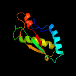
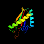
1 c2l26A_
100.0
46
PDB header: membrane proteinChain: A: PDB Molecule: uncharacterized protein rv0899/mt0922;PDBTitle: rv0899 from mycobacterium tuberculosis contains two separated domains
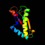
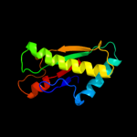
2 c2k1sA_
100.0
100
PDB header: lipoproteinChain: A: PDB Molecule: inner membrane lipoprotein yiad;PDBTitle: solution nmr structure of the folded c-terminal fragment of yiad from2 escherichia coli. northeast structural genomics consortium target3 er553.
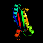
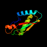
3 c3khnB_
100.0
22
PDB header: structural genomics, unknown functionChain: B: PDB Molecule: motb protein, putative;PDBTitle: crystal structure of putative motb like protein dvu_22282 from desulfovibrio vulgaris.
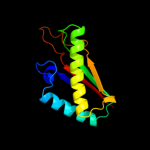
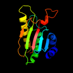
4 c2kgwA_
100.0
47
PDB header: membrane proteinChain: A: PDB Molecule: outer membrane protein a;PDBTitle: solution structure of the carboxy-terminal domain of ompatb, a pore2 forming protein from mycobacterium tuberculosis
5 d2aizp1
100.0
30
Fold: Bacillus chorismate mutase-likeSuperfamily: OmpA-likeFamily: OmpA-like6 c3td4D_
100.0
46
PDB header: membrane protein,peptide binding proteinChain: D: PDB Molecule: outer membrane protein omp38;PDBTitle: crystal structure of ompa-like domain from acinetobacter baumannii in2 complex with diaminopimelate
7 d1r1ma_
100.0
27
Fold: Bacillus chorismate mutase-likeSuperfamily: OmpA-likeFamily: OmpA-like8 c1r1mA_
100.0
27
PDB header: membrane proteinChain: A: PDB Molecule: outer membrane protein class 4;PDBTitle: structure of the ompa-like domain of rmpm from neisseria2 meningitidis
9 d2hqsc1
100.0
28
Fold: Bacillus chorismate mutase-likeSuperfamily: OmpA-likeFamily: OmpA-like10 c3cyqM_
100.0
30
PDB header: membrane proteinChain: M: PDB Molecule: chemotaxis protein motb;PDBTitle: the crystal structure of the complex of the c-terminal domain of2 helicobacter pylori motb (residues 125-256) with n-acetylmuramic acid
11 c2zvyB_
100.0
21
PDB header: membrane proteinChain: B: PDB Molecule: chemotaxis protein motb;PDBTitle: structure of the periplasmic domain of motb from salmonella2 (crystal form ii)
12 c3ldtA_
100.0
28
PDB header: membrane proteinChain: A: PDB Molecule: outer membrane protein, ompa family protein;PDBTitle: crystal structure of an outer membrane protein(ompa)from2 legionella pneumophila
13 c3oonA_
99.9
28
PDB header: membrane proteinChain: A: PDB Molecule: outer membrane protein (tpn50);PDBTitle: the structure of an outer membrance protein from borrelia burgdorferi2 b31
14 c2zovA_
99.9
24
PDB header: membrane proteinChain: A: PDB Molecule: chemotaxis protein motb;PDBTitle: structure of the periplasmic domain of motb from salmonella2 (crystal form i)
15 c2zf8A_
99.8
25
PDB header: structural proteinChain: A: PDB Molecule: component of sodium-driven polar flagellar motor;PDBTitle: crystal structure of moty
16 c2r6hC_
60.8
25
PDB header: oxidoreductaseChain: C: PDB Molecule: nadh:ubiquinone oxidoreductase, na translocating, fPDBTitle: crystal structure of the domain comprising the nad binding and the fad2 binding regions of the nadh:ubiquinone oxidoreductase, na3 translocating, f subunit from porphyromonas gingivalis
17 c2qw5B_
55.6
11
PDB header: isomeraseChain: B: PDB Molecule: xylose isomerase-like tim barrel;PDBTitle: crystal structure of a putative sugar phosphate isomerase/epimerase2 (ava4194) from anabaena variabilis atcc 29413 at 1.78 a resolution
18 d1gvha3
51.9
24
Fold: Ferredoxin reductase-like, C-terminal NADP-linked domainSuperfamily: Ferredoxin reductase-like, C-terminal NADP-linked domainFamily: Flavohemoglobin, C-terminal domain19 c1gvhA_
51.9
25
PDB header: oxidoreductaseChain: A: PDB Molecule: flavohemoprotein;PDBTitle: the x-ray structure of ferric escherichia coli2 flavohemoglobin reveals an unespected geometry of the3 distal heme pocket
20 d1tiaa_
49.4
21
Fold: alpha/beta-HydrolasesSuperfamily: alpha/beta-HydrolasesFamily: Fungal lipases21 c3cqyA_
not modelled
44.1
18
PDB header: transferaseChain: A: PDB Molecule: anhydro-n-acetylmuramic acid kinase;PDBTitle: crystal structure of a functionally unknown protein (so_1313) from2 shewanella oneidensis mr-1
22 d1tiba_
not modelled
42.9
25
Fold: alpha/beta-HydrolasesSuperfamily: alpha/beta-HydrolasesFamily: Fungal lipases23 d1cqxa3
not modelled
38.9
25
Fold: Ferredoxin reductase-like, C-terminal NADP-linked domainSuperfamily: Ferredoxin reductase-like, C-terminal NADP-linked domainFamily: Flavohemoglobin, C-terminal domain24 c3ngmB_
not modelled
38.6
25
PDB header: hydrolaseChain: B: PDB Molecule: extracellular lipase;PDBTitle: crystal structure of lipase from gibberella zeae
25 c1cqxB_
not modelled
33.4
25
PDB header: lipid binding proteinChain: B: PDB Molecule: flavohemoprotein;PDBTitle: crystal structure of the flavohemoglobin from alcaligenes eutrophus at2 1.75 a resolution
26 c2gpjA_
not modelled
33.3
9
PDB header: fad-binding proteinChain: A: PDB Molecule: siderophore-interacting protein;PDBTitle: crystal structure of a siderophore-interacting protein (sputcn32_0076)2 from shewanella putrefaciens cn-32 at 2.20 a resolution
27 c3qbwA_
not modelled
32.6
21
PDB header: transferaseChain: A: PDB Molecule: anhydro-n-acetylmuramic acid kinase;PDBTitle: crystal structure of pseudomonas aeruginosa 1,6-anhydro-n-2 actetylmuramic acid kinase (anmk) bound to adenosine diphosphate
28 c3g7nA_
not modelled
32.6
20
PDB header: hydrolaseChain: A: PDB Molecule: lipase;PDBTitle: crystal structure of a triacylglycerol lipase from2 penicillium expansum at 1.3
29 d1krha2
not modelled
31.3
23
Fold: Ferredoxin reductase-like, C-terminal NADP-linked domainSuperfamily: Ferredoxin reductase-like, C-terminal NADP-linked domainFamily: Aromatic dioxygenase reductase-like30 c3o0dF_
not modelled
30.9
25
PDB header: hydrolaseChain: F: PDB Molecule: triacylglycerol lipase;PDBTitle: crystal structure of lip2 lipase from yarrowia lipolytica at 1.7 a2 resolution
31 c1tvcA_
not modelled
30.7
11
PDB header: oxidoreductaseChain: A: PDB Molecule: methane monooxygenase component c;PDBTitle: fad and nadh binding domain of methane monooxygenase2 reductase from methylococcus capsulatus (bath)
32 d1tvca2
not modelled
29.9
11
Fold: Ferredoxin reductase-like, C-terminal NADP-linked domainSuperfamily: Ferredoxin reductase-like, C-terminal NADP-linked domainFamily: Aromatic dioxygenase reductase-like33 d1uzhc1
not modelled
26.3
15
Fold: RuBisCO, small subunitSuperfamily: RuBisCO, small subunitFamily: RuBisCO, small subunit34 d1uwca_
not modelled
25.2
25
Fold: alpha/beta-HydrolasesSuperfamily: alpha/beta-HydrolasesFamily: Fungal lipases35 d1rbli_
not modelled
25.0
22
Fold: RuBisCO, small subunitSuperfamily: RuBisCO, small subunitFamily: RuBisCO, small subunit36 d1bxni_
not modelled
24.7
16
Fold: RuBisCO, small subunitSuperfamily: RuBisCO, small subunitFamily: RuBisCO, small subunit37 c1amuB_
not modelled
23.7
14
PDB header: peptide synthetaseChain: B: PDB Molecule: gramicidin synthetase 1;PDBTitle: phenylalanine activating domain of gramicidin synthetase 12 in a complex with amp and phenylalanine
38 c3pe3D_
not modelled
22.4
18
PDB header: transferaseChain: D: PDB Molecule: udp-n-acetylglucosamine--peptide n-PDBTitle: structure of human o-glcnac transferase and its complex with a peptide2 substrate
39 d1svdm1
not modelled
21.2
24
Fold: RuBisCO, small subunitSuperfamily: RuBisCO, small subunitFamily: RuBisCO, small subunit40 c3pa8A_
not modelled
20.6
16
PDB header: toxin/peptide inhibitorChain: A: PDB Molecule: toxin b;PDBTitle: structure of the c. difficile tcdb cysteine protease domain in complex2 with a peptide inhibitor
41 c3iteB_
not modelled
20.2
17
PDB header: ligaseChain: B: PDB Molecule: sidn siderophore synthetase;PDBTitle: the third adenylation domain of the fungal sidn non-2 ribosomal peptide synthetase
42 d2axla1
not modelled
20.1
16
Fold: DNA/RNA-binding 3-helical bundleSuperfamily: "Winged helix" DNA-binding domainFamily: RecQ helicase DNA-binding domain-like43 d8ruci_
not modelled
16.2
31
Fold: RuBisCO, small subunitSuperfamily: RuBisCO, small subunitFamily: RuBisCO, small subunit44 d1xdpa4
not modelled
15.0
18
Fold: Phospholipase D/nucleaseSuperfamily: Phospholipase D/nucleaseFamily: Polyphosphate kinase C-terminal domain45 c1krhA_
not modelled
14.9
23
PDB header: oxidoreductaseChain: A: PDB Molecule: benzoate 1,2-dioxygenase reductase;PDBTitle: x-ray stucture of benzoate dioxygenase reductase
46 d2o8ra4
not modelled
14.9
24
Fold: Phospholipase D/nucleaseSuperfamily: Phospholipase D/nucleaseFamily: Polyphosphate kinase C-terminal domain47 c1jrjA_
not modelled
14.8
17
PDB header: hormone/growth factorChain: A: PDB Molecule: exendin-4;PDBTitle: solution structure of exendin-4 in 30-vol% trifluoroethanol
48 d1bwvs_
not modelled
14.5
19
Fold: RuBisCO, small subunitSuperfamily: RuBisCO, small subunitFamily: RuBisCO, small subunit49 d1ir1s_
not modelled
14.3
22
Fold: RuBisCO, small subunitSuperfamily: RuBisCO, small subunitFamily: RuBisCO, small subunit50 d1wdds_
not modelled
14.3
27
Fold: RuBisCO, small subunitSuperfamily: RuBisCO, small subunitFamily: RuBisCO, small subunit51 d1jmxa1
not modelled
14.3
18
Fold: Cytochrome cSuperfamily: Cytochrome cFamily: Quinohemoprotein amine dehydrogenase A chain, domains 1 and 252 d1i4ua_
not modelled
14.2
22
Fold: LipocalinsSuperfamily: LipocalinsFamily: Retinol binding protein-like53 d1r3na1
not modelled
13.9
8
Fold: Phosphorylase/hydrolase-likeSuperfamily: Zn-dependent exopeptidasesFamily: Bacterial dinuclear zinc exopeptidases54 c3ds8A_
not modelled
13.4
15
PDB header: structural genomics, unknown functionChain: A: PDB Molecule: lin2722 protein;PDBTitle: the crysatl structure of the gene lin2722 products from listeria2 innocua
55 d1d4oa_
not modelled
13.4
16
Fold: DHS-like NAD/FAD-binding domainSuperfamily: DHS-like NAD/FAD-binding domainFamily: Transhydrogenase domain III (dIII)56 c1pt9B_
not modelled
12.6
16
PDB header: oxidoreductaseChain: B: PDB Molecule: nad(p) transhydrogenase, mitochondrial;PDBTitle: crystal structure analysis of the diii component of transhydrogenase2 with a thio-nicotinamide nucleotide analogue
57 c3og9A_
not modelled
12.4
12
PDB header: hydrolaseChain: A: PDB Molecule: protein yahd a copper inducible hydrolase;PDBTitle: structure of yahd with malic acid
58 c3a9lB_
not modelled
12.2
22
PDB header: hydrolaseChain: B: PDB Molecule: poly-gamma-glutamate hydrolase;PDBTitle: structure of bacteriophage poly-gamma-glutamate hydrolase
59 c1nauA_
not modelled
12.1
18
PDB header: hormone/growth factorChain: A: PDB Molecule: glucagon;PDBTitle: nmr solution structure of the glucagon antagonist [deshis1,2 desphe6, glu9]glucagon amide in the presence of3 perdeuterated dodecylphosphocholine micelles
60 d3tgla_
not modelled
12.1
25
Fold: alpha/beta-HydrolasesSuperfamily: alpha/beta-HydrolasesFamily: Fungal lipases61 c2bgjB_
not modelled
12.0
13
PDB header: oxidoreductaseChain: B: PDB Molecule: ferredoxin-nadp(h) reductase;PDBTitle: x-ray structure of the ferredoxin-nadp(h) reductase from2 rhodobacter capsulatus at 2.1 angstroms
62 c3iibA_
not modelled
12.0
15
PDB header: hydrolaseChain: A: PDB Molecule: peptidase m28;PDBTitle: crystal structure of peptidase m28 precursor (yp_926796.1) from2 shewanella amazonensis sb2b at 1.70 a resolution
63 c3l7pA_
not modelled
11.7
21
PDB header: transcriptionChain: A: PDB Molecule: putative nitrogen regulatory protein pii;PDBTitle: crystal structure of smu.1657c, putative nitrogen regulatory protein2 pii from streptococcus mutans
64 d1tkja1
not modelled
10.9
17
Fold: Phosphorylase/hydrolase-likeSuperfamily: Zn-dependent exopeptidasesFamily: Bacterial dinuclear zinc exopeptidases65 d1lgya_
not modelled
10.8
20
Fold: alpha/beta-HydrolasesSuperfamily: alpha/beta-HydrolasesFamily: Fungal lipases66 c3ca8B_
not modelled
10.7
12
PDB header: structural genomics, unknown functionChain: B: PDB Molecule: protein ydcf;PDBTitle: crystal structure of escherichia coli ydcf, an s-adenosyl-l-methionine2 utilizing enzyme
67 c2rd5D_
not modelled
10.5
20
PDB header: protein bindingChain: D: PDB Molecule: pii protein;PDBTitle: structural basis for the regulation of n-acetylglutamate kinase by pii2 in arabidopsis thaliana
68 d1saza1
not modelled
10.2
16
Fold: Ribonuclease H-like motifSuperfamily: Actin-like ATPase domainFamily: Acetokinase-like69 c1gplA_
not modelled
10.0
18
PDB header: serine esteraseChain: A: PDB Molecule: rp2 lipase;PDBTitle: rp2 lipase
70 c2ckcA_
not modelled
9.9
23
PDB header: hydrolaseChain: A: PDB Molecule: chromodomain-helicase-dna-binding protein 7;PDBTitle: solution structures of the brk domains of the human chromo2 helicase domain 7 and 8, reveals structural similarity3 with gyf domain suggesting a role in protein interaction
71 d2ckca1
not modelled
9.9
23
Fold: GYF/BRK domain-likeSuperfamily: BRK domain-likeFamily: BRK domain-like72 c2ogsA_
not modelled
9.9
24
PDB header: hydrolaseChain: A: PDB Molecule: thermostable carboxylesterase est50;PDBTitle: crystal structure of the geobacillus stearothermophilus2 carboxylesterase est55 at ph 6.2
73 c3b9eA_
not modelled
9.9
15
PDB header: hydrolaseChain: A: PDB Molecule: chitinase a;PDBTitle: crystal structure of inactive mutant e315m chitinase a from2 vibrio harveyi
74 c2oghA_
not modelled
9.9
27
PDB header: translationChain: A: PDB Molecule: eukaryotic translation initiation factor eif-1;PDBTitle: solution structure of yeast eif1
75 d1e32a3
not modelled
9.8
18
Fold: Cdc48 domain 2-likeSuperfamily: Cdc48 domain 2-likeFamily: Cdc48 domain 2-like76 d1amua_
not modelled
9.8
13
Fold: Acetyl-CoA synthetase-likeSuperfamily: Acetyl-CoA synthetase-likeFamily: Acetyl-CoA synthetase-like77 d1pbya1
not modelled
9.7
11
Fold: Cytochrome cSuperfamily: Cytochrome cFamily: Quinohemoprotein amine dehydrogenase A chain, domains 1 and 278 d1ej7s_
not modelled
9.3
23
Fold: RuBisCO, small subunitSuperfamily: RuBisCO, small subunitFamily: RuBisCO, small subunit79 d2v0ea1
not modelled
9.2
25
Fold: GYF/BRK domain-likeSuperfamily: BRK domain-likeFamily: BRK domain-like80 d2nvma1
not modelled
9.0
13
Fold: XisI-likeSuperfamily: XisI-likeFamily: XisI-like81 d1wfya_
not modelled
9.0
9
Fold: beta-Grasp (ubiquitin-like)Superfamily: Ubiquitin-likeFamily: Ras-binding domain, RBD82 c3eyiB_
not modelled
8.9
33
PDB header: dna binding protein/dnaChain: B: PDB Molecule: z-dna-binding protein 1;PDBTitle: the crystal structure of the second z-dna binding domain of2 human dai (zbp1) in complex with z-dna
83 d1gk8i_
not modelled
8.8
13
Fold: RuBisCO, small subunitSuperfamily: RuBisCO, small subunitFamily: RuBisCO, small subunit84 c3mxoB_
not modelled
8.8
15
PDB header: hydrolaseChain: B: PDB Molecule: serine/threonine-protein phosphatase pgam5, mitochondrial;PDBTitle: crystal structure oh human phosphoglycerate mutase family member 52 (pgam5)
85 d1uzdc1
not modelled
8.6
13
Fold: RuBisCO, small subunitSuperfamily: RuBisCO, small subunitFamily: RuBisCO, small subunit86 c1f8uA_
not modelled
8.5
18
PDB header: hydrolase/toxinChain: A: PDB Molecule: acetylcholinesterase;PDBTitle: crystal structure of mutant e202q of human acetylcholinesterase2 complexed with green mamba venom peptide fasciculin-ii
87 d1f8ua_
not modelled
8.5
18
Fold: alpha/beta-HydrolasesSuperfamily: alpha/beta-HydrolasesFamily: Acetylcholinesterase-like88 c2apnA_
not modelled
8.5
17
PDB header: structural genomics, unknown functionChain: A: PDB Molecule: protein hi1723;PDBTitle: hi1723 solution structure
89 c1hplB_
not modelled
8.3
12
PDB header: hydrolase(carboxylic esterase)Chain: B: PDB Molecule: lipase;PDBTitle: horse pancreatic lipase. the crystal structure at 2.32 angstroms resolution
90 c2el7A_
not modelled
7.9
12
PDB header: ligaseChain: A: PDB Molecule: tryptophanyl-trna synthetase;PDBTitle: crystal structure of tryptophanyl-trna synthetase from thermus2 thermophilus
91 d2nlva1
not modelled
7.9
33
Fold: XisI-likeSuperfamily: XisI-likeFamily: XisI-like92 d1u5ta2
not modelled
7.8
19
Fold: DNA/RNA-binding 3-helical bundleSuperfamily: "Winged helix" DNA-binding domainFamily: Vacuolar sorting protein domain93 d1o57a1
not modelled
7.7
11
Fold: DNA/RNA-binding 3-helical bundleSuperfamily: "Winged helix" DNA-binding domainFamily: N-terminal domain of Bacillus PurR94 d2v6ai1
not modelled
7.6
13
Fold: RuBisCO, small subunitSuperfamily: RuBisCO, small subunitFamily: RuBisCO, small subunit95 d1qy7a_
not modelled
7.4
14
Fold: Ferredoxin-likeSuperfamily: GlnB-likeFamily: Prokaryotic signal transducing protein96 c3b64A_
not modelled
7.3
25
PDB header: cytokineChain: A: PDB Molecule: macrophage migration inhibitory factor-likePDBTitle: macrophage migration inhibitory factor (mif) from2 /leishmania major
97 d1rp1a2
not modelled
7.3
10
Fold: alpha/beta-HydrolasesSuperfamily: alpha/beta-HydrolasesFamily: Pancreatic lipase, N-terminal domain98 c2pfuA_
not modelled
7.2
11
PDB header: transport proteinChain: A: PDB Molecule: biopolymer transport exbd protein;PDBTitle: nmr strcuture determination of the periplasmic domain of exbd from2 e.coli
99 d2ha2a1
not modelled
7.0
21
Fold: alpha/beta-HydrolasesSuperfamily: alpha/beta-HydrolasesFamily: Acetylcholinesterase-like