| 1 | c3gbtA_
|
|
 |
100.0 |
21 |
PDB header:transferase
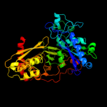
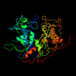
Chain: A: PDB Molecule:gluconate kinase;
PDBTitle: crystal structure of gluconate kinase from lactobacillus acidophilus
|
| 2 | c3jvpA_
|
|
 |
100.0 |
22 |
PDB header:transferase
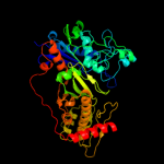
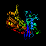
Chain: A: PDB Molecule:ribulokinase;
PDBTitle: crystal structure of ribulokinase from bacillus halodurans
|
| 3 | c3ifrB_
|
|
 |
100.0 |
24 |
PDB header:transferase
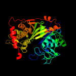
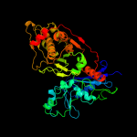
Chain: B: PDB Molecule:carbohydrate kinase, fggy;
PDBTitle: the crystal structure of xylulose kinase from rhodospirillum rubrum
|
| 4 | c3gg4B_
|
|
 |
100.0 |
18 |
PDB header:transferase
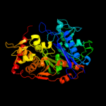
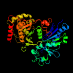
Chain: B: PDB Molecule:glycerol kinase;
PDBTitle: the crystal structure of glycerol kinase from yersinia2 pseudotuberculosis
|
| 5 | c3flcX_
|
|
 |
100.0 |
21 |
PDB header:transferase
Chain: X: PDB Molecule:glycerol kinase;
PDBTitle: crystal structure of the his-tagged h232r mutant of glycerol kinase2 from enterococcus casseliflavus with glycerol
|
| 6 | c2d4wA_
|
|
 |
100.0 |
21 |
PDB header:transferase
Chain: A: PDB Molecule:glycerol kinase;
PDBTitle: crystal structure of glycerol kinase from cellulomonas sp.2 nt3060
|
| 7 | c2nlxA_
|
|
 |
100.0 |
22 |
PDB header:transferase
Chain: A: PDB Molecule:xylulose kinase;
PDBTitle: crystal structure of the apo e. coli xylulose kinase
|
| 8 | c3hz6A_
|
|
 |
100.0 |
21 |
PDB header:transferase
Chain: A: PDB Molecule:xylulokinase;
PDBTitle: crystal structure of xylulokinase from chromobacterium violaceum
|
| 9 | c2zf5O_
|
|
 |
100.0 |
22 |
PDB header:transferase
Chain: O: PDB Molecule:glycerol kinase;
PDBTitle: crystal structure of highly thermostable glycerol kinase from a2 hyperthermophilic archaeon
|
| 10 | c3g25B_
|
|
 |
100.0 |
17 |
PDB header:transferase
Chain: B: PDB Molecule:glycerol kinase;
PDBTitle: 1.9 angstrom crystal structure of glycerol kinase (glpk) from2 staphylococcus aureus in complex with glycerol.
|
| 11 | c2dpnB_
|
|
 |
100.0 |
21 |
PDB header:transferase
Chain: B: PDB Molecule:glycerol kinase;
PDBTitle: crystal structure of the glycerol kinase from thermus2 thermophilus hb8
|
| 12 | c2w40C_
|
|
 |
100.0 |
20 |
PDB header:transferase
Chain: C: PDB Molecule:glycerol kinase, putative;
PDBTitle: crystal structure of plasmodium falciparum glycerol kinase2 with bound glycerol
|
| 13 | c1glbG_
|
|
 |
100.0 |
19 |
PDB header:phosphotransferase
Chain: G: PDB Molecule:glycerol kinase;
PDBTitle: structure of the regulatory complex of escherichia coli iiiglc with2 glycerol kinase
|
| 14 | c3ezwD_
|
|
 |
100.0 |
20 |
PDB header:transferase
Chain: D: PDB Molecule:glycerol kinase;
PDBTitle: crystal structure of a hyperactive escherichia coli glycerol kinase2 mutant gly230 --> asp obtained using microfluidic crystallization3 devices
|
| 15 | c1xupO_
|
|
 |
100.0 |
21 |
PDB header:transferase
Chain: O: PDB Molecule:glycerol kinase;
PDBTitle: enterococcus casseliflavus glycerol kinase complexed with glycerol
|
| 16 | c2cgkB_
|
|
 |
100.0 |
16 |
PDB header:transferase
Chain: B: PDB Molecule:l-rhamnulose kinase;
PDBTitle: crystal structure of l-rhamnulose kinase from escherichia2 coli in an open uncomplexed conformation.
|
| 17 | c3i8bA_
|
|
 |
100.0 |
18 |
PDB header:transferase
Chain: A: PDB Molecule:xylulose kinase;
PDBTitle: the crystal structure of xylulose kinase from2 bifidobacterium adolescentis
|
| 18 | c3h6eB_
|
|
 |
100.0 |
17 |
PDB header:transferase
Chain: B: PDB Molecule:carbohydrate kinase, fggy;
PDBTitle: the crystal structure of a carbohydrate kinase from novosphingobium2 aromaticivorans
|
| 19 | d2p3ra1
|
|
 |
100.0 |
22 |
Fold:Ribonuclease H-like motif
Superfamily:Actin-like ATPase domain
Family:Glycerol kinase |
| 20 | d1r59o1
|
|
 |
100.0 |
22 |
Fold:Ribonuclease H-like motif
Superfamily:Actin-like ATPase domain
Family:Glycerol kinase |
| 21 | d2p3ra2 |
|
not modelled |
100.0 |
18 |
Fold:Ribonuclease H-like motif
Superfamily:Actin-like ATPase domain
Family:Glycerol kinase |
| 22 | d1r59o2 |
|
not modelled |
100.0 |
21 |
Fold:Ribonuclease H-like motif
Superfamily:Actin-like ATPase domain
Family:Glycerol kinase |
| 23 | d1huxa_ |
|
not modelled |
99.4 |
16 |
Fold:Ribonuclease H-like motif
Superfamily:Actin-like ATPase domain
Family:BadF/BadG/BcrA/BcrD-like |
| 24 | c3h1qB_ |
|
not modelled |
99.4 |
17 |
PDB header:structural protein
Chain: B: PDB Molecule:ethanolamine utilization protein eutj;
PDBTitle: crystal structure of ethanolamine utilization protein eutj from2 carboxydothermus hydrogenoformans
|
| 25 | c2e2pA_ |
|
not modelled |
99.2 |
18 |
PDB header:transferase
Chain: A: PDB Molecule:hexokinase;
PDBTitle: crystal structure of sulfolobus tokodaii hexokinase in2 complex with adp
|
| 26 | c3eo3B_ |
|
not modelled |
99.0 |
20 |
PDB header:isomerase, transferase
Chain: B: PDB Molecule:bifunctional udp-n-acetylglucosamine 2-epimerase/n-
PDBTitle: crystal structure of the n-acetylmannosamine kinase domain of human2 gne protein
|
| 27 | d2ewsa1 |
|
not modelled |
98.9 |
16 |
Fold:Ribonuclease H-like motif
Superfamily:Actin-like ATPase domain
Family:Fumble-like |
| 28 | c2ch5D_ |
|
not modelled |
98.9 |
15 |
PDB header:transferase
Chain: D: PDB Molecule:nagk protein;
PDBTitle: crystal structure of human n-acetylglucosamine kinase in2 complex with n-acetylglucosamine
|
| 29 | d1zc6a1 |
|
not modelled |
98.8 |
18 |
Fold:Ribonuclease H-like motif
Superfamily:Actin-like ATPase domain
Family:BadF/BadG/BcrA/BcrD-like |
| 30 | c3enoB_ |
|
not modelled |
98.8 |
12 |
PDB header:hydrolase/unknown function
Chain: B: PDB Molecule:putative o-sialoglycoprotein endopeptidase;
PDBTitle: crystal structure of pyrococcus furiosus pcc1 in complex2 with thermoplasma acidophilum kae1
|
| 31 | c2ivoC_ |
|
not modelled |
98.8 |
14 |
PDB header:hydrolase
Chain: C: PDB Molecule:up1;
PDBTitle: structure of up1 protein
|
| 32 | c1z05A_ |
|
not modelled |
98.8 |
14 |
PDB header:transcription
Chain: A: PDB Molecule:transcriptional regulator, rok family;
PDBTitle: crystal structure of the rok family transcriptional regulator, homolog2 of e.coli mlc protein.
|
| 33 | c2qm1D_ |
|
not modelled |
98.8 |
24 |
PDB header:transferase
Chain: D: PDB Molecule:glucokinase;
PDBTitle: crystal structure of glucokinase from enterococcus faecalis
|
| 34 | c3r8eA_ |
|
not modelled |
98.7 |
21 |
PDB header:transferase
Chain: A: PDB Molecule:hypothetical sugar kinase;
PDBTitle: crystal structure of a hypothetical sugar kinase (chu_1875) from2 cytophaga hutchinsonii atcc 33406 at 1.65 a resolution
|
| 35 | c1e4gT_ |
|
not modelled |
98.7 |
15 |
PDB header:bacterial cell division
Chain: T: PDB Molecule:cell division protein ftsa;
PDBTitle: ftsa (atp-bound form) from thermotoga maritima
|
| 36 | c1z6rC_ |
|
not modelled |
98.7 |
15 |
PDB header:transcription
Chain: C: PDB Molecule:mlc protein;
PDBTitle: crystal structure of mlc from escherichia coli
|
| 37 | c3cqyA_ |
|
not modelled |
98.5 |
15 |
PDB header:transferase
Chain: A: PDB Molecule:anhydro-n-acetylmuramic acid kinase;
PDBTitle: crystal structure of a functionally unknown protein (so_1313) from2 shewanella oneidensis mr-1
|
| 38 | d1z05a3 |
|
not modelled |
98.4 |
15 |
Fold:Ribonuclease H-like motif
Superfamily:Actin-like ATPase domain
Family:ROK |
| 39 | d2ch5a2 |
|
not modelled |
98.3 |
17 |
Fold:Ribonuclease H-like motif
Superfamily:Actin-like ATPase domain
Family:BadF/BadG/BcrA/BcrD-like |
| 40 | c1zc6A_ |
|
not modelled |
98.3 |
17 |
PDB header:structural genomics, unknown function
Chain: A: PDB Molecule:probable n-acetylglucosamine kinase;
PDBTitle: crystal structure of putative n-acetylglucosamine kinase from2 chromobacterium violaceum. northeast structural genomics target3 cvr23.
|
| 41 | d2hoea3 |
|
not modelled |
98.3 |
8 |
Fold:Ribonuclease H-like motif
Superfamily:Actin-like ATPase domain
Family:ROK |
| 42 | c3en9B_ |
|
not modelled |
98.2 |
18 |
PDB header:hydrolase
Chain: B: PDB Molecule:o-sialoglycoprotein endopeptidase/protein kinase;
PDBTitle: structure of the methanococcus jannaschii kae1-bud32 fusion2 protein
|
| 43 | c1dkgD_ |
|
not modelled |
98.0 |
17 |
PDB header:complex (hsp24/hsp70)
Chain: D: PDB Molecule:molecular chaperone dnak;
PDBTitle: crystal structure of the nucleotide exchange factor grpe2 bound to the atpase domain of the molecular chaperone dnak
|
| 44 | d1z6ra2 |
|
not modelled |
98.0 |
16 |
Fold:Ribonuclease H-like motif
Superfamily:Actin-like ATPase domain
Family:ROK |
| 45 | c2ap1A_ |
|
not modelled |
98.0 |
16 |
PDB header:transferase
Chain: A: PDB Molecule:putative regulator protein;
PDBTitle: crystal structure of the putative regulatory protein
|
| 46 | c1ig8A_ |
|
not modelled |
97.9 |
15 |
PDB header:transferase
Chain: A: PDB Molecule:hexokinase pii;
PDBTitle: crystal structure of yeast hexokinase pii with the correct2 amino acid sequence
|
| 47 | c3d2fC_ |
|
not modelled |
97.9 |
14 |
PDB header:chaperone
Chain: C: PDB Molecule:heat shock protein homolog sse1;
PDBTitle: crystal structure of a complex of sse1p and hsp70
|
| 48 | c2v7zA_ |
|
not modelled |
97.8 |
12 |
PDB header:chaperone
Chain: A: PDB Molecule:heat shock cognate 71 kda protein;
PDBTitle: crystal structure of the 70-kda heat shock cognate protein2 from rattus norvegicus in post-atp hydrolysis state
|
| 49 | c1v4sA_ |
|
not modelled |
97.8 |
15 |
PDB header:transferase
Chain: A: PDB Molecule:glucokinase isoform 2;
PDBTitle: crystal structure of human glucokinase
|
| 50 | d1dkgd2 |
|
not modelled |
97.8 |
17 |
Fold:Ribonuclease H-like motif
Superfamily:Actin-like ATPase domain
Family:Actin/HSP70 |
| 51 | d1woqa1 |
|
not modelled |
97.8 |
14 |
Fold:Ribonuclease H-like motif
Superfamily:Actin-like ATPase domain
Family:ROK |
| 52 | c2v7yA_ |
|
not modelled |
97.8 |
15 |
PDB header:chaperone
Chain: A: PDB Molecule:chaperone protein dnak;
PDBTitle: crystal structure of the molecular chaperone dnak from2 geobacillus kaustophilus hta426 in post-atp hydrolysis3 state
|
| 53 | d2ap1a2 |
|
not modelled |
97.7 |
19 |
Fold:Ribonuclease H-like motif
Superfamily:Actin-like ATPase domain
Family:ROK |
| 54 | c1hpmA_ |
|
not modelled |
97.7 |
12 |
PDB header:hydrolase (acting on acid anhydrides)
Chain: A: PDB Molecule:44k atpase fragment (n-terminal) of 7o kd heat-
PDBTitle: how potassium affects the activity of the molecular2 chaperone hsc70. ii. potassium binds specifically in the3 atpase active site
|
| 55 | c3htvA_ |
|
not modelled |
97.7 |
21 |
PDB header:transferase
Chain: A: PDB Molecule:d-allose kinase;
PDBTitle: crystal structure of d-allose kinase (np_418508.1) from escherichia2 coli k12 at 1.95 a resolution
|
| 56 | d1jcea2 |
|
not modelled |
97.6 |
15 |
Fold:Ribonuclease H-like motif
Superfamily:Actin-like ATPase domain
Family:Actin/HSP70 |
| 57 | d1q18a1 |
|
not modelled |
97.6 |
16 |
Fold:Ribonuclease H-like motif
Superfamily:Actin-like ATPase domain
Family:Glucokinase |
| 58 | d1sz2a1 |
|
not modelled |
97.6 |
15 |
Fold:Ribonuclease H-like motif
Superfamily:Actin-like ATPase domain
Family:Glucokinase |
| 59 | d1bupa2 |
|
not modelled |
97.5 |
12 |
Fold:Ribonuclease H-like motif
Superfamily:Actin-like ATPase domain
Family:Actin/HSP70 |
| 60 | c3vgkB_ |
|
not modelled |
97.5 |
23 |
PDB header:transferase
Chain: B: PDB Molecule:glucokinase;
PDBTitle: crystal structure of a rok family glucokinase from streptomyces2 griseus
|
| 61 | c2gupA_ |
|
not modelled |
97.5 |
20 |
PDB header:transferase
Chain: A: PDB Molecule:rok family protein;
PDBTitle: structural genomics, the crystal structure of a rok family protein2 from streptococcus pneumoniae tigr4 in complex with sucrose
|
| 62 | c2hoeA_ |
|
not modelled |
97.5 |
8 |
PDB header:transferase
Chain: A: PDB Molecule:n-acetylglucosamine kinase;
PDBTitle: crystal structure of n-acetylglucosamine kinase (tm1224) from2 thermotoga maritima at 2.46 a resolution
|
| 63 | d2e8aa2 |
|
not modelled |
97.5 |
13 |
Fold:Ribonuclease H-like motif
Superfamily:Actin-like ATPase domain
Family:Actin/HSP70 |
| 64 | d1e4ft1 |
|
not modelled |
97.5 |
16 |
Fold:Ribonuclease H-like motif
Superfamily:Actin-like ATPase domain
Family:Actin/HSP70 |
| 65 | c3iucC_ |
|
not modelled |
97.5 |
15 |
PDB header:chaperone
Chain: C: PDB Molecule:heat shock 70kda protein 5 (glucose-regulated
PDBTitle: crystal structure of the human 70kda heat shock protein 52 (bip/grp78) atpase domain in complex with adp
|
| 66 | c1jcgA_ |
|
not modelled |
97.4 |
16 |
PDB header:structural protein
Chain: A: PDB Molecule:rod shape-determining protein mreb;
PDBTitle: mreb from thermotoga maritima, amppnp
|
| 67 | c1sazA_ |
|
not modelled |
97.4 |
11 |
PDB header:transferase
Chain: A: PDB Molecule:probable butyrate kinase 2;
PDBTitle: membership in the askha superfamily: enzymological2 properties and crystal structure of butyrate kinase 2 from3 thermotoga maritima
|
| 68 | c2khoA_ |
|
not modelled |
97.4 |
19 |
PDB header:chaperone
Chain: A: PDB Molecule:heat shock protein 70;
PDBTitle: nmr-rdc / xray structure of e. coli hsp70 (dnak) chaperone2 (1-605) complexed with adp and substrate
|
| 69 | c1qhaA_ |
|
not modelled |
97.4 |
13 |
PDB header:transferase
Chain: A: PDB Molecule:protein (hexokinase);
PDBTitle: human hexokinase type i complexed with atp analogue amp-pnp
|
| 70 | d2aa4a1 |
|
not modelled |
97.3 |
14 |
Fold:Ribonuclease H-like motif
Superfamily:Actin-like ATPase domain
Family:ROK |
| 71 | c1bdgA_ |
|
not modelled |
97.3 |
13 |
PDB header:hexokinase
Chain: A: PDB Molecule:hexokinase;
PDBTitle: hexokinase from schistosoma mansoni complexed with glucose
|
| 72 | c2aa4B_ |
|
not modelled |
97.3 |
14 |
PDB header:transferase
Chain: B: PDB Molecule:putative n-acetylmannosamine kinase;
PDBTitle: crystal structure of escherichia coli putative n-2 acetylmannosamine kinase, new york structural genomics3 consortium
|
| 73 | c3mcpA_ |
|
not modelled |
97.2 |
12 |
PDB header:transferase
Chain: A: PDB Molecule:glucokinase;
PDBTitle: crystal structure of glucokinase (bdi_1628) from parabacteroides2 distasonis atcc 8503 at 3.00 a resolution
|
| 74 | c1xc3A_ |
|
not modelled |
97.1 |
17 |
PDB header:transferase
Chain: A: PDB Molecule:putative fructokinase;
PDBTitle: structure of a putative fructokinase from bacillus subtilis
|
| 75 | d2gupa1 |
|
not modelled |
97.0 |
19 |
Fold:Ribonuclease H-like motif
Superfamily:Actin-like ATPase domain
Family:ROK |
| 76 | c1woqB_ |
|
not modelled |
97.0 |
14 |
PDB header:transferase
Chain: B: PDB Molecule:inorganic polyphosphate/atp-glucomannokinase;
PDBTitle: crystal structure of inorganic polyphosphate/atp-glucomannokinase from2 arthrobacter sp. strain km at 1.8 a resolution
|
| 77 | c3p4iA_ |
|
not modelled |
96.9 |
16 |
PDB header:transferase
Chain: A: PDB Molecule:acetate kinase;
PDBTitle: crystal structure of acetate kinase from mycobacterium avium
|
| 78 | c3tsuA_ |
|
not modelled |
96.9 |
13 |
PDB header:transferase
Chain: A: PDB Molecule:transcriptional regulatory protein;
PDBTitle: crystal structure of e. coli hypf with amp-pnp and carbamoyl phosphate
|
| 79 | d1xc3a1 |
|
not modelled |
96.8 |
16 |
Fold:Ribonuclease H-like motif
Superfamily:Actin-like ATPase domain
Family:ROK |
| 80 | c3qbwA_ |
|
not modelled |
96.8 |
16 |
PDB header:transferase
Chain: A: PDB Molecule:anhydro-n-acetylmuramic acid kinase;
PDBTitle: crystal structure of pseudomonas aeruginosa 1,6-anhydro-n-2 actetylmuramic acid kinase (anmk) bound to adenosine diphosphate
|
| 81 | d1e4ft2 |
|
not modelled |
96.6 |
18 |
Fold:Ribonuclease H-like motif
Superfamily:Actin-like ATPase domain
Family:Actin/HSP70 |
| 82 | c3khyA_ |
|
not modelled |
96.4 |
17 |
PDB header:transferase
Chain: A: PDB Molecule:propionate kinase;
PDBTitle: crystal structure of a propionate kinase from francisella2 tularensis subsp. tularensis schu s4
|
| 83 | c1tuuA_ |
|
not modelled |
96.4 |
18 |
PDB header:transferase
Chain: A: PDB Molecule:acetate kinase;
PDBTitle: acetate kinase crystallized with atpgs
|
| 84 | c2q2rA_ |
|
not modelled |
96.4 |
7 |
PDB header:transferase
Chain: A: PDB Molecule:glucokinase 1, putative;
PDBTitle: trypanosoma cruzi glucokinase in complex with beta-d-glucose and adp
|
| 85 | c3lm2B_ |
|
not modelled |
96.4 |
15 |
PDB header:transferase
Chain: B: PDB Molecule:putative kinase;
PDBTitle: crystal structure of putative kinase. (17743352) from agrobacterium2 tumefaciens str. c58 (dupont) at 1.70 a resolution
|
| 86 | c2ychA_ |
|
not modelled |
96.4 |
17 |
PDB header:cell cycle
Chain: A: PDB Molecule:competence protein pilm;
PDBTitle: pilm-piln type iv pilus biogenesis complex
|
| 87 | d1bg3a3 |
|
not modelled |
96.3 |
11 |
Fold:Ribonuclease H-like motif
Superfamily:Actin-like ATPase domain
Family:Hexokinase |
| 88 | d1bdga1 |
|
not modelled |
96.1 |
13 |
Fold:Ribonuclease H-like motif
Superfamily:Actin-like ATPase domain
Family:Hexokinase |
| 89 | c2d0oA_ |
|
not modelled |
96.0 |
17 |
PDB header:chaperone
Chain: A: PDB Molecule:diol dehydratase-reactivating factor large
PDBTitle: strcuture of diol dehydratase-reactivating factor complexed2 with adp and mg2+
|
| 90 | d1ig8a1 |
|
not modelled |
95.9 |
19 |
Fold:Ribonuclease H-like motif
Superfamily:Actin-like ATPase domain
Family:Hexokinase |
| 91 | d1v4sa1 |
|
not modelled |
95.8 |
13 |
Fold:Ribonuclease H-like motif
Superfamily:Actin-like ATPase domain
Family:Hexokinase |
| 92 | c1zbsA_ |
|
not modelled |
95.7 |
16 |
PDB header:structural genomics, unknown function
Chain: A: PDB Molecule:hypothetical protein pg1100;
PDBTitle: crystal structure of the putative n-acetylglucosamine kinase (pg1100)2 from porphyromonas gingivalis, northeast structural genomics target3 pgr18
|
| 93 | d3bzka5 |
|
not modelled |
95.7 |
22 |
Fold:Ribonuclease H-like motif
Superfamily:Ribonuclease H-like
Family:Tex RuvX-like domain-like |
| 94 | c3t69A_ |
|
not modelled |
95.5 |
23 |
PDB header:transferase
Chain: A: PDB Molecule:putative 2-dehydro-3-deoxygalactonokinase;
PDBTitle: crystal structure of a putative 2-dehydro-3-deoxygalactonokinase2 protein from sinorhizobium meliloti
|
| 95 | d1bg3a1 |
|
not modelled |
95.5 |
13 |
Fold:Ribonuclease H-like motif
Superfamily:Actin-like ATPase domain
Family:Hexokinase |
| 96 | c3hm8D_ |
|
not modelled |
95.2 |
11 |
PDB header:transferase
Chain: D: PDB Molecule:hexokinase-3;
PDBTitle: crystal structure of the c-terminal hexokinase domain of human hk3
|
| 97 | d1czan3 |
|
not modelled |
95.2 |
10 |
Fold:Ribonuclease H-like motif
Superfamily:Actin-like ATPase domain
Family:Hexokinase |
| 98 | d1czan1 |
|
not modelled |
94.9 |
15 |
Fold:Ribonuclease H-like motif
Superfamily:Actin-like ATPase domain
Family:Hexokinase |
| 99 | d2i7na2 |
|
not modelled |
94.9 |
17 |
Fold:Ribonuclease H-like motif
Superfamily:Actin-like ATPase domain
Family:Fumble-like |
| 100 | d1ig8a2 |
|
not modelled |
94.3 |
17 |
Fold:Ribonuclease H-like motif
Superfamily:Actin-like ATPase domain
Family:Hexokinase |
| 101 | d2zgya2 |
|
not modelled |
94.0 |
16 |
Fold:Ribonuclease H-like motif
Superfamily:Actin-like ATPase domain
Family:Actin/HSP70 |
| 102 | d1g99a1 |
|
not modelled |
93.7 |
20 |
Fold:Ribonuclease H-like motif
Superfamily:Actin-like ATPase domain
Family:Acetokinase-like |
| 103 | d1saza2 |
|
not modelled |
93.6 |
11 |
Fold:Ribonuclease H-like motif
Superfamily:Actin-like ATPase domain
Family:Acetokinase-like |
| 104 | d1u6za2 |
|
not modelled |
93.6 |
11 |
Fold:Ribonuclease H-like motif
Superfamily:Actin-like ATPase domain
Family:Ppx/GppA phosphatase |
| 105 | c1t6dB_ |
|
not modelled |
93.3 |
12 |
PDB header:hydrolase
Chain: B: PDB Molecule:exopolyphosphatase;
PDBTitle: miras phasing of the aquifex aeolicus ppx/gppa phosphatase: crystal2 structure of the type ii variant
|
| 106 | d2ch5a1 |
|
not modelled |
93.2 |
16 |
Fold:Ribonuclease H-like motif
Superfamily:Actin-like ATPase domain
Family:BadF/BadG/BcrA/BcrD-like |
| 107 | d2i7pa1 |
|
not modelled |
93.1 |
17 |
Fold:Ribonuclease H-like motif
Superfamily:Actin-like ATPase domain
Family:Fumble-like |
| 108 | c1mwmA_ |
|
not modelled |
92.7 |
17 |
PDB header:structural protein
Chain: A: PDB Molecule:parm;
PDBTitle: parm from plasmid r1 adp form
|
| 109 | d2d0oa2 |
|
not modelled |
92.5 |
23 |
Fold:Ribonuclease H-like motif
Superfamily:Actin-like ATPase domain
Family:ATPase domain of dehydratase reactivase alpha subunit |
| 110 | d2e1za1 |
|
not modelled |
92.5 |
16 |
Fold:Ribonuclease H-like motif
Superfamily:Actin-like ATPase domain
Family:Acetokinase-like |
| 111 | c2oceA_ |
|
not modelled |
92.5 |
20 |
PDB header:structural genomics, unknown function
Chain: A: PDB Molecule:hypothetical protein pa5201;
PDBTitle: crystal structure of tex family protein pa5201 from2 pseudomonas aeruginosa
|
| 112 | c3dwlB_ |
|
not modelled |
92.0 |
9 |
PDB header:structural protein
Chain: B: PDB Molecule:actin-related protein 3;
PDBTitle: crystal structure of fission yeast arp2/3 complex lacking the arp22 subunit
|
| 113 | d1t6ca1 |
|
not modelled |
91.9 |
12 |
Fold:Ribonuclease H-like motif
Superfamily:Actin-like ATPase domain
Family:Ppx/GppA phosphatase |
| 114 | d2e8aa1 |
|
not modelled |
91.9 |
32 |
Fold:Ribonuclease H-like motif
Superfamily:Actin-like ATPase domain
Family:Actin/HSP70 |
| 115 | c2h3gX_ |
|
not modelled |
91.7 |
21 |
PDB header:biosynthetic protein
Chain: X: PDB Molecule:biosynthetic protein;
PDBTitle: structure of the type iii pantothenate kinase (coax) from bacillus2 anthracis
|
| 116 | c1zxoB_ |
|
not modelled |
91.5 |
13 |
PDB header:unknown function
Chain: B: PDB Molecule:conserved hypothetical protein q8a1p1;
PDBTitle: x-ray crystal structure of protein q8a1p1 from bacteroides2 thetaiotaomicron. northeast structural genomics consortium3 target btr25.
|
| 117 | c3aapA_ |
|
not modelled |
91.3 |
14 |
PDB header:hydrolase
Chain: A: PDB Molecule:ectonucleoside triphosphate diphosphohydrolase i;
PDBTitle: crystal structure of lp1ntpdase from legionella pneumophila
|
| 118 | c3mdqA_ |
|
not modelled |
90.7 |
10 |
PDB header:hydrolase
Chain: A: PDB Molecule:exopolyphosphatase;
PDBTitle: crystal structure of an exopolyphosphatase (chu_0316) from cytophaga2 hutchinsonii atcc 33406 at 1.50 a resolution
|
| 119 | d1bdga2 |
|
not modelled |
90.7 |
16 |
Fold:Ribonuclease H-like motif
Superfamily:Actin-like ATPase domain
Family:Hexokinase |
| 120 | d3bexa1 |
|
not modelled |
90.5 |
20 |
Fold:Ribonuclease H-like motif
Superfamily:Actin-like ATPase domain
Family:CoaX-like |