1 c1efpC_
100.0
29
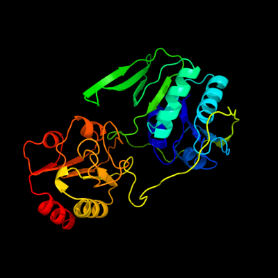
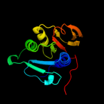
PDB header: electron transportChain: C: PDB Molecule: protein (electron transfer flavoprotein);PDBTitle: electron transfer flavoprotein (etf) from paracoccus2 denitrificans
2 d1efva2
100.0
43
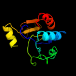
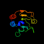
Fold: DHS-like NAD/FAD-binding domainSuperfamily: DHS-like NAD/FAD-binding domainFamily: C-terminal domain of the electron transfer flavoprotein alpha subunit3 d1efpa2
100.0
45
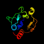
Fold: DHS-like NAD/FAD-binding domainSuperfamily: DHS-like NAD/FAD-binding domainFamily: C-terminal domain of the electron transfer flavoprotein alpha subunit4 d3clsd2
100.0
37
Fold: DHS-like NAD/FAD-binding domainSuperfamily: DHS-like NAD/FAD-binding domainFamily: C-terminal domain of the electron transfer flavoprotein alpha subunit5 d3clsd1
100.0
18
Fold: Adenine nucleotide alpha hydrolase-likeSuperfamily: Adenine nucleotide alpha hydrolases-likeFamily: ETFP subunits6 c1o94D_
100.0
18
PDB header: electron transportChain: D: PDB Molecule: electron transfer flavoprotein alpha-subunit;PDBTitle: ternary complex between trimethylamine dehydrogenase and2 electron transferring flavoprotein
7 c3ih5A_
100.0
22
PDB header: electron transportChain: A: PDB Molecule: electron transfer flavoprotein alpha-subunit;PDBTitle: crystal structure of electron transfer flavoprotein alpha-2 subunit from bacteroides thetaiotaomicron
8 d1efva1
100.0
17
Fold: Adenine nucleotide alpha hydrolase-likeSuperfamily: Adenine nucleotide alpha hydrolases-likeFamily: ETFP subunits9 c1t9gR_
100.0
16
PDB header: oxidoreductase, electron transportChain: R: PDB Molecule: electron transfer flavoprotein alpha-subunit,PDBTitle: structure of the human mcad:etf complex
10 d1efpa1
100.0
16
Fold: Adenine nucleotide alpha hydrolase-likeSuperfamily: Adenine nucleotide alpha hydrolases-likeFamily: ETFP subunits11 c3fetA_
100.0
19
PDB header: electron transportChain: A: PDB Molecule: electron transfer flavoprotein subunit alpha relatedPDBTitle: crystal structure of the electron transfer flavoprotein subunit alpha2 related protein ta0212 from thermoplasma acidophilum
12 d1efpb_
99.9
14
Fold: Adenine nucleotide alpha hydrolase-likeSuperfamily: Adenine nucleotide alpha hydrolases-likeFamily: ETFP subunits13 d3clsc1
99.9
11
Fold: Adenine nucleotide alpha hydrolase-likeSuperfamily: Adenine nucleotide alpha hydrolases-likeFamily: ETFP subunits14 d1efvb_
99.9
17
Fold: Adenine nucleotide alpha hydrolase-likeSuperfamily: Adenine nucleotide alpha hydrolases-likeFamily: ETFP subunits15 d1o94c_
99.8
14
Fold: Adenine nucleotide alpha hydrolase-likeSuperfamily: Adenine nucleotide alpha hydrolases-likeFamily: ETFP subunits16 d1t9ba1
98.8
18
Fold: DHS-like NAD/FAD-binding domainSuperfamily: DHS-like NAD/FAD-binding domainFamily: Pyruvate oxidase and decarboxylase, middle domain17 d1ozha1
98.7
19
Fold: DHS-like NAD/FAD-binding domainSuperfamily: DHS-like NAD/FAD-binding domainFamily: Pyruvate oxidase and decarboxylase, middle domain18 d2ez9a1
98.4
17
Fold: DHS-like NAD/FAD-binding domainSuperfamily: DHS-like NAD/FAD-binding domainFamily: Pyruvate oxidase and decarboxylase, middle domain19 d2ji7a1
98.3
17
Fold: DHS-like NAD/FAD-binding domainSuperfamily: DHS-like NAD/FAD-binding domainFamily: Pyruvate oxidase and decarboxylase, middle domain20 d2ihta1
98.1
24
Fold: DHS-like NAD/FAD-binding domainSuperfamily: DHS-like NAD/FAD-binding domainFamily: Pyruvate oxidase and decarboxylase, middle domain21 c1ozhD_
not modelled
97.8
18
PDB header: lyaseChain: D: PDB Molecule: acetolactate synthase, catabolic;PDBTitle: the crystal structure of klebsiella pneumoniae acetolactate2 synthase with enzyme-bound cofactor and with an unusual3 intermediate.
22 d1ybha1
not modelled
97.7
21
Fold: DHS-like NAD/FAD-binding domainSuperfamily: DHS-like NAD/FAD-binding domainFamily: Pyruvate oxidase and decarboxylase, middle domain23 d1q6za1
not modelled
97.7
16
Fold: DHS-like NAD/FAD-binding domainSuperfamily: DHS-like NAD/FAD-binding domainFamily: Pyruvate oxidase and decarboxylase, middle domain24 c2q27B_
not modelled
97.5
13
PDB header: lyaseChain: B: PDB Molecule: oxalyl-coa decarboxylase;PDBTitle: crystal structure of oxalyl-coa decarboxylase from escherichia coli
25 d2djia1
not modelled
97.5
21
Fold: DHS-like NAD/FAD-binding domainSuperfamily: DHS-like NAD/FAD-binding domainFamily: Pyruvate oxidase and decarboxylase, middle domain26 c1powA_
not modelled
97.4
17
PDB header: oxidoreductase(oxygen as acceptor)Chain: A: PDB Molecule: pyruvate oxidase;PDBTitle: the refined structures of a stabilized mutant and of wild-type2 pyruvate oxidase from lactobacillus plantarum
27 c2djiA_
not modelled
97.3
19
PDB header: oxidoreductaseChain: A: PDB Molecule: pyruvate oxidase;PDBTitle: crystal structure of pyruvate oxidase from aerococcus2 viridans containing fad
28 c2pgnA_
not modelled
97.3
20
PDB header: hydrolaseChain: A: PDB Molecule: cyclohexane-1,2-dione hydrolase (cdh);PDBTitle: the crystal structure of fad and thdp-dependent cyclohexane-1,2-dione2 hydrolase in complex with cyclohexane-1,2-dione
29 c3lq1A_
not modelled
97.3
12
PDB header: transferaseChain: A: PDB Molecule: 2-succinyl-5-enolpyruvyl-6-hydroxy-3-cyclohexene-PDBTitle: crystal structure of 2-succinyl-6-hydroxy-2,4-cyclohexadiene2 1-carboxylic acid synthase/2-oxoglutarate decarboxylase3 from listeria monocytogenes str. 4b f2365
30 c2ji6B_
not modelled
97.3
18
PDB header: lyaseChain: B: PDB Molecule: oxalyl-coa decarboxylase;PDBTitle: x-ray structure of oxalyl-coa decarboxylase in complex with2 3-deaza-thdp and oxalyl-coa
31 c1t9dB_
not modelled
97.1
19
PDB header: transferaseChain: B: PDB Molecule: acetolactate synthase, mitochondrial;PDBTitle: crystal structure of yeast acetohydroxyacid synthase in2 complex with a sulfonylurea herbicide, metsulfuron methyl
32 c1upaC_
not modelled
96.8
20
PDB header: synthaseChain: C: PDB Molecule: carboxyethylarginine synthase;PDBTitle: carboxyethylarginine synthase from streptomyces2 clavuligerus (semet structure)
33 c3eyaE_
not modelled
96.7
24
PDB header: oxidoreductaseChain: E: PDB Molecule: pyruvate dehydrogenase [cytochrome];PDBTitle: structural basis for membrane binding and catalytic2 activation of the peripheral membrane enzyme pyruvate3 oxidase from escherichia coli
34 c2ag1A_
not modelled
96.7
15
PDB header: lyaseChain: A: PDB Molecule: benzaldehyde lyase;PDBTitle: crystal structure of benzaldehyde lyase (bal)- semet
35 c1yi1A_
not modelled
96.7
20
PDB header: transferaseChain: A: PDB Molecule: acetolactate synthase;PDBTitle: crystal structure of arabidopsis thaliana acetohydroxyacid synthase in2 complex with a sulfonylurea herbicide, tribenuron methyl
36 c2x7jA_
not modelled
96.3
14
PDB header: transferaseChain: A: PDB Molecule: 2-succinyl-5-enolpyruvyl-6-hydroxy-3-cyclohexenePDBTitle: structure of the menaquinone biosynthesis protein mend from2 bacillus subtilis
37 c2panF_
not modelled
96.3
18
PDB header: lyaseChain: F: PDB Molecule: glyoxylate carboligase;PDBTitle: crystal structure of e. coli glyoxylate carboligase
38 d1uana_
not modelled
96.3
15
Fold: LmbE-likeSuperfamily: LmbE-likeFamily: LmbE-like39 d1zpda1
not modelled
96.2
21
Fold: DHS-like NAD/FAD-binding domainSuperfamily: DHS-like NAD/FAD-binding domainFamily: Pyruvate oxidase and decarboxylase, middle domain40 c2v3wC_
not modelled
96.0
16
PDB header: lyaseChain: C: PDB Molecule: benzoylformate decarboxylase;PDBTitle: crystal structure of the benzoylformate decarboxylase2 variant l461a from pseudomonas putida
41 d1m2ka_
not modelled
95.9
26
Fold: DHS-like NAD/FAD-binding domainSuperfamily: DHS-like NAD/FAD-binding domainFamily: Sir2 family of transcriptional regulators42 d1yc5a1
not modelled
95.8
15
Fold: DHS-like NAD/FAD-binding domainSuperfamily: DHS-like NAD/FAD-binding domainFamily: Sir2 family of transcriptional regulators43 d1pvda1
not modelled
95.8
13
Fold: DHS-like NAD/FAD-binding domainSuperfamily: DHS-like NAD/FAD-binding domainFamily: Pyruvate oxidase and decarboxylase, middle domain44 c2ixdB_
not modelled
95.6
19
PDB header: hydrolaseChain: B: PDB Molecule: lmbe-related protein;PDBTitle: crystal structure of the putative deacetylase bc1534 from2 bacilus cereus
45 c1jscA_
not modelled
95.2
19
PDB header: lyaseChain: A: PDB Molecule: acetohydroxy-acid synthase;PDBTitle: crystal structure of the catalytic subunit of yeast2 acetohydroxyacid synthase: a target for herbicidal3 inhibitors
46 d2b4ya1
not modelled
95.0
18
Fold: DHS-like NAD/FAD-binding domainSuperfamily: DHS-like NAD/FAD-binding domainFamily: Sir2 family of transcriptional regulators47 c3k35D_
not modelled
94.8
27
PDB header: hydrolaseChain: D: PDB Molecule: nad-dependent deacetylase sirtuin-6;PDBTitle: crystal structure of human sirt6
48 c3glsC_
not modelled
94.7
27
PDB header: hydrolaseChain: C: PDB Molecule: nad-dependent deacetylase sirtuin-3,PDBTitle: crystal structure of human sirt3
49 c1zpdA_
not modelled
94.6
18
PDB header: alcohol fermentationChain: A: PDB Molecule: pyruvate decarboxylase;PDBTitle: pyruvate decarboxylase from zymomonas mobilis
50 d1ma3a_
not modelled
94.6
15
Fold: DHS-like NAD/FAD-binding domainSuperfamily: DHS-like NAD/FAD-binding domainFamily: Sir2 family of transcriptional regulators51 c2jlaD_
not modelled
94.2
14
PDB header: transferaseChain: D: PDB Molecule: 2-succinyl-5-enolpyruvyl-6-hydroxy-3-cyclohexenePDBTitle: crystal structure of e.coli mend, 2-succinyl-5-enolpyruvyl-2 6-hydroxy-3-cyclohexadiene-1-carboxylate synthase - semet3 protein
52 c2dzdB_
not modelled
94.1
13
PDB header: ligaseChain: B: PDB Molecule: pyruvate carboxylase;PDBTitle: crystal structure of the biotin carboxylase domain of2 pyruvate carboxylase
53 c3pkiF_
not modelled
94.0
27
PDB header: hydrolaseChain: F: PDB Molecule: nad-dependent deacetylase sirtuin-6;PDBTitle: human sirt6 crystal structure in complex with adp ribose
54 c2vbiF_
not modelled
93.9
17
PDB header: lyaseChain: F: PDB Molecule: pyruvate decarboxylase;PDBTitle: holostructure of pyruvate decarboxylase from acetobacter2 pasteurianus
55 d1q1aa_
not modelled
93.9
13
Fold: DHS-like NAD/FAD-binding domainSuperfamily: DHS-like NAD/FAD-binding domainFamily: Sir2 family of transcriptional regulators56 c1q14A_
not modelled
92.8
13
PDB header: hydrolaseChain: A: PDB Molecule: hst2 protein;PDBTitle: structure and autoregulation of the yeast hst2 homolog of sir2
57 d1ovma1
not modelled
92.6
9
Fold: DHS-like NAD/FAD-binding domainSuperfamily: DHS-like NAD/FAD-binding domainFamily: Pyruvate oxidase and decarboxylase, middle domain58 c1ulzA_
not modelled
92.4
13
PDB header: ligaseChain: A: PDB Molecule: pyruvate carboxylase n-terminal domain;PDBTitle: crystal structure of the biotin carboxylase subunit of pyruvate2 carboxylase
59 d1s5pa_
not modelled
92.1
21
Fold: DHS-like NAD/FAD-binding domainSuperfamily: DHS-like NAD/FAD-binding domainFamily: Sir2 family of transcriptional regulators60 c3ouzA_
not modelled
92.1
8
PDB header: ligaseChain: A: PDB Molecule: biotin carboxylase;PDBTitle: crystal structure of biotin carboxylase-adp complex from campylobacter2 jejuni
61 c2w93A_
not modelled
91.5
19
PDB header: lyaseChain: A: PDB Molecule: pyruvate decarboxylase isozyme 1;PDBTitle: crystal structure of the saccharomyces cerevisiae pyruvate2 decarboxylase variant e477q in complex with the surrogate3 pyruvamide
62 d1j8fa_
not modelled
91.1
30
Fold: DHS-like NAD/FAD-binding domainSuperfamily: DHS-like NAD/FAD-binding domainFamily: Sir2 family of transcriptional regulators63 c3jwpA_
not modelled
90.9
25
PDB header: transcriptionChain: A: PDB Molecule: transcriptional regulatory protein sir2 homologue;PDBTitle: crystal structure of plasmodium falciparum sir2a (pf13_0152) in2 complex with amp
64 c2p10D_
not modelled
89.5
11
PDB header: hydrolaseChain: D: PDB Molecule: mll9387 protein;PDBTitle: crystal structure of a putative phosphonopyruvate hydrolase (mll9387)2 from mesorhizobium loti maff303099 at 2.15 a resolution
65 c2hjhB_
not modelled
89.2
19
PDB header: hydrolaseChain: B: PDB Molecule: nad-dependent histone deacetylase sir2;PDBTitle: crystal structure of the sir2 deacetylase
66 d1tq8a_
not modelled
88.5
15
Fold: Adenine nucleotide alpha hydrolase-likeSuperfamily: Adenine nucleotide alpha hydrolases-likeFamily: Universal stress protein-like67 c1ovmC_
not modelled
87.8
11
PDB header: lyaseChain: C: PDB Molecule: indole-3-pyruvate decarboxylase;PDBTitle: crystal structure of indolepyruvate decarboxylase from2 enterobacter cloacae
68 c3dfmA_
not modelled
87.5
14
PDB header: hydrolaseChain: A: PDB Molecule: teicoplanin pseudoaglycone deacetylase orf2;PDBTitle: the crystal structure of the zinc inhibited form of2 teicoplanin deacetylase orf2
69 c2vbgB_
not modelled
87.0
21
PDB header: lyaseChain: B: PDB Molecule: branched-chain alpha-ketoacid decarboxylase;PDBTitle: the complex structure of the branched-chain keto acid2 decarboxylase (kdca) from lactococcus lactis with 2r-1-3 hydroxyethyl-deazathdp
70 c3g8cB_
not modelled
85.9
8
PDB header: ligaseChain: B: PDB Molecule: biotin carboxylase;PDBTitle: crystal stucture of biotin carboxylase in complex with2 biotin, bicarbonate, adp and mg ion
71 c1nvmG_
not modelled
85.6
14
PDB header: lyase/oxidoreductaseChain: G: PDB Molecule: 4-hydroxy-2-oxovalerate aldolase;PDBTitle: crystal structure of a bifunctional aldolase-dehydrogenase :2 sequestering a reactive and volatile intermediate
72 c2nxwB_
not modelled
84.9
22
PDB header: lyaseChain: B: PDB Molecule: phenyl-3-pyruvate decarboxylase;PDBTitle: crystal structure of phenylpyruvate decarboxylase of azospirillum2 brasilense
73 d1q74a_
not modelled
84.7
16
Fold: LmbE-likeSuperfamily: LmbE-likeFamily: LmbE-like74 c3dfiA_
not modelled
83.8
14
PDB header: hydrolaseChain: A: PDB Molecule: pseudoaglycone deacetylase dbv21;PDBTitle: the crystal structure of antimicrobial reagent a40926 pseudoaglycone2 deacetylase dbv21
75 c3bg5C_
not modelled
82.4
14
PDB header: ligaseChain: C: PDB Molecule: pyruvate carboxylase;PDBTitle: crystal structure of staphylococcus aureus pyruvate2 carboxylase
76 c3ot5D_
not modelled
82.1
14
PDB header: isomeraseChain: D: PDB Molecule: udp-n-acetylglucosamine 2-epimerase;PDBTitle: 2.2 angstrom resolution crystal structure of putative udp-n-2 acetylglucosamine 2-epimerase from listeria monocytogenes
77 c2gpwC_
not modelled
81.9
9
PDB header: ligaseChain: C: PDB Molecule: biotin carboxylase;PDBTitle: crystal structure of the biotin carboxylase subunit, f363a2 mutant, of acetyl-coa carboxylase from escherichia coli.
78 c2vpqA_
not modelled
81.5
13
PDB header: ligaseChain: A: PDB Molecule: acetyl-coa carboxylase;PDBTitle: crystal structure of biotin carboxylase from s. aureus2 complexed with amppnp
79 c3cf4G_
not modelled
80.5
16
PDB header: oxidoreductaseChain: G: PDB Molecule: acetyl-coa decarboxylase/synthase epsilon subunit;PDBTitle: structure of the codh component of the m. barkeri acds complex
80 d1x94a_
not modelled
80.2
18
Fold: SIS domainSuperfamily: SIS domainFamily: mono-SIS domain81 c2x3yA_
not modelled
78.7
13
PDB header: isomeraseChain: A: PDB Molecule: phosphoheptose isomerase;PDBTitle: crystal structure of gmha from burkholderia pseudomallei
82 d1qh8a_
not modelled
76.9
14
Fold: Chelatase-likeSuperfamily: "Helical backbone" metal receptorFamily: Nitrogenase iron-molybdenum protein83 c2dy0A_
not modelled
72.0
14
PDB header: transferaseChain: A: PDB Molecule: adenine phosphoribosyltransferase;PDBTitle: crystal structure of project jw0458 from escherichia coli
84 d1qh8b_
not modelled
71.4
15
Fold: Chelatase-likeSuperfamily: "Helical backbone" metal receptorFamily: Nitrogenase iron-molybdenum protein85 d1l1qa_
not modelled
69.7
19
Fold: PRTase-likeSuperfamily: PRTase-likeFamily: Phosphoribosyltransferases (PRTases)86 c3gmiA_
not modelled
69.6
7
PDB header: structural genomics, unknown functionChain: A: PDB Molecule: upf0348 protein mj0951;PDBTitle: crystal structure of a protein of unknown function from2 methanocaldococcus jannaschii
87 d1m1na_
not modelled
69.6
12
Fold: Chelatase-likeSuperfamily: "Helical backbone" metal receptorFamily: Nitrogenase iron-molybdenum protein88 c3fxaA_
not modelled
68.2
16
PDB header: sugar binding proteinChain: A: PDB Molecule: sis domain protein;PDBTitle: crystal structure of a putative sugar-phosphate isomerase2 (lmof2365_0531) from listeria monocytogenes str. 4b f2365 at 1.60 a3 resolution
89 d1nvma2
not modelled
67.1
14
Fold: TIM beta/alpha-barrelSuperfamily: AldolaseFamily: HMGL-like90 d1tk9a_
not modelled
66.6
18
Fold: SIS domainSuperfamily: SIS domainFamily: mono-SIS domain91 c2pn1A_
not modelled
65.9
15
PDB header: ligaseChain: A: PDB Molecule: carbamoylphosphate synthase large subunit;PDBTitle: crystal structure of carbamoylphosphate synthase large subunit (split2 gene in mj) (zp_00538348.1) from exiguobacterium sp. 255-15 at 2.00 a3 resolution
92 c1q7tA_
not modelled
64.8
18
PDB header: hydrolaseChain: A: PDB Molecule: hypothetical protein rv1170;PDBTitle: rv1170 (mshb) from mycobacterium tuberculosis
93 d1miob_
not modelled
63.2
15
Fold: Chelatase-likeSuperfamily: "Helical backbone" metal receptorFamily: Nitrogenase iron-molybdenum protein94 c1kjjA_
not modelled
61.0
14
PDB header: transferaseChain: A: PDB Molecule: phosphoribosylglycinamide formyltransferase 2;PDBTitle: crystal structure of glycniamide ribonucleotide2 transformylase in complex with mg-atp-gamma-s
95 c3bo9B_
not modelled
60.1
11
PDB header: oxidoreductaseChain: B: PDB Molecule: putative nitroalkan dioxygenase;PDBTitle: crystal structure of putative nitroalkan dioxygenase (tm0800) from2 thermotoga maritima at 2.71 a resolution
96 c2xhzC_
not modelled
60.0
19
PDB header: isomeraseChain: C: PDB Molecule: arabinose 5-phosphate isomerase;PDBTitle: probing the active site of the sugar isomerase domain from e. coli2 arabinose-5-phosphate isomerase via x-ray crystallography
97 c3n6rK_
not modelled
59.7
12
PDB header: ligaseChain: K: PDB Molecule: propionyl-coa carboxylase, alpha subunit;PDBTitle: crystal structure of the holoenzyme of propionyl-coa carboxylase (pcc)
98 d1a9xa4
not modelled
58.4
13
Fold: PreATP-grasp domainSuperfamily: PreATP-grasp domainFamily: BC N-terminal domain-like99 c3u9sE_
not modelled
58.2
11
PDB header: ligaseChain: E: PDB Molecule: methylcrotonyl-coa carboxylase, alpha-subunit;PDBTitle: crystal structure of p. aeruginosa 3-methylcrotonyl-coa carboxylase2 (mcc) 750 kd holoenzyme, coa complex
100 c3shoA_
not modelled
56.8
22
PDB header: transcription regulatorChain: A: PDB Molecule: transcriptional regulator, rpir family;PDBTitle: crystal structure of rpir transcription factor from sphaerobacter2 thermophilus (sugar isomerase domain)
101 c3ivuB_
not modelled
54.1
13
PDB header: transferaseChain: B: PDB Molecule: homocitrate synthase, mitochondrial;PDBTitle: homocitrate synthase lys4 bound to 2-og
102 c2gjlA_
not modelled
53.7
12
PDB header: oxidoreductaseChain: A: PDB Molecule: hypothetical protein pa1024;PDBTitle: crystal structure of 2-nitropropane dioxygenase
103 c2ze5A_
not modelled
53.7
22
PDB header: transferaseChain: A: PDB Molecule: isopentenyl transferase;PDBTitle: crystal structure of adenosine phosphate-isopentenyltransferase
104 c1m6vE_
not modelled
53.4
13
PDB header: ligaseChain: E: PDB Molecule: carbamoyl phosphate synthetase large chain;PDBTitle: crystal structure of the g359f (small subunit) point mutant of2 carbamoyl phosphate synthetase
105 d1y0ba1
not modelled
52.6
14
Fold: PRTase-likeSuperfamily: PRTase-likeFamily: Phosphoribosyltransferases (PRTases)106 c3iupB_
not modelled
52.4
17
PDB header: oxidoreductaseChain: B: PDB Molecule: putative nadph:quinone oxidoreductase;PDBTitle: crystal structure of putative nadph:quinone oxidoreductase2 (yp_296108.1) from ralstonia eutropha jmp134 at 1.70 a resolution
107 d2iyva1
not modelled
51.4
28
Fold: P-loop containing nucleoside triphosphate hydrolasesSuperfamily: P-loop containing nucleoside triphosphate hydrolasesFamily: Shikimate kinase (AroK)108 c2dwcB_
not modelled
50.0
15
PDB header: transferaseChain: B: PDB Molecule: 433aa long hypothetical phosphoribosylglycinamide formylPDBTitle: crystal structure of probable phosphoribosylglycinamide formyl2 transferase from pyrococcus horikoshii ot3 complexed with adp
109 c1nriA_
not modelled
49.3
14
PDB header: structural genomics, unknown functionChain: A: PDB Molecule: hypothetical protein hi0754;PDBTitle: crystal structure of putative phosphosugar isomerase hi0754 from2 haemophilus influenzae
110 d1nria_
not modelled
49.3
14
Fold: SIS domainSuperfamily: SIS domainFamily: mono-SIS domain111 d1rqba2
not modelled
48.5
14
Fold: TIM beta/alpha-barrelSuperfamily: AldolaseFamily: HMGL-like112 c3bg3B_
not modelled
48.2
17
PDB header: ligaseChain: B: PDB Molecule: pyruvate carboxylase, mitochondrial;PDBTitle: crystal structure of human pyruvate carboxylase (missing2 the biotin carboxylase domain at the n-terminus)
113 c3krtC_
not modelled
48.2
12
PDB header: oxidoreductaseChain: C: PDB Molecule: crotonyl coa reductase;PDBTitle: crystal structure of putative crotonyl coa reductase from streptomyces2 coelicolor a3(2)
114 d1g2qa_
not modelled
47.8
11
Fold: PRTase-likeSuperfamily: PRTase-likeFamily: Phosphoribosyltransferases (PRTases)115 c1rr2A_
not modelled
47.4
11
PDB header: transferaseChain: A: PDB Molecule: transcarboxylase 5s subunit;PDBTitle: propionibacterium shermanii transcarboxylase 5s subunit bound to 2-2 ketobutyric acid
116 c1gsoA_
not modelled
46.7
10
PDB header: ligaseChain: A: PDB Molecule: protein (glycinamide ribonucleotide synthetase);PDBTitle: glycinamide ribonucleotide synthetase (gar-syn) from e.2 coli.
117 d1qb7a_
not modelled
44.5
17
Fold: PRTase-likeSuperfamily: PRTase-likeFamily: Phosphoribosyltransferases (PRTases)118 d1a9xa3
not modelled
43.6
15
Fold: PreATP-grasp domainSuperfamily: PreATP-grasp domainFamily: BC N-terminal domain-like119 c3ga2A_
not modelled
43.0
13
PDB header: hydrolaseChain: A: PDB Molecule: endonuclease v;PDBTitle: crystal structure of the endonuclease_v (bsu36170) from2 bacillus subtilis, northeast structural genomics3 consortium target sr624
120 d1v4va_
not modelled
42.5
13
Fold: UDP-Glycosyltransferase/glycogen phosphorylaseSuperfamily: UDP-Glycosyltransferase/glycogen phosphorylaseFamily: UDP-N-acetylglucosamine 2-epimerase