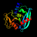
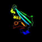
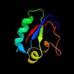
1 c3gocB_
100.0
35
PDB header: hydrolaseChain: B: PDB Molecule: endonuclease v;PDBTitle: crystal structure of the endonuclease v (sav1684) from streptomyces2 avermitilis. northeast structural genomics consortium target svr196
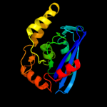
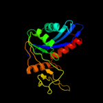
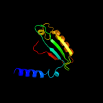
2 c3ga2A_
100.0
26
PDB header: hydrolaseChain: A: PDB Molecule: endonuclease v;PDBTitle: crystal structure of the endonuclease_v (bsu36170) from2 bacillus subtilis, northeast structural genomics3 consortium target sr624
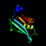
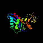
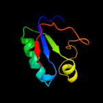
3 c2w36B_
100.0
38
PDB header: hydrolaseChain: B: PDB Molecule: endonuclease v;PDBTitle: structures of endonuclease v with dna reveal initiation of2 deaminated adenine repair
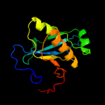
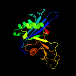
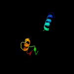
4 c2qh9B_
98.5
15
PDB header: structural genomics, unknown functionChain: B: PDB Molecule: upf0215 protein af_1433;PDBTitle: the crystal structure of a protein of unknown function from2 archaeoglobus fulgidus dsm 4304
5 c2nrzB_
97.9
26
PDB header: hydrolaseChain: B: PDB Molecule: uvrabc system protein c;PDBTitle: crystal structure of the c-terminal half of uvrc bound to2 its catalytic divalent cation
6 c3c65A_
97.7
19
PDB header: hydrolaseChain: A: PDB Molecule: uvrabc system protein c;PDBTitle: crystal structure of bacillus stearothermophilus uvrc 5'2 endonuclease domain
7 c2nrrA_
96.2
27
PDB header: hydrolaseChain: A: PDB Molecule: uvrabc system protein c;PDBTitle: crystal structure of the c-terminal rnaseh endonuclase2 domain of uvrc
8 c1u04A_
94.4
14
PDB header: hydrolase/gene regulationChain: A: PDB Molecule: hypothetical protein pf0537;PDBTitle: crystal structure of full length argonaute from pyrococcus furiosus
9 c2f8sA_
93.2
16
PDB header: rna binding protein/rnaChain: A: PDB Molecule: argonaute protein;PDBTitle: crystal structure of aa-ago with externally-bound sirna
10 d1u04a2
85.8
14
Fold: Ribonuclease H-like motifSuperfamily: Ribonuclease H-likeFamily: PIWI domain11 d1yvua2
85.4
15
Fold: Ribonuclease H-like motifSuperfamily: Ribonuclease H-likeFamily: PIWI domain12 d3bzka5
81.3
20
Fold: Ribonuclease H-like motifSuperfamily: Ribonuclease H-likeFamily: Tex RuvX-like domain-like13 c3taiB_
71.3
14
PDB header: hydrolaseChain: B: PDB Molecule: dna double-strand break repair protein nura;PDBTitle: crystal structure of nura
14 c2zkqb_
60.7
27
PDB header: ribosomal protein/rnaChain: B: PDB Molecule: rna expansion segment es3;PDBTitle: structure of a mammalian ribosomal 40s subunit within an2 80s complex obtained by docking homology models of the rna3 and proteins into an 8.7 a cryo-em map
15 c3bchA_
58.8
24
PDB header: cell adhesion, ribosomal proteinChain: A: PDB Molecule: 40s ribosomal protein sa;PDBTitle: crystal structure of the human laminin receptor precursor
16 d1efva2
56.4
20
Fold: DHS-like NAD/FAD-binding domainSuperfamily: DHS-like NAD/FAD-binding domainFamily: C-terminal domain of the electron transfer flavoprotein alpha subunit17 c2oceA_
54.7
17
PDB header: structural genomics, unknown functionChain: A: PDB Molecule: hypothetical protein pa5201;PDBTitle: crystal structure of tex family protein pa5201 from2 pseudomonas aeruginosa
18 c1efpC_
49.6
17
PDB header: electron transportChain: C: PDB Molecule: protein (electron transfer flavoprotein);PDBTitle: electron transfer flavoprotein (etf) from paracoccus2 denitrificans
19 c3bbnB_
47.0
23
PDB header: ribosomeChain: B: PDB Molecule: ribosomal protein s2;PDBTitle: homology model for the spinach chloroplast 30s subunit2 fitted to 9.4a cryo-em map of the 70s chlororibosome.
20 c2obnA_
46.2
14
PDB header: unknown functionChain: A: PDB Molecule: hypothetical protein;PDBTitle: crystal structure of a duf1611 family protein (ava_3511) from anabaena2 variabilis atcc 29413 at 2.30 a resolution
21 d1efpa2
not modelled
45.5
17
Fold: DHS-like NAD/FAD-binding domainSuperfamily: DHS-like NAD/FAD-binding domainFamily: C-terminal domain of the electron transfer flavoprotein alpha subunit22 d1vi6a_
not modelled
44.6
29
Fold: Flavodoxin-likeSuperfamily: Ribosomal protein S2Family: Ribosomal protein S223 c3rggD_
not modelled
41.6
11
PDB header: lyaseChain: D: PDB Molecule: phosphoribosylaminoimidazole carboxylase, pure protein;PDBTitle: crystal structure of treponema denticola pure bound to air
24 c3cvoA_
not modelled
36.8
31
PDB header: transferaseChain: A: PDB Molecule: methyltransferase-like protein of unknown function;PDBTitle: crystal structure of a methyltransferase-like protein (spo2022) from2 silicibacter pomeroyi dss-3 at 1.80 a resolution
25 d1kcfa2
not modelled
35.7
15
Fold: Ribonuclease H-like motifSuperfamily: Ribonuclease H-likeFamily: Mitochondrial resolvase ydc2 catalytic domain26 c3izbA_
not modelled
35.3
29
PDB header: ribosomeChain: A: PDB Molecule: 40s ribosomal protein rps0 (s2p);PDBTitle: localization of the small subunit ribosomal proteins into a 6.1 a2 cryo-em map of saccharomyces cerevisiae translating 80s ribosome
27 c2qmoA_
not modelled
32.8
22
PDB header: ligaseChain: A: PDB Molecule: dethiobiotin synthetase;PDBTitle: crystal structure of dethiobiotin synthetase (biod) from helicobacter2 pylori
28 c2etjA_
not modelled
31.8
57
PDB header: hydrolaseChain: A: PDB Molecule: ribonuclease hii;PDBTitle: crystal structure of ribonuclease hii (ec 3.1.26.4) (rnase hii)2 (tm0915) from thermotoga maritima at 1.74 a resolution
29 d2etja1
not modelled
31.8
57
Fold: Ribonuclease H-like motifSuperfamily: Ribonuclease H-likeFamily: Ribonuclease H30 c1jb0M_
not modelled
31.2
50
PDB header: photosynthesisChain: M: PDB Molecule: photosystem 1 reaction centre subunit xii;PDBTitle: crystal structure of photosystem i: a photosynthetic reaction center2 and core antenna system from cyanobacteria
31 d1jb0m_
not modelled
31.2
50
Fold: Single transmembrane helixSuperfamily: Subunit XII of photosystem I reaction centre, PsaMFamily: Subunit XII of photosystem I reaction centre, PsaM32 c3c6aA_
not modelled
29.2
16
PDB header: viral proteinChain: A: PDB Molecule: terminase large subunit;PDBTitle: crystal structure of the rb49 gp17 nuclease domain
33 d2uubb1
not modelled
29.0
23
Fold: Flavodoxin-likeSuperfamily: Ribosomal protein S2Family: Ribosomal protein S234 d3clsd2
not modelled
28.7
20
Fold: DHS-like NAD/FAD-binding domainSuperfamily: DHS-like NAD/FAD-binding domainFamily: C-terminal domain of the electron transfer flavoprotein alpha subunit35 c1xuzA_
not modelled
28.5
24
PDB header: biosynthetic proteinChain: A: PDB Molecule: polysialic acid capsule biosynthesis protein siac;PDBTitle: crystal structure analysis of sialic acid synthase (neub)from2 neisseria meningitidis, bound to mn2+, phosphoenolpyruvate, and n-3 acetyl mannosaminitol
36 c2f1rA_
not modelled
26.3
24
PDB header: biosynthetic proteinChain: A: PDB Molecule: molybdopterin-guanine dinucleotide biosynthesisPDBTitle: crystal structure of molybdopterin-guanine biosynthesis2 protein b (mobb)
37 d2gy9b1
not modelled
25.2
16
Fold: Flavodoxin-likeSuperfamily: Ribosomal protein S2Family: Ribosomal protein S238 c1s1hB_
not modelled
24.4
30
PDB header: ribosomeChain: B: PDB Molecule: 40s ribosomal protein s0-a;PDBTitle: structure of the ribosomal 80s-eef2-sordarin complex from2 yeast obtained by docking atomic models for rna and protein3 components into a 11.7 a cryo-em map. this file, 1s1h,4 contains 40s subunit. the 60s ribosomal subunit is in file5 1s1i.
39 d2zdra2
not modelled
21.7
27
Fold: TIM beta/alpha-barrelSuperfamily: AldolaseFamily: NeuB-like40 c3iz6A_
not modelled
19.8
26
PDB header: ribosomeChain: A: PDB Molecule: 40s ribosomal protein sa (s2p);PDBTitle: localization of the small subunit ribosomal proteins into a 5.5 a2 cryo-em map of triticum aestivum translating 80s ribosome
41 d2foka4
not modelled
19.8
33
Fold: Restriction endonuclease-likeSuperfamily: Restriction endonuclease-likeFamily: Restriction endonuclease FokI, C-terminal (catalytic) domain42 d1io2a_
not modelled
19.7
28
Fold: Ribonuclease H-like motifSuperfamily: Ribonuclease H-likeFamily: Ribonuclease H43 c2xznB_
not modelled
16.6
26
PDB header: ribosomeChain: B: PDB Molecule: rps0e;PDBTitle: crystal structure of the eukaryotic 40s ribosomal2 subunit in complex with initiation factor 1. this file3 contains the 40s subunit and initiation factor for4 molecule 2
44 d2g0ta1
not modelled
14.6
28
Fold: P-loop containing nucleoside triphosphate hydrolasesSuperfamily: P-loop containing nucleoside triphosphate hydrolasesFamily: Nitrogenase iron protein-like45 c1fokA_
not modelled
13.4
26
PDB header: hydrolase/dnaChain: A: PDB Molecule: protein (foki restriction endonucleas);PDBTitle: structure of restriction endonuclease foki bound to dna
46 c3jvpA_
not modelled
13.2
18
PDB header: transferaseChain: A: PDB Molecule: ribulokinase;PDBTitle: crystal structure of ribulokinase from bacillus halodurans
47 d1qcza_
not modelled
12.7
19
Fold: Flavodoxin-likeSuperfamily: N5-CAIR mutase (phosphoribosylaminoimidazole carboxylase, PurE)Family: N5-CAIR mutase (phosphoribosylaminoimidazole carboxylase, PurE)48 c2ivoC_
not modelled
12.5
16
PDB header: hydrolaseChain: C: PDB Molecule: up1;PDBTitle: structure of up1 protein
49 c1glbG_
not modelled
12.3
17
PDB header: phosphotransferaseChain: G: PDB Molecule: glycerol kinase;PDBTitle: structure of the regulatory complex of escherichia coli iiiglc with2 glycerol kinase
50 d2p3ra1
not modelled
12.0
17
Fold: Ribonuclease H-like motifSuperfamily: Actin-like ATPase domainFamily: Glycerol kinase51 c3kgkA_
not modelled
11.7
31
PDB header: chaperoneChain: A: PDB Molecule: arsenical resistance operon trans-acting repressor arsd;PDBTitle: crystal structure of arsd
52 d1o66a_
not modelled
11.6
35
Fold: TIM beta/alpha-barrelSuperfamily: Phosphoenolpyruvate/pyruvate domainFamily: Ketopantoate hydroxymethyltransferase PanB53 c2ygkA_
not modelled
11.2
16
PDB header: hydrolaseChain: A: PDB Molecule: nura;PDBTitle: crystal structure of the nura nuclease from sulfolobus solfataricus
54 c3ktbD_
not modelled
10.6
15
PDB header: transcription regulatorChain: D: PDB Molecule: arsenical resistance operon trans-acting repressor;PDBTitle: crystal structure of arsenical resistance operon trans-acting2 repressor from bacteroides vulgatus atcc 8482
55 d1k38a_
not modelled
10.0
10
Fold: beta-lactamase/transpeptidase-likeSuperfamily: beta-lactamase/transpeptidase-likeFamily: beta-Lactamase/D-ala carboxypeptidase56 d1g6sa_
not modelled
8.8
11
Fold: IF3-likeSuperfamily: EPT/RTPC-likeFamily: Enolpyruvate transferase, EPT57 d2d0oa2
not modelled
8.6
35
Fold: Ribonuclease H-like motifSuperfamily: Actin-like ATPase domainFamily: ATPase domain of dehydratase reactivase alpha subunit58 d1k1sa1
not modelled
8.6
21
Fold: Lesion bypass DNA polymerase (Y-family), little finger domainSuperfamily: Lesion bypass DNA polymerase (Y-family), little finger domainFamily: Lesion bypass DNA polymerase (Y-family), little finger domain59 d2ayia1
not modelled
8.4
11
Fold: Thermophilic metalloprotease-likeSuperfamily: Thermophilic metalloprotease-likeFamily: Thermophilic metalloprotease (M29)60 d1w9ha1
not modelled
8.4
17
Fold: Ribonuclease H-like motifSuperfamily: Ribonuclease H-likeFamily: PIWI domain61 c3ezkB_
not modelled
8.0
11
PDB header: hydrolaseChain: B: PDB Molecule: dna packaging protein gp17;PDBTitle: bacteriophage t4 gp17 motor assembly based on crystal2 structures and cryo-em reconstructions
62 c3oirA_
not modelled
7.9
12
PDB header: transport proteinChain: A: PDB Molecule: sulfate transporter sulfate transporter family protein;PDBTitle: crystal structure of sulfate transporter family protein from wolinella2 succinogenes
63 c2dcqA_
not modelled
7.7
19
PDB header: unknown functionChain: A: PDB Molecule: putative protein at4g01050;PDBTitle: fully automated nmr structure determination of the2 rhodanese homology domain at4g01050(175-295) from3 arabidopsis thaliana
64 d1o4va_
not modelled
7.7
15
Fold: Flavodoxin-likeSuperfamily: N5-CAIR mutase (phosphoribosylaminoimidazole carboxylase, PurE)Family: N5-CAIR mutase (phosphoribosylaminoimidazole carboxylase, PurE)65 d1pswa_
not modelled
7.7
13
Fold: UDP-Glycosyltransferase/glycogen phosphorylaseSuperfamily: UDP-Glycosyltransferase/glycogen phosphorylaseFamily: ADP-heptose LPS heptosyltransferase II66 d1nija1
not modelled
7.6
16
Fold: P-loop containing nucleoside triphosphate hydrolasesSuperfamily: P-loop containing nucleoside triphosphate hydrolasesFamily: Nitrogenase iron protein-like67 d1gzga_
not modelled
7.6
24
Fold: TIM beta/alpha-barrelSuperfamily: AldolaseFamily: 5-aminolaevulinate dehydratase, ALAD (porphobilinogen synthase)68 d2b7ta1
not modelled
7.6
9
Fold: dsRBD-likeSuperfamily: dsRNA-binding domain-likeFamily: Double-stranded RNA-binding domain (dsRBD)69 c1zu4A_
not modelled
7.4
19
PDB header: protein transportChain: A: PDB Molecule: ftsy;PDBTitle: crystal structure of ftsy from mycoplasma mycoides- space2 group p21212
70 c2wcvI_
not modelled
7.4
27
PDB header: isomeraseChain: I: PDB Molecule: l-fucose mutarotase;PDBTitle: crystal structure of bacterial fucu
71 c3orsD_
not modelled
7.3
17
PDB header: isomerase,biosynthetic proteinChain: D: PDB Molecule: n5-carboxyaminoimidazole ribonucleotide mutase;PDBTitle: crystal structure of n5-carboxyaminoimidazole ribonucleotide mutase2 from staphylococcus aureus
72 c3cf4G_
not modelled
7.3
21
PDB header: oxidoreductaseChain: G: PDB Molecule: acetyl-coa decarboxylase/synthase epsilon subunit;PDBTitle: structure of the codh component of the m. barkeri acds complex
73 c3fmfA_
not modelled
7.1
13
PDB header: ligaseChain: A: PDB Molecule: dethiobiotin synthetase;PDBTitle: crystal structure of mycobacterium tuberculosis dethiobiotin2 synthetase complexed with 7,8 diaminopelargonic acid carbamate
74 c2iuwA_
not modelled
7.1
16
PDB header: oxidoreductaseChain: A: PDB Molecule: alkylated repair protein alkb homolog 3;PDBTitle: crystal structure of human abh3 in complex with iron ion2 and 2-oxoglutarate
75 c3trhI_
not modelled
7.0
29
PDB header: lyaseChain: I: PDB Molecule: phosphoribosylaminoimidazole carboxylasePDBTitle: structure of a phosphoribosylaminoimidazole carboxylase catalytic2 subunit (pure) from coxiella burnetii
76 c3gbtA_
not modelled
6.9
18
PDB header: transferaseChain: A: PDB Molecule: gluconate kinase;PDBTitle: crystal structure of gluconate kinase from lactobacillus acidophilus
77 c1u8xX_
not modelled
6.8
15
PDB header: hydrolaseChain: X: PDB Molecule: maltose-6'-phosphate glucosidase;PDBTitle: crystal structure of glva from bacillus subtilis, a metal-requiring,2 nad-dependent 6-phospho-alpha-glucosidase
78 c2a6aB_
not modelled
6.7
22
PDB header: hydrolaseChain: B: PDB Molecule: hypothetical protein tm0874;PDBTitle: crystal structure of glycoprotein endopeptidase (tm0874) from2 thermotoga maritima at 2.50 a resolution
79 c2odaB_
not modelled
6.5
32
PDB header: protein bindingChain: B: PDB Molecule: hypothetical protein pspto_2114;PDBTitle: crystal structure of pspto_2114
80 d1xo1a2
not modelled
6.5
31
Fold: PIN domain-likeSuperfamily: PIN domain-likeFamily: 5' to 3' exonuclease catalytic domain81 c3ezfA_
not modelled
6.2
18
PDB header: biosynthetic proteinChain: A: PDB Molecule: para;PDBTitle: partition protein
82 d2iuwa1
not modelled
6.1
20
Fold: Double-stranded beta-helixSuperfamily: Clavaminate synthase-likeFamily: AlkB-like83 c3n53B_
not modelled
6.0
39
PDB header: transcriptionChain: B: PDB Molecule: response regulator receiver modulated diguanylate cyclase;PDBTitle: crystal structure of a response regulator receiver modulated2 diguanylate cyclase from pelobacter carbinolicus
84 d1rf6a_
not modelled
6.0
17
Fold: IF3-likeSuperfamily: EPT/RTPC-likeFamily: Enolpyruvate transferase, EPT85 c3iabB_
not modelled
5.9
30
PDB header: hydrolase/rnaChain: B: PDB Molecule: ribonucleases p/mrp protein subunit pop7;PDBTitle: crystal structure of rnase p /rnase mrp proteins pop6, pop72 in a complex with the p3 domain of rnase mrp rna
86 d1xmpa_
not modelled
5.9
17
Fold: Flavodoxin-likeSuperfamily: N5-CAIR mutase (phosphoribosylaminoimidazole carboxylase, PurE)Family: N5-CAIR mutase (phosphoribosylaminoimidazole carboxylase, PurE)87 d1jx4a1
not modelled
5.9
14
Fold: Lesion bypass DNA polymerase (Y-family), little finger domainSuperfamily: Lesion bypass DNA polymerase (Y-family), little finger domainFamily: Lesion bypass DNA polymerase (Y-family), little finger domain88 d1oy0a_
not modelled
5.9
22
Fold: TIM beta/alpha-barrelSuperfamily: Phosphoenolpyruvate/pyruvate domainFamily: Ketopantoate hydroxymethyltransferase PanB89 c3odhB_
not modelled
5.7
22
PDB header: hydrolase/dnaChain: B: PDB Molecule: okrai endonuclease;PDBTitle: structure of okrai/dna complex
90 c2d0bA_
not modelled
5.7
10
PDB header: hydrolaseChain: A: PDB Molecule: ribonuclease hiii;PDBTitle: crystal structure of bst-rnase hiii in complex with mg2+
91 d1osna_
not modelled
5.6
26
Fold: P-loop containing nucleoside triphosphate hydrolasesSuperfamily: P-loop containing nucleoside triphosphate hydrolasesFamily: Nucleotide and nucleoside kinases92 c1qeyA_
not modelled
5.5
28
PDB header: gene regulationChain: A: PDB Molecule: protein (regulatory protein mnt);PDBTitle: nmr structure determination of the tetramerization domain2 of the mnt repressor: an asymmetric a-helical assembly in3 slow exchange
93 c1qeyD_
not modelled
5.5
28
PDB header: gene regulationChain: D: PDB Molecule: protein (regulatory protein mnt);PDBTitle: nmr structure determination of the tetramerization domain2 of the mnt repressor: an asymmetric a-helical assembly in3 slow exchange
94 c1qeyB_
not modelled
5.5
28
PDB header: gene regulationChain: B: PDB Molecule: protein (regulatory protein mnt);PDBTitle: nmr structure determination of the tetramerization domain2 of the mnt repressor: an asymmetric a-helical assembly in3 slow exchange
95 c1qeyC_
not modelled
5.5
28
PDB header: gene regulationChain: C: PDB Molecule: protein (regulatory protein mnt);PDBTitle: nmr structure determination of the tetramerization domain2 of the mnt repressor: an asymmetric a-helical assembly in3 slow exchange
96 d1bdoa_
not modelled
5.4
23
Fold: Barrel-sandwich hybridSuperfamily: Single hybrid motifFamily: Biotinyl/lipoyl-carrier proteins and domains97 c3g25B_
not modelled
5.4
18
PDB header: transferaseChain: B: PDB Molecule: glycerol kinase;PDBTitle: 1.9 angstrom crystal structure of glycerol kinase (glpk) from2 staphylococcus aureus in complex with glycerol.
98 d1u11a_
not modelled
5.3
15
Fold: Flavodoxin-likeSuperfamily: N5-CAIR mutase (phosphoribosylaminoimidazole carboxylase, PurE)Family: N5-CAIR mutase (phosphoribosylaminoimidazole carboxylase, PurE)99 c2fw9A_
not modelled
5.3
17
PDB header: lyaseChain: A: PDB Molecule: n5-carboxyaminoimidazole ribonucleotide mutase;PDBTitle: structure of pure (n5-carboxyaminoimidazole ribonucleotide mutase)2 h59f from the acidophilic bacterium acetobacter aceti, at ph 8