1 c1jccC_
99.8
87
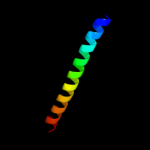
PDB header: membrane proteinChain: C: PDB Molecule: major outer membrane lipoprotein;PDBTitle: crystal structure of a novel alanine-zipper trimer at 1.7 a2 resolution, v13a,l16a,v20a,l23a,v27a,m30a,v34a mutations
2 c1t8zA_
99.6
72
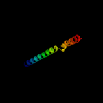
PDB header: membrane proteinChain: A: PDB Molecule: major outer membrane lipoprotein;PDBTitle: atomic structure of a novel tryptophan-zipper pentamer
3 c2gr7C_
78.2
25
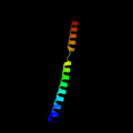
PDB header: membrane proteinChain: C: PDB Molecule: adhesin;PDBTitle: hia 992-1098
4 d2gr7a1
78.2
25
Fold: Pili subunitsSuperfamily: Pili subunitsFamily: YadA C-terminal domain-like5 c2pohA_
67.8
15
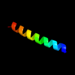
PDB header: viral proteinChain: A: PDB Molecule: head completion protein;PDBTitle: structure of phage p22 tail needle gp26
6 c1avyA_
53.9
29
PDB header: coiled coilChain: A: PDB Molecule: fibritin;PDBTitle: fibritin deletion mutant m (bacteriophage t4)
7 c1gk6B_
53.9
24
PDB header: vimentinChain: B: PDB Molecule: vimentin;PDBTitle: human vimentin coil 2b fragment linked to gcn4 leucine2 zipper (z2b)
8 c2ykqC_
53.3
13
PDB header: rna-binding proteinChain: C: PDB Molecule: line-1 orf1p;PDBTitle: structure of the human line-1 orf1p trimer
9 c1fosF_
51.0
30
PDB header: transcription/dnaChain: F: PDB Molecule: c-jun proto-oncogene protein;PDBTitle: two human c-fos:c-jun:dna complexes
10 c1x8yA_
49.3
22
PDB header: structural proteinChain: A: PDB Molecule: lamin a/c;PDBTitle: human lamin coil 2b
11 c1coiA_
46.8
43
PDB header: alpha-helical bundleChain: A: PDB Molecule: coil-vald;PDBTitle: designed trimeric coiled coil-vald
12 c2xzrA_
44.0
9
PDB header: cell adhesionChain: A: PDB Molecule: immunoglobulin-binding protein eibd;PDBTitle: escherichia coli immunoglobulin-binding protein eibd 391-438 fused2 to gcn4 adaptors
13 c1gk4A_
40.5
16
PDB header: vimentinChain: A: PDB Molecule: vimentin;PDBTitle: human vimentin coil 2b fragment (cys2)
14 c3movB_
40.4
18
PDB header: structural proteinChain: B: PDB Molecule: lamin-b1;PDBTitle: crystal structure of human lamin-b1 coil 2 segment
15 c1ij2C_
39.5
30
PDB header: transcriptionChain: C: PDB Molecule: general control protein gcn4;PDBTitle: gcn4-pvtl coiled-coil trimer with threonine at the a(16)2 position
16 c1rb6C_
39.2
30
PDB header: dna binding proteinChain: C: PDB Molecule: general control protein gcn4;PDBTitle: antiparallel trimer of gcn4-leucine zipper core mutant as2 n16a tetragonal form
17 c3k7zA_
39.2
30
PDB header: dna binding proteinChain: A: PDB Molecule: general control protein gcn4;PDBTitle: gcn4-leucine zipper core mutant as n16a trigonal automatic2 solution
18 c1swiA_
39.2
30
PDB header: leucine zipperChain: A: PDB Molecule: gcn4p1;PDBTitle: gcn4-leucine zipper core mutant as n16a complexed with2 benzene
19 c1rb1B_
39.2
30
PDB header: dna binding proteinChain: B: PDB Molecule: general control protein gcn4;PDBTitle: gcn4-leucine zipper core mutant as n16a trigonal automatic2 solution
20 c3k7zB_
39.2
30
PDB header: dna binding proteinChain: B: PDB Molecule: general control protein gcn4;PDBTitle: gcn4-leucine zipper core mutant as n16a trigonal automatic2 solution
21 c1rb1A_
not modelled
39.2
30
PDB header: dna binding proteinChain: A: PDB Molecule: general control protein gcn4;PDBTitle: gcn4-leucine zipper core mutant as n16a trigonal automatic2 solution
22 d1k4ta1
not modelled
38.7
31
Fold: Long alpha-hairpinSuperfamily: Eukaryotic DNA topoisomerase I, dispensable insert domainFamily: Eukaryotic DNA topoisomerase I, dispensable insert domain23 c1ij3C_
not modelled
37.9
30
PDB header: transcriptionChain: C: PDB Molecule: general control protein gcn4;PDBTitle: gcn4-pvsl coiled-coil trimer with serine at the a(16)2 position
24 c1ij3B_
not modelled
37.9
30
PDB header: transcriptionChain: B: PDB Molecule: general control protein gcn4;PDBTitle: gcn4-pvsl coiled-coil trimer with serine at the a(16)2 position
25 c1ij2B_
not modelled
37.5
30
PDB header: transcriptionChain: B: PDB Molecule: general control protein gcn4;PDBTitle: gcn4-pvtl coiled-coil trimer with threonine at the a(16)2 position
26 c2o7hF_
not modelled
34.4
30
PDB header: transcriptionChain: F: PDB Molecule: general control protein gcn4;PDBTitle: crystal structure of trimeric coiled coil gcn4 leucine zipper
27 c1ce0B_
not modelled
33.7
17
PDB header: hiv-1 envelope proteinChain: B: PDB Molecule: protein (leucine zipper model h38-p1);PDBTitle: trimerization specificity in hiv-1 gp41: analysis with a2 gcn4 leucine zipper model
28 c3m9bK_
not modelled
32.1
24
PDB header: chaperoneChain: K: PDB Molecule: proteasome-associated atpase;PDBTitle: crystal structure of the amino terminal coiled coil domain and the2 inter domain of the mycobacterium tuberculosis proteasomal atpase mpa
29 c3u59C_
not modelled
31.7
11
PDB header: contractile proteinChain: C: PDB Molecule: tropomyosin beta chain;PDBTitle: n-terminal 98-aa fragment of smooth muscle tropomyosin beta
30 c3na7A_
not modelled
29.7
24
PDB header: gene regulation, chaperoneChain: A: PDB Molecule: hp0958;PDBTitle: 2.2 angstrom structure of the hp0958 protein from helicobacter pylori2 ccug 17874
31 c2qihA_
not modelled
28.1
18
PDB header: cell adhesionChain: A: PDB Molecule: protein uspa1;PDBTitle: crystal structure of 527-665 fragment of uspa1 protein from2 moraxella catarrhalis
32 c1junB_
not modelled
27.7
28
PDB header: transcription regulationChain: B: PDB Molecule: c-jun homodimer;PDBTitle: nmr study of c-jun homodimer
33 c2efrB_
not modelled
27.6
23
PDB header: contractile proteinChain: B: PDB Molecule: general control protein gcn4 and tropomyosin 1 alpha chain;PDBTitle: crystal structure of the c-terminal tropomyosin fragment with n- and2 c-terminal extensions of the leucine zipper at 1.8 angstroms3 resolution
34 c1ztaA_
not modelled
26.8
30
PDB header: dna-binding motifChain: A: PDB Molecule: leucine zipper monomer;PDBTitle: the solution structure of a leucine-zipper motif peptide
35 c2wpqA_
not modelled
25.9
13
PDB header: membrane proteinChain: A: PDB Molecule: trimeric autotransporter adhesin fragment;PDBTitle: salmonella enterica sada 479-519 fused to gcn4 adaptors (2 sadak3, in-register fusion)
36 c1ci6B_
not modelled
25.0
31
PDB header: transcriptionChain: B: PDB Molecule: transcription factor c/ebp beta;PDBTitle: transcription factor atf4-c/ebp beta bzip heterodimer
37 c2diuA_
not modelled
23.2
46
PDB header: rna binding proteinChain: A: PDB Molecule: kiaa0430 protein;PDBTitle: solution structure of the rrm domain of kiaa0430 protein
38 c1gd2G_
not modelled
23.0
16
PDB header: transcription/dnaChain: G: PDB Molecule: transcription factor pap1;PDBTitle: crystal structure of bzip transcription factor pap1 bound2 to dna
39 c3okqA_
not modelled
22.9
32
PDB header: protein bindingChain: A: PDB Molecule: bud site selection protein 6;PDBTitle: crystal structure of a core domain of yeast actin nucleation cofactor2 bud6
40 c1deqD_
not modelled
22.6
18
PDB header: PDB COMPND: 41 c1ei3E_
not modelled
22.4
4
PDB header: PDB COMPND: 42 c2ki0A_
not modelled
22.0
33
PDB header: de novo proteinChain: A: PDB Molecule: ds119;PDBTitle: nmr structure of a de novo designed beta alpha beta
43 c3bvhE_
not modelled
20.5
25
PDB header: blood clottingChain: E: PDB Molecule: fibrinogen beta chain;PDBTitle: crystal structure of recombinant gammad364a fibrinogen fragment d with2 the peptide ligand gly-pro-arg-pro-amide
44 c2hpcF_
not modelled
20.2
20
PDB header: blood clottingChain: F: PDB Molecule: fibrinogen, gamma polypeptide;PDBTitle: crystal structure of fragment d from human fibrinogen complexed with2 gly-pro-arg-pro-amide.
45 c3emoA_
not modelled
19.7
19
PDB header: membrane protein/cell adhesionChain: A: PDB Molecule: hia (adhesin);PDBTitle: crystal structure of transmembrane hia 973-1098
46 c3sjbC_
not modelled
19.5
24
PDB header: hydrolase/transport proteinChain: C: PDB Molecule: golgi to er traffic protein 1;PDBTitle: crystal structure of s. cerevisiae get3 in the open state in complex2 with get1 cytosolic domain
47 c2gl2B_
not modelled
19.4
26
PDB header: cell adhesionChain: B: PDB Molecule: adhesion a;PDBTitle: crystal structure of the tetra muntant (t66g,r67g,f68g,2 y69g) of bacterial adhesin fada
48 c2x7aB_
not modelled
18.4
24
PDB header: immune systemChain: B: PDB Molecule: bone marrow stromal antigen 2;PDBTitle: structural basis of hiv-1 tethering to membranes by the2 bst2-tetherin ectodomain
49 c1deqO_
not modelled
18.0
6
PDB header: PDB COMPND: 50 c2wt7B_
not modelled
18.0
22
PDB header: transcriptionChain: B: PDB Molecule: transcription factor mafb;PDBTitle: crystal structure of the bzip heterodimeric complex2 mafb:cfos bound to dna
51 c2js5B_
not modelled
18.0
21
PDB header: structural genomics, unknown functionChain: B: PDB Molecule: uncharacterized protein;PDBTitle: nmr structure of protein q60c73_metca. northeast structural2 genomics consortium target mcr1
52 c3bvhC_
not modelled
18.0
19
PDB header: blood clottingChain: C: PDB Molecule: fibrinogen gamma chain;PDBTitle: crystal structure of recombinant gammad364a fibrinogen fragment d with2 the peptide ligand gly-pro-arg-pro-amide
53 c2w83C_
not modelled
17.2
22
PDB header: protein transportChain: C: PDB Molecule: c-jun-amino-terminal kinase-interacting proteinPDBTitle: crystal structure of the arf6 gtpase in complex with a2 specific effector, jip4
54 c2fxmB_
not modelled
16.0
23
PDB header: contractile proteinChain: B: PDB Molecule: myosin heavy chain, cardiac muscle beta isoform;PDBTitle: structure of the human beta-myosin s2 fragment
55 c2d3eD_
not modelled
15.8
15
PDB header: contractile proteinChain: D: PDB Molecule: general control protein gcn4 and tropomyosin 1PDBTitle: crystal structure of the c-terminal fragment of rabbit2 skeletal alpha-tropomyosin
56 c2e43A_
not modelled
15.5
14
PDB header: transcription/dnaChain: A: PDB Molecule: ccaat/enhancer-binding protein beta;PDBTitle: crystal structure of c/ebpbeta bzip homodimer k269a mutant2 bound to a high affinity dna fragment
57 c3swfA_
not modelled
15.5
15
PDB header: transport proteinChain: A: PDB Molecule: cgmp-gated cation channel alpha-1;PDBTitle: cnga1 621-690 containing clz domain
58 c2npsD_
not modelled
15.3
15
PDB header: transport proteinChain: D: PDB Molecule: syntaxin-6;PDBTitle: crystal structure of the early endosomal snare complex
59 c1j1dF_
not modelled
15.0
14
PDB header: contractile proteinChain: F: PDB Molecule: troponin i;PDBTitle: crystal structure of the 46kda domain of human cardiac2 troponin in the ca2+ saturated form
60 c3hnwB_
not modelled
14.8
9
PDB header: structural genomics, unknown functionChain: B: PDB Molecule: uncharacterized protein;PDBTitle: crystal structure of a basic coiled-coil protein of unknown function2 from eubacterium eligens atcc 27750
61 c2xdjF_
not modelled
14.1
10
PDB header: unknown functionChain: F: PDB Molecule: uncharacterized protein ybgf;PDBTitle: crystal structure of the n-terminal domain of e.coli ybgf
62 c2xv5A_
not modelled
13.9
21
PDB header: structural proteinChain: A: PDB Molecule: lamin-a/c;PDBTitle: human lamin a coil 2b fragment
63 c3ghgD_
not modelled
13.9
22
PDB header: blood clottingChain: D: PDB Molecule: fibrinogen alpha chain;PDBTitle: crystal structure of human fibrinogen
64 c1z56B_
not modelled
13.7
18
PDB header: ligaseChain: B: PDB Molecule: ligase interacting factor 1;PDBTitle: co-crystal structure of lif1p-lig4p
65 c3ipdB_
not modelled
13.6
16
PDB header: exocytosisChain: B: PDB Molecule: syntaxin-1a;PDBTitle: helical extension of the neuronal snare complex into the2 membrane, spacegroup i 21 21 21
66 c1ci6A_
not modelled
13.5
22
PDB header: transcriptionChain: A: PDB Molecule: transcription factor atf-4;PDBTitle: transcription factor atf4-c/ebp beta bzip heterodimer
67 c1fosE_
not modelled
13.4
11
PDB header: transcription/dnaChain: E: PDB Molecule: p55-c-fos proto-oncogene protein;PDBTitle: two human c-fos:c-jun:dna complexes
68 c2zdiC_
not modelled
13.2
12
PDB header: chaperoneChain: C: PDB Molecule: prefoldin subunit alpha;PDBTitle: crystal structure of prefoldin from pyrococcus horikoshii2 ot3
69 c3sjaG_
not modelled
13.0
27
PDB header: hydrolase/transport proteinChain: G: PDB Molecule: golgi to er traffic protein 1;PDBTitle: crystal structure of s. cerevisiae get3 in the open state in complex2 with get1 cytosolic domain
70 c1j1eC_
not modelled
12.6
16
PDB header: contractile proteinChain: C: PDB Molecule: troponin i;PDBTitle: crystal structure of the 52kda domain of human cardiac2 troponin in the ca2+ saturated form
71 c1n73A_
not modelled
12.5
8
PDB header: blood clottingChain: A: PDB Molecule: fibrin alpha-1 chain;PDBTitle: fibrin d-dimer, lamprey complexed with the peptide ligand: gly-his-2 arg-pro-amide
72 c1bb1B_
not modelled
12.2
23
PDB header: de novo protein designChain: B: PDB Molecule: designed, thermostable heterotrimeric coiledPDBTitle: crystal structure of a designed, thermostable2 heterotrimeric coiled coil
73 c3eukC_
not modelled
12.2
26
PDB header: cell cycleChain: C: PDB Molecule: chromosome partition protein mukb, linker;PDBTitle: crystal structure of muke-mukf(residues 292-443)-mukb(head2 domain)-atpgammas complex, asymmetric dimer
74 c2wukD_
not modelled
11.5
14
PDB header: cell cycleChain: D: PDB Molecule: septum site-determining protein diviva;PDBTitle: diviva n-terminal domain, f17a mutant
75 c2j7aC_
not modelled
11.2
56
PDB header: oxidoreductaseChain: C: PDB Molecule: cytochrome c quinol dehydrogenase nrfh;PDBTitle: crystal structure of cytochrome c nitrite reductase nrfha2 complex from desulfovibrio vulgaris
76 c1j1eB_
not modelled
11.0
16
PDB header: contractile proteinChain: B: PDB Molecule: troponin t;PDBTitle: crystal structure of the 52kda domain of human cardiac2 troponin in the ca2+ saturated form
77 c3ntnB_
not modelled
10.9
21
PDB header: membrane proteinChain: B: PDB Molecule: uspa1;PDBTitle: crystal structure of uspa1 head and neck domain from moraxella2 catarrhalis
78 c1qoyA_
not modelled
10.8
23
PDB header: toxinChain: A: PDB Molecule: hemolysin e;PDBTitle: e.coli hemolysin e (hlye, clya, shea)
79 d1vb8a_
not modelled
10.8
50
Fold: Knottins (small inhibitors, toxins, lectins)Superfamily: CyclotidesFamily: Cycloviolacin80 c1vb8A_
not modelled
10.8
50
PDB header: plant proteinChain: A: PDB Molecule: viola hederacea root peptide 1;PDBTitle: solution structure of vhr1, the first cyclotide from root2 tissue
81 c2dw3A_
not modelled
10.7
50
PDB header: photosynthesisChain: A: PDB Molecule: intrinsic membrane protein pufx;PDBTitle: solution structure of the rhodobacter sphaeroides pufx2 membrane protein
82 c3swyB_
not modelled
10.7
22
PDB header: transport proteinChain: B: PDB Molecule: cyclic nucleotide-gated cation channel alpha-3;PDBTitle: cnga3 626-672 containing clz domain
83 c1t2kD_
not modelled
10.6
22
PDB header: transcription/dnaChain: D: PDB Molecule: cyclic-amp-dependent transcription factor atf-2;PDBTitle: structure of the dna binding domains of irf3, atf-2 and jun2 bound to dna
84 c1by0A_
not modelled
10.3
32
PDB header: rna binding proteinChain: A: PDB Molecule: protein (hepatitis delta antigen);PDBTitle: n-terminal leucine-repeat region of hepatitis delta antigen
85 c2akfC_
not modelled
10.3
35
PDB header: protein bindingChain: C: PDB Molecule: coronin-1a;PDBTitle: crystal structure of the coiled-coil domain of coronin 1
86 c2akfA_
not modelled
10.3
35
PDB header: protein bindingChain: A: PDB Molecule: coronin-1a;PDBTitle: crystal structure of the coiled-coil domain of coronin 1
87 c2akfB_
not modelled
10.3
35
PDB header: protein bindingChain: B: PDB Molecule: coronin-1a;PDBTitle: crystal structure of the coiled-coil domain of coronin 1
88 c3ghgK_
not modelled
10.3
8
PDB header: blood clottingChain: K: PDB Molecule: fibrinogen beta chain;PDBTitle: crystal structure of human fibrinogen
89 c3n27A_
not modelled
10.2
38
PDB header: viral proteinChain: A: PDB Molecule: fusion glycoprotein f0, linker, fusion glycoprotein f0;PDBTitle: molecular basis of the inhibition of henipa viruses
90 c3hizB_
not modelled
9.7
13
PDB header: transferase/oncoproteinChain: B: PDB Molecule: phosphatidylinositol 3-kinase regulatory subunitPDBTitle: crystal structure of p110alpha h1047r mutant in complex with2 nish2 of p85alpha
91 c1nbjA_
not modelled
9.6
42
PDB header: plant proteinChain: A: PDB Molecule: cycloviolacin o1;PDBTitle: high-resolution solution structure of cycloviolacin o1
92 d1nbja_
not modelled
9.6
42
Fold: Knottins (small inhibitors, toxins, lectins)Superfamily: CyclotidesFamily: Cycloviolacin93 c3iynR_
not modelled
9.6
22
PDB header: virusChain: R: PDB Molecule: hexon-associated protein;PDBTitle: 3.6-angstrom cryoem structure of human adenovirus type 5
94 c2hpcH_
not modelled
9.6
25
PDB header: blood clottingChain: H: PDB Molecule: fibrinogen beta chain;PDBTitle: crystal structure of fragment d from human fibrinogen complexed with2 gly-pro-arg-pro-amide.
95 c3u1aC_
not modelled
9.3
13
PDB header: contractile proteinChain: C: PDB Molecule: smooth muscle tropomyosin alpha;PDBTitle: n-terminal 81-aa fragment of smooth muscle tropomyosin alpha
96 c1sfcD_
not modelled
9.2
15
PDB header: transport proteinChain: D: PDB Molecule: protein (snap-25b);PDBTitle: neuronal synaptic fusion complex
97 c1n7sB_
not modelled
8.9
20
PDB header: transport proteinChain: B: PDB Molecule: syntaxin 1a;PDBTitle: high resolution structure of a truncated neuronal snare2 complex
98 c3dbzB_
not modelled
8.7
28
PDB header: sugar binding proteinChain: B: PDB Molecule: pulmonary surfactant-associated protein d;PDBTitle: human surfactant protein d
99 c1m7lA_
not modelled
8.5
26
PDB header: sugar binding proteinChain: A: PDB Molecule: pulmonary surfactant-associated protein d;PDBTitle: solution structure of the coiled-coil trimerization domain2 from lung surfactant protein d