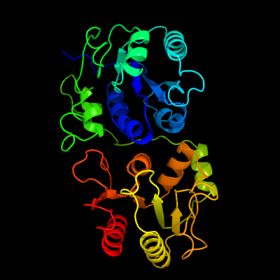
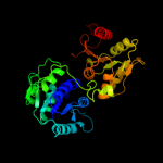
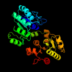
1 c3tovB_
100.0
14
PDB header: transferaseChain: B: PDB Molecule: glycosyl transferase family 9;PDBTitle: the crystal structure of the glycosyl transferase family 9 from2 veillonella parvula dsm 2008
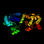
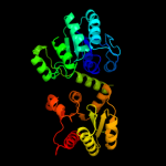
2 d1pswa_
100.0
13
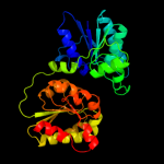
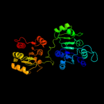
Fold: UDP-Glycosyltransferase/glycogen phosphorylaseSuperfamily: UDP-Glycosyltransferase/glycogen phosphorylaseFamily: ADP-heptose LPS heptosyltransferase II3 c2h1fB_
100.0
12
PDB header: transferaseChain: B: PDB Molecule: lipopolysaccharide heptosyltransferase-1;PDBTitle: e. coli heptosyltransferase waac with adp
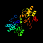
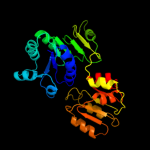
4 d1v4va_
99.2
12
Fold: UDP-Glycosyltransferase/glycogen phosphorylaseSuperfamily: UDP-Glycosyltransferase/glycogen phosphorylaseFamily: UDP-N-acetylglucosamine 2-epimerase5 c3ot5D_
99.1
11
PDB header: isomeraseChain: D: PDB Molecule: udp-n-acetylglucosamine 2-epimerase;PDBTitle: 2.2 angstrom resolution crystal structure of putative udp-n-2 acetylglucosamine 2-epimerase from listeria monocytogenes
6 d1o6ca_
98.6
10
Fold: UDP-Glycosyltransferase/glycogen phosphorylaseSuperfamily: UDP-Glycosyltransferase/glycogen phosphorylaseFamily: UDP-N-acetylglucosamine 2-epimerase7 d1f6da_
98.6
10
Fold: UDP-Glycosyltransferase/glycogen phosphorylaseSuperfamily: UDP-Glycosyltransferase/glycogen phosphorylaseFamily: UDP-N-acetylglucosamine 2-epimerase8 c3dzcA_
98.4
12
PDB header: isomeraseChain: A: PDB Molecule: udp-n-acetylglucosamine 2-epimerase;PDBTitle: 2.35 angstrom resolution structure of wecb (vc0917), a udp-n-2 acetylglucosamine 2-epimerase from vibrio cholerae.
9 c2xcuC_
97.8
11
PDB header: transferaseChain: C: PDB Molecule: 3-deoxy-d-manno-2-octulosonic acid transferase;PDBTitle: membrane-embedded monofunctional glycosyltransferase waaa of aquifex2 aeolicus, comlex with cmp
10 c3hbmA_
97.3
12
PDB header: hydrolaseChain: A: PDB Molecule: udp-sugar hydrolase;PDBTitle: crystal structure of pseg from campylobacter jejuni
11 c2vsnB_
97.2
9
PDB header: transferaseChain: B: PDB Molecule: xcogt;PDBTitle: structure and topological arrangement of an o-glcnac2 transferase homolog: insight into molecular control of3 intracellular glycosylation
12 d1pn3a_
97.1
11
Fold: UDP-Glycosyltransferase/glycogen phosphorylaseSuperfamily: UDP-Glycosyltransferase/glycogen phosphorylaseFamily: Gtf glycosyltransferase13 c3c4vB_
96.7
15
PDB header: transferaseChain: B: PDB Molecule: predicted glycosyltransferases;PDBTitle: structure of the retaining glycosyltransferase msha:the2 first step in mycothiol biosynthesis. organism:3 corynebacterium glutamicum : complex with udp and 1l-ins-1-4 p.
14 c2o6lA_
96.3
16
PDB header: transferaseChain: A: PDB Molecule: udp-glucuronosyltransferase 2b7;PDBTitle: crystal structure of the udp-glucuronic acid binding domain2 of the human drug metabolizing udp-glucuronosyltransferase3 2b7
15 c2gejA_
96.1
13
PDB header: transferaseChain: A: PDB Molecule: phosphatidylinositol mannosyltransferase (pima);PDBTitle: crystal structure of phosphatidylinositol mannosyltransferase (pima)2 from mycobacterium smegmatis in complex with gdp-man
16 c2xmpB_
95.9
10
PDB header: sugar binding proteinChain: B: PDB Molecule: trehalose-synthase tret;PDBTitle: crystal structure of trehalose synthase tret mutant e326a2 from p.horishiki in complex with udp
17 c3othB_
95.5
14
PDB header: transferase/antibioticChain: B: PDB Molecule: calg1;PDBTitle: crystal structure of calg1, calicheamicin glycostyltransferase, tdp2 and calicheamicin alpha3i bound form
18 c3okaA_
95.3
13
PDB header: transferaseChain: A: PDB Molecule: gdp-mannose-dependent alpha-(1-6)-phosphatidylinositolPDBTitle: crystal structure of corynebacterium glutamicum pimb' in complex with2 gdp-man (triclinic crystal form)
19 d1rrva_
95.3
13
Fold: UDP-Glycosyltransferase/glycogen phosphorylaseSuperfamily: UDP-Glycosyltransferase/glycogen phosphorylaseFamily: Gtf glycosyltransferase20 d1f0ka_
95.3
9
Fold: UDP-Glycosyltransferase/glycogen phosphorylaseSuperfamily: UDP-Glycosyltransferase/glycogen phosphorylaseFamily: Peptidoglycan biosynthesis glycosyltransferase MurG21 c2x6rA_
not modelled
95.2
10
PDB header: isomeraseChain: A: PDB Molecule: trehalose-synthase tret;PDBTitle: crystal structure of trehalose synthase tret from p.2 horikoshi produced by soaking in trehalose
22 c3iaaB_
not modelled
95.2
15
PDB header: transferaseChain: B: PDB Molecule: calg2;PDBTitle: crystal structure of calg2, calicheamicin glycosyltransferase, tdp2 bound form
23 c3pe3D_
not modelled
95.1
11
PDB header: transferaseChain: D: PDB Molecule: udp-n-acetylglucosamine--peptide n-PDBTitle: structure of human o-glcnac transferase and its complex with a peptide2 substrate
24 c2r60A_
not modelled
95.1
14
PDB header: transferaseChain: A: PDB Molecule: glycosyl transferase, group 1;PDBTitle: structure of apo sucrose phosphate synthase (sps) of2 halothermothrix orenii
25 c2jjmH_
not modelled
94.1
11
PDB header: transferaseChain: H: PDB Molecule: glycosyl transferase, group 1 family protein;PDBTitle: crystal structure of a family gt4 glycosyltransferase from2 bacillus anthracis orf ba1558.
26 c3q3hA_
not modelled
91.7
8
PDB header: transferaseChain: A: PDB Molecule: hmw1c-like glycosyltransferase;PDBTitle: crystal structure of the actinobacillus pleuropneumoniae hmw1c2 glycosyltransferase in complex with udp-glc
27 d2c1xa1
not modelled
91.1
9
Fold: UDP-Glycosyltransferase/glycogen phosphorylaseSuperfamily: UDP-Glycosyltransferase/glycogen phosphorylaseFamily: UDPGT-like28 c3qhpB_
not modelled
90.9
12
PDB header: transferaseChain: B: PDB Molecule: type 1 capsular polysaccharide biosynthesis protein jPDBTitle: crystal structure of the catalytic domain of cholesterol-alpha-2 glucosyltransferase from helicobacter pylori
29 c2q6vA_
not modelled
90.2
14
PDB header: transferaseChain: A: PDB Molecule: glucuronosyltransferase gumk;PDBTitle: crystal structure of gumk in complex with udp
30 d2f9fa1
not modelled
90.1
16
Fold: UDP-Glycosyltransferase/glycogen phosphorylaseSuperfamily: UDP-Glycosyltransferase/glycogen phosphorylaseFamily: Glycosyl transferases group 131 c3ehdA_
not modelled
89.4
28
PDB header: structural genomics, unknown functionChain: A: PDB Molecule: uncharacterized conserved protein;PDBTitle: crystal structure of conserved protein from enterococcus faecalis v583
32 c2x0dA_
not modelled
89.3
11
PDB header: transferaseChain: A: PDB Molecule: wsaf;PDBTitle: apo structure of wsaf
33 d1o6da_
not modelled
88.6
8
Fold: alpha/beta knotSuperfamily: alpha/beta knotFamily: YbeA-like34 d1vh0a_
not modelled
88.1
8
Fold: alpha/beta knotSuperfamily: alpha/beta knotFamily: YbeA-like35 c2khzB_
not modelled
87.4
18
PDB header: nuclear proteinChain: B: PDB Molecule: c-myc-responsive protein rcl;PDBTitle: solution structure of rcl
36 c3d0qB_
not modelled
87.4
11
PDB header: transferaseChain: B: PDB Molecule: protein calg3;PDBTitle: crystal structure of calg3 from micromonospora echinospora determined2 in space group i222
37 d1to0a_
not modelled
87.2
10
Fold: alpha/beta knotSuperfamily: alpha/beta knotFamily: YbeA-like38 c2p6pB_
not modelled
86.7
12
PDB header: transferaseChain: B: PDB Molecule: glycosyl transferase;PDBTitle: x-ray crystal structure of c-c bond-forming dtdp-d-olivose-transferase2 urdgt2
39 c2iyaB_
not modelled
86.1
10
PDB header: transferaseChain: B: PDB Molecule: oleandomycin glycosyltransferase;PDBTitle: the crystal structure of macrolide glycosyltransferases: a2 blueprint for antibiotic engineering
40 d2f62a1
not modelled
85.8
32
Fold: Flavodoxin-likeSuperfamily: N-(deoxy)ribosyltransferase-likeFamily: N-deoxyribosyltransferase41 c3ia7A_
not modelled
85.3
11
PDB header: transferaseChain: A: PDB Molecule: calg4;PDBTitle: crystal structure of calg4, the calicheamicin glycosyltransferase
42 d1ns5a_
not modelled
84.7
7
Fold: alpha/beta knotSuperfamily: alpha/beta knotFamily: YbeA-like43 d1iira_
not modelled
84.6
10
Fold: UDP-Glycosyltransferase/glycogen phosphorylaseSuperfamily: UDP-Glycosyltransferase/glycogen phosphorylaseFamily: Gtf glycosyltransferase44 c3oy2A_
not modelled
84.4
9
PDB header: viral protein,transferaseChain: A: PDB Molecule: glycosyltransferase b736l;PDBTitle: crystal structure of a putative glycosyltransferase from paramecium2 bursaria chlorella virus ny2a
45 d2g8la1
82.5
9
Fold: AF1104-likeSuperfamily: AF1104-likeFamily: AF1104-like46 c3gjzB_
not modelled
81.6
13
PDB header: immune systemChain: B: PDB Molecule: microcin immunity protein mccf;PDBTitle: crystal structure of microcin immunity protein mccf from bacillus2 anthracis str. ames
47 d1s2da_
not modelled
81.4
19
Fold: Flavodoxin-likeSuperfamily: N-(deoxy)ribosyltransferase-likeFamily: N-deoxyribosyltransferase48 d1f8ya_
not modelled
81.2
22
Fold: Flavodoxin-likeSuperfamily: N-(deoxy)ribosyltransferase-likeFamily: N-deoxyribosyltransferase49 c2iyfA_
not modelled
78.5
8
PDB header: transferaseChain: A: PDB Molecule: oleandomycin glycosyltransferase;PDBTitle: the crystal structure of macrolide glycosyltransferases: a2 blueprint for antibiotic engineering
50 d1to6a_
not modelled
78.0
16
Fold: Glycerate kinase ISuperfamily: Glycerate kinase IFamily: Glycerate kinase I51 c3l7mC_
not modelled
76.2
16
PDB header: structural proteinChain: C: PDB Molecule: teichoic acid biosynthesis protein f;PDBTitle: structure of the wall teichoic acid polymerase tagf, h548a
52 d1w6ta1
not modelled
75.4
16
Fold: TIM beta/alpha-barrelSuperfamily: Enolase C-terminal domain-likeFamily: Enolase53 c2qzsA_
not modelled
73.3
11
PDB header: transferaseChain: A: PDB Molecule: glycogen synthase;PDBTitle: crystal structure of wild-type e.coli gs in complex with adp2 and glucose(wtgsb)
54 d2bisa1
not modelled
73.2
12
Fold: UDP-Glycosyltransferase/glycogen phosphorylaseSuperfamily: UDP-Glycosyltransferase/glycogen phosphorylaseFamily: Glycosyl transferases group 155 d2iw1a1
not modelled
72.4
14
Fold: UDP-Glycosyltransferase/glycogen phosphorylaseSuperfamily: UDP-Glycosyltransferase/glycogen phosphorylaseFamily: Glycosyl transferases group 156 c3uj2C_
not modelled
69.1
20
PDB header: lyaseChain: C: PDB Molecule: enolase 1;PDBTitle: crystal structure of an enolase from anaerostipes caccae (efi target2 efi-502054) with bound mg and sulfate
57 d1iyxa1
not modelled
66.5
19
Fold: TIM beta/alpha-barrelSuperfamily: Enolase C-terminal domain-likeFamily: Enolase58 c3pdiG_
not modelled
66.0
11
PDB header: protein bindingChain: G: PDB Molecule: nitrogenase mofe cofactor biosynthesis protein nife;PDBTitle: precursor bound nifen
59 c2h31A_
not modelled
65.0
8
PDB header: ligase, lyaseChain: A: PDB Molecule: multifunctional protein ade2;PDBTitle: crystal structure of human paics, a bifunctional carboxylase and2 synthetase in purine biosynthesis
60 c2fymA_
not modelled
63.3
19
PDB header: lyaseChain: A: PDB Molecule: enolase;PDBTitle: crystal structure of e. coli enolase complexed with the2 minimal binding segment of rnase e.
61 c3cwcB_
not modelled
62.0
21
PDB header: transferaseChain: B: PDB Molecule: putative glycerate kinase 2;PDBTitle: crystal structure of putative glycerate kinase 2 from salmonella2 typhimurium lt2
62 d2fyma1
not modelled
59.3
18
Fold: TIM beta/alpha-barrelSuperfamily: Enolase C-terminal domain-likeFamily: Enolase63 c3rggD_
not modelled
56.6
13
PDB header: lyaseChain: D: PDB Molecule: phosphoribosylaminoimidazole carboxylase, pure protein;PDBTitle: crystal structure of treponema denticola pure bound to air
64 d1ybha1
not modelled
55.6
10
Fold: DHS-like NAD/FAD-binding domainSuperfamily: DHS-like NAD/FAD-binding domainFamily: Pyruvate oxidase and decarboxylase, middle domain65 d1rzua_
not modelled
55.5
13
Fold: UDP-Glycosyltransferase/glycogen phosphorylaseSuperfamily: UDP-Glycosyltransferase/glycogen phosphorylaseFamily: Glycosyl transferases group 166 d2acva1
not modelled
54.4
8
Fold: UDP-Glycosyltransferase/glycogen phosphorylaseSuperfamily: UDP-Glycosyltransferase/glycogen phosphorylaseFamily: UDPGT-like67 c3tqpA_
not modelled
54.3
17
PDB header: lyaseChain: A: PDB Molecule: enolase;PDBTitle: structure of an enolase (eno) from coxiella burnetii
68 c2jzcA_
not modelled
52.8
12
PDB header: transferaseChain: A: PDB Molecule: udp-n-acetylglucosamine transferase subunitPDBTitle: nmr solution structure of alg13: the sugar donor subunit of2 a yeast n-acetylglucosamine transferase. northeast3 structural genomics consortium target yg1
69 d1m1na_
not modelled
50.7
6
Fold: Chelatase-likeSuperfamily: "Helical backbone" metal receptorFamily: Nitrogenase iron-molybdenum protein70 c2xdqB_
not modelled
49.7
11
PDB header: oxidoreductaseChain: B: PDB Molecule: light-independent protochlorophyllide reductase subunit b;PDBTitle: dark operative protochlorophyllide oxidoreductase (chln-2 chlb)2 complex
71 c3l8mA_
not modelled
49.0
16
PDB header: transferaseChain: A: PDB Molecule: probable thiamine pyrophosphokinase;PDBTitle: crystal structure of a probable thiamine pyrophosphokinase2 from staphylococcus saprophyticus subsp. saprophyticus.3 northeast structural genomics consortium target id syr86
72 c2h9aA_
not modelled
40.8
13
PDB header: oxidoreductaseChain: A: PDB Molecule: carbon monoxide dehydrogenase corrinoid/iron-PDBTitle: corrinoid iron-sulfur protein
73 c3k94A_
not modelled
40.2
17
PDB header: transferaseChain: A: PDB Molecule: thiamin pyrophosphokinase;PDBTitle: crystal structure of thiamin pyrophosphokinase from geobacillus2 thermodenitrificans, northeast structural genomics consortium target3 gtr2
74 d1qh8a_
not modelled
38.9
10
Fold: Chelatase-likeSuperfamily: "Helical backbone" metal receptorFamily: Nitrogenase iron-molybdenum protein75 c3qtpB_
not modelled
38.8
14
PDB header: lyaseChain: B: PDB Molecule: enolase 1;PDBTitle: crystal structure analysis of entamoeba histolytica enolase
76 c2axqA_
not modelled
36.4
12
PDB header: oxidoreductaseChain: A: PDB Molecule: saccharopine dehydrogenase;PDBTitle: apo histidine-tagged saccharopine dehydrogenase (l-glu2 forming) from saccharomyces cerevisiae
77 d2ptza1
not modelled
33.3
17
Fold: TIM beta/alpha-barrelSuperfamily: Enolase C-terminal domain-likeFamily: Enolase78 c3qn3B_
not modelled
32.6
17
PDB header: lyaseChain: B: PDB Molecule: enolase;PDBTitle: phosphopyruvate hydratase from campylobacter jejuni.
79 c3ouzA_
not modelled
32.3
13
PDB header: ligaseChain: A: PDB Molecule: biotin carboxylase;PDBTitle: crystal structure of biotin carboxylase-adp complex from campylobacter2 jejuni
80 d1w3ia_
not modelled
32.2
7
Fold: TIM beta/alpha-barrelSuperfamily: AldolaseFamily: Class I aldolase81 d1z6na1
not modelled
31.8
12
Fold: Thioredoxin foldSuperfamily: Thioredoxin-likeFamily: Thioltransferase82 d2bfwa1
not modelled
31.6
10
Fold: UDP-Glycosyltransferase/glycogen phosphorylaseSuperfamily: UDP-Glycosyltransferase/glycogen phosphorylaseFamily: Glycosyl transferases group 183 d1t9ba1
not modelled
31.2
7
Fold: DHS-like NAD/FAD-binding domainSuperfamily: DHS-like NAD/FAD-binding domainFamily: Pyruvate oxidase and decarboxylase, middle domain84 c3cq9C_
not modelled
31.2
14
PDB header: transferaseChain: C: PDB Molecule: uncharacterized protein lp_1622;PDBTitle: crystal structure of the lp_1622 protein from lactobacillus2 plantarum. northeast structural genomics consortium target3 lpr114
85 c3lm8D_
not modelled
30.4
21
PDB header: transferaseChain: D: PDB Molecule: thiamine pyrophosphokinase;PDBTitle: crystal structure of thiamine pyrophosphokinase from2 bacillus subtilis, northeast structural genomics consortium3 target sr677
86 c3afoB_
not modelled
28.9
18
PDB header: transferaseChain: B: PDB Molecule: nadh kinase pos5;PDBTitle: crystal structure of yeast nadh kinase complexed with nadh
87 c3cf4G_
not modelled
28.0
16
PDB header: oxidoreductaseChain: G: PDB Molecule: acetyl-coa decarboxylase/synthase epsilon subunit;PDBTitle: structure of the codh component of the m. barkeri acds complex
88 d2ez9a1
not modelled
28.0
13
Fold: DHS-like NAD/FAD-binding domainSuperfamily: DHS-like NAD/FAD-binding domainFamily: Pyruvate oxidase and decarboxylase, middle domain89 c3aerB_
not modelled
26.7
11
PDB header: oxidoreductaseChain: B: PDB Molecule: light-independent protochlorophyllide reductase subunit b;PDBTitle: structure of the light-independent protochlorophyllide reductase2 catalyzing a key reduction for greening in the dark
90 d1m1nb_
not modelled
26.2
13
Fold: Chelatase-likeSuperfamily: "Helical backbone" metal receptorFamily: Nitrogenase iron-molybdenum protein91 c3d3aA_
not modelled
26.0
6
PDB header: hydrolaseChain: A: PDB Molecule: beta-galactosidase;PDBTitle: crystal structure of a beta-galactosidase from bacteroides2 thetaiotaomicron
92 c2j0wA_
not modelled
25.5
22
PDB header: transferaseChain: A: PDB Molecule: lysine-sensitive aspartokinase 3;PDBTitle: crystal structure of e. coli aspartokinase iii in complex2 with aspartate and adp (r-state)
93 c1zrsB_
not modelled
25.3
14
PDB header: hydrolaseChain: B: PDB Molecule: hypothetical protein;PDBTitle: wild-type ld-carboxypeptidase
94 c3b8kA_
not modelled
24.9
14
PDB header: transferaseChain: A: PDB Molecule: dihydrolipoyllysine-residue acetyltransferase;PDBTitle: structure of the truncated human dihydrolipoyl2 acetyltransferase (e2)
95 d2fgea3
not modelled
24.5
16
Fold: LuxS/MPP-like metallohydrolaseSuperfamily: LuxS/MPP-like metallohydrolaseFamily: MPP-like96 c2hlhA_
not modelled
24.1
14
PDB header: transferaseChain: A: PDB Molecule: nodulation fucosyltransferase;PDBTitle: crystal structure of fucosyltransferase nodz from bradyrhizobium
97 c3aerC_
not modelled
24.0
6
PDB header: oxidoreductaseChain: C: PDB Molecule: light-independent protochlorophyllide reductase subunit n;PDBTitle: structure of the light-independent protochlorophyllide reductase2 catalyzing a key reduction for greening in the dark
98 d2h1qa1
not modelled
23.5
9
Fold: PLP-dependent transferase-likeSuperfamily: Dhaf3308-likeFamily: Dhaf3308-like99 c3thdD_
not modelled
23.3
6
PDB header: hydrolaseChain: D: PDB Molecule: beta-galactosidase;PDBTitle: crystal structure of human beta-galactosidase in complex with 1-2 deoxygalactonojirimycin
100 c3hbjA_
not modelled
23.2
10
PDB header: transferaseChain: A: PDB Molecule: flavonoid 3-o-glucosyltransferase;PDBTitle: structure of ugt78g1 complexed with udp
101 d2jdid3
not modelled
23.1
8
Fold: P-loop containing nucleoside triphosphate hydrolasesSuperfamily: P-loop containing nucleoside triphosphate hydrolasesFamily: RecA protein-like (ATPase-domain)102 d2djia1
not modelled
22.9
9
Fold: DHS-like NAD/FAD-binding domainSuperfamily: DHS-like NAD/FAD-binding domainFamily: Pyruvate oxidase and decarboxylase, middle domain103 c3ihkC_
not modelled
22.8
15
PDB header: transferaseChain: C: PDB Molecule: thiamin pyrophosphokinase;PDBTitle: crystal structure of thiamin pyrophosphokinase from2 s.mutans, northeast structural genomics consortium target3 smr83
104 c3otrC_
not modelled
22.0
15
PDB header: lyaseChain: C: PDB Molecule: enolase;PDBTitle: 2.75 angstrom crystal structure of enolase 1 from toxoplasma gondii
105 d1miob_
not modelled
21.8
17
Fold: Chelatase-likeSuperfamily: "Helical backbone" metal receptorFamily: Nitrogenase iron-molybdenum protein106 c3rhzB_
not modelled
21.3
16
PDB header: transferaseChain: B: PDB Molecule: nucleotide sugar synthetase-like protein;PDBTitle: structure and functional analysis of a new subfamily of2 glycosyltransferases required for glycosylation of serine-rich3 streptococcal adhesions