1 d1ga8a_
100.0
19
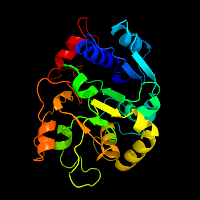
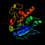
Fold: Nucleotide-diphospho-sugar transferasesSuperfamily: Nucleotide-diphospho-sugar transferasesFamily: Galactosyltransferase LgtC2 c1ga8A_
100.0
19
PDB header: transferaseChain: A: PDB Molecule: galactosyl transferase lgtc;PDBTitle: crystal structure of galacosyltransferase lgtc in complex2 with donor and acceptor sugar analogs.
3 c3tztB_
100.0
24
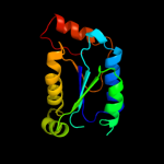
PDB header: transferaseChain: B: PDB Molecule: glycosyl transferase family 8;PDBTitle: the structure of a protein in glycosyl transferase family 8 from2 anaerococcus prevotii.
4 c1zcyA_
100.0
17
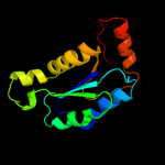
PDB header: transferaseChain: A: PDB Molecule: glycogenin-1;PDBTitle: apo form of a mutant of glycogenin in which asp159 is replaced by ser
5 d1ll2a_
100.0
17
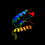
Fold: Nucleotide-diphospho-sugar transferasesSuperfamily: Nucleotide-diphospho-sugar transferasesFamily: Glycogenin6 c1zctB_
100.0
16
PDB header: transferaseChain: B: PDB Molecule: glycogenin-1;PDBTitle: structure of glycogenin truncated at residue 270 in a2 complex with udp
7 c1xhbA_
95.3
13
PDB header: transferaseChain: A: PDB Molecule: polypeptide n-acetylgalactosaminyltransferase 1;PDBTitle: the crystal structure of udp-galnac: polypeptide alpha-n-2 acetylgalactosaminyltransferase-t1
8 d1o7qa_
92.7
15
Fold: Nucleotide-diphospho-sugar transferasesSuperfamily: Nucleotide-diphospho-sugar transferasesFamily: alpha-1,3-galactosyltransferase-like9 c2d7iA_
88.9
10
PDB header: transferaseChain: A: PDB Molecule: polypeptide n-acetylgalactosaminyltransferase 10;PDBTitle: crsytal structure of pp-galnac-t10 with udp, galnac and mn2+
10 c2ffuA_
85.2
15
PDB header: transferaseChain: A: PDB Molecule: polypeptide n-acetylgalactosaminyltransferase 2;PDBTitle: crystal structure of human ppgalnact-2 complexed with udp2 and ea2
11 d1fo8a_
70.8
11
Fold: Nucleotide-diphospho-sugar transferasesSuperfamily: Nucleotide-diphospho-sugar transferasesFamily: N-acetylglucosaminyltransferase I12 d2py5a2
67.9
20
Fold: DNA/RNA polymerasesSuperfamily: DNA/RNA polymerasesFamily: DNA polymerase I13 c2ex3I_
57.2
18
PDB header: transferase/replicationChain: I: PDB Molecule: dna polymerase;PDBTitle: bacteriophage phi29 dna polymerase bound to terminal protein
14 d1lzia_
45.3
12
Fold: Nucleotide-diphospho-sugar transferasesSuperfamily: Nucleotide-diphospho-sugar transferasesFamily: alpha-1,3-galactosyltransferase-like15 d1w7pd2
42.8
15
Fold: DNA/RNA-binding 3-helical bundleSuperfamily: "Winged helix" DNA-binding domainFamily: Vacuolar sorting protein domain16 c2npiB_
30.2
16
PDB header: transcriptionChain: B: PDB Molecule: protein clp1;PDBTitle: clp1-atp-pcf11 complex
17 c2z86D_
26.5
6
PDB header: transferaseChain: D: PDB Molecule: chondroitin synthase;PDBTitle: crystal structure of chondroitin polymerase from2 escherichia coli strain k4 (k4cp) complexed with udp-glcua3 and udp
18 c2p73A_
24.2
19
PDB header: transferaseChain: A: PDB Molecule: putative glycosyltransferase (mannosyltransferase) involvedPDBTitle: crystal structure of a glycosyltransferase involved in the2 glycosylation of the major capsid of pbcv-1
19 d1qg8a_
23.4
20
Fold: Nucleotide-diphospho-sugar transferasesSuperfamily: Nucleotide-diphospho-sugar transferasesFamily: Spore coat polysaccharide biosynthesis protein SpsA20 c3lmaC_
23.3
12
PDB header: membrane proteinChain: C: PDB Molecule: stage v sporulation protein ad (spovad);PDBTitle: crystal structure of the stage v sporulation protein ad2 (spovad) from bacillus licheniformis. northeast structural3 genomics consortium target bir6.
21 d1ibia1
not modelled
22.6
64
Fold: Glucocorticoid receptor-like (DNA-binding domain)Superfamily: Glucocorticoid receptor-like (DNA-binding domain)Family: LIM domain22 d1s4na_
not modelled
18.4
16
Fold: Nucleotide-diphospho-sugar transferasesSuperfamily: Nucleotide-diphospho-sugar transferasesFamily: Glycolipid 2-alpha-mannosyltransferase23 c2w3zA_
not modelled
18.0
10
PDB header: hydrolaseChain: A: PDB Molecule: putative deacetylase;PDBTitle: structure of a streptococcus mutans ce4 esterase
24 d1acoa2
not modelled
16.7
14
Fold: Aconitase iron-sulfur domainSuperfamily: Aconitase iron-sulfur domainFamily: Aconitase iron-sulfur domain25 c3ew8A_
not modelled
15.5
30
PDB header: hydrolaseChain: A: PDB Molecule: histone deacetylase 8;PDBTitle: crystal structure analysis of human hdac8 d101l variant
26 d2c71a1
not modelled
12.6
14
Fold: 7-stranded beta/alpha barrelSuperfamily: Glycoside hydrolase/deacetylaseFamily: NodB-like polysaccharide deacetylase27 c3maxB_
not modelled
12.5
35
PDB header: hydrolaseChain: B: PDB Molecule: histone deacetylase 2;PDBTitle: crystal structure of human hdac2 complexed with an n-(2-aminophenyl)2 benzamide
28 c2ktvA_
not modelled
11.7
18
PDB header: translationChain: A: PDB Molecule: eukaryotic peptide chain release factor subunit 1;PDBTitle: human erf1 c-domain, "open" conformer
29 c4a69A_
not modelled
11.6
30
PDB header: transcriptionChain: A: PDB Molecule: histone deacetylase 3,;PDBTitle: structure of hdac3 bound to corepressor and inositol tetraphosphate
30 d1xhba2
not modelled
11.3
15
Fold: Nucleotide-diphospho-sugar transferasesSuperfamily: Nucleotide-diphospho-sugar transferasesFamily: Polypeptide N-acetylgalactosaminyltransferase 1, N-terminal domain31 c3ckvA_
not modelled
11.0
12
PDB header: unknown functionChain: A: PDB Molecule: putative uncharacterized protein;PDBTitle: crystal structure of a mycobacterial protein
32 c3m9qA_
not modelled
10.7
20
PDB header: dna binding proteinChain: A: PDB Molecule: protein male-specific lethal-3;PDBTitle: drosophila msl3 chromodomain
33 c1hf2A_
not modelled
10.6
17
PDB header: cell division proteinChain: A: PDB Molecule: septum site-determining protein minc;PDBTitle: crystal structure of the bacterial cell-division inhibitor2 minc from t. maritima
34 d1u5tb1
not modelled
10.3
13
Fold: DNA/RNA-binding 3-helical bundleSuperfamily: "Winged helix" DNA-binding domainFamily: Vacuolar sorting protein domain35 c3e20C_
not modelled
10.1
24
PDB header: translationChain: C: PDB Molecule: eukaryotic peptide chain release factor subunit 1;PDBTitle: crystal structure of s.pombe erf1/erf3 complex
36 c3m9pA_
not modelled
9.5
40
PDB header: dna binding protein/dnaChain: A: PDB Molecule: male-specific lethal 3 homolog;PDBTitle: human msl3 chromodomain bound to dna and h4k20me1 peptide
37 d1hf2a1
not modelled
8.9
17
Fold: Single-stranded right-handed beta-helixSuperfamily: Cell-division inhibitor MinC, C-terminal domainFamily: Cell-division inhibitor MinC, C-terminal domain38 d1t64a_
not modelled
6.9
18
Fold: Arginase/deacetylaseSuperfamily: Arginase/deacetylaseFamily: Histone deacetylase, HDAC39 d1dt9a2
not modelled
6.6
18
Fold: Bacillus chorismate mutase-likeSuperfamily: L30e-likeFamily: ERF1/Dom34 C-terminal domain-like40 c1w17A_
not modelled
6.4
11
PDB header: hydrolaseChain: A: PDB Molecule: probable polysaccharide deacetylase pdaa;PDBTitle: structure of bacillus subtilis pdaa, a family 42 carbohydrate esterase.
41 d1c3pa_
not modelled
6.1
29
Fold: Arginase/deacetylaseSuperfamily: Arginase/deacetylaseFamily: Histone deacetylase, HDAC42 c3menC_
not modelled
5.9
35
PDB header: hydrolaseChain: C: PDB Molecule: acetylpolyamine aminohydrolase;PDBTitle: crystal structure of acetylpolyamine aminohydrolase from burkholderia2 pseudomallei, iodide soak
43 c2zmeB_
not modelled
5.8
26
PDB header: protein transportChain: B: PDB Molecule: vacuolar protein-sorting-associated protein 36;PDBTitle: integrated structural and functional model of the human escrt-ii2 complex
44 d1ctla1
not modelled
5.8
55
Fold: Glucocorticoid receptor-like (DNA-binding domain)Superfamily: Glucocorticoid receptor-like (DNA-binding domain)Family: LIM domain45 d2f5ka1
not modelled
5.6
47
Fold: SH3-like barrelSuperfamily: Chromo domain-likeFamily: Chromo barrel domain46 d2g3ra2
not modelled
5.5
56
Fold: SH3-like barrelSuperfamily: Tudor/PWWP/MBTFamily: Tudor domain47 c3bcvA_
not modelled
5.4
12
PDB header: transferaseChain: A: PDB Molecule: putative glycosyltransferase protein;PDBTitle: crystal structure of a putative glycosyltransferase from bacteroides2 fragilis
48 c3ir9A_
not modelled
5.4
16
PDB header: structural genomics, unknown functionChain: A: PDB Molecule: peptide chain release factor subunit 1;PDBTitle: c-terminal domain of peptide chain release factor from2 methanosarcina mazei.
49 c1omxB_
not modelled
5.3
11
PDB header: transferaseChain: B: PDB Molecule: alpha-1,4-n-acetylhexosaminyltransferase extl2;PDBTitle: crystal structure of mouse alpha-1,4-n-2 acetylhexosaminyltransferase (extl2)