1 c2v9dB_
100.0
57
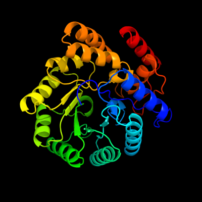
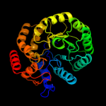
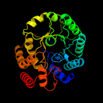
PDB header: lyaseChain: B: PDB Molecule: yage;PDBTitle: crystal structure of yage, a prophage protein belonging to2 the dihydrodipicolinic acid synthase family from e. coli3 k12
2 c3fluD_
100.0
32
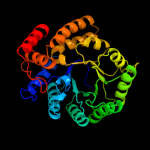
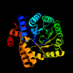
PDB header: lyaseChain: D: PDB Molecule: dihydrodipicolinate synthase;PDBTitle: crystal structure of dihydrodipicolinate synthase from the pathogen2 neisseria meningitidis
3 c3g0sA_
100.0
28
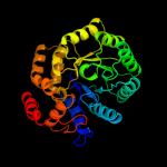
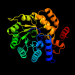
PDB header: lyaseChain: A: PDB Molecule: dihydrodipicolinate synthase;PDBTitle: dihydrodipicolinate synthase from salmonella typhimurium lt2
4 d2a6na1
100.0
29
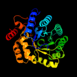
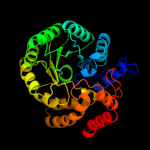
Fold: TIM beta/alpha-barrelSuperfamily: AldolaseFamily: Class I aldolase5 d1xxxa1
100.0
26
Fold: TIM beta/alpha-barrelSuperfamily: AldolaseFamily: Class I aldolase6 d1o5ka_
100.0
29
Fold: TIM beta/alpha-barrelSuperfamily: AldolaseFamily: Class I aldolase7 c3si9B_
100.0
31
PDB header: lyaseChain: B: PDB Molecule: dihydrodipicolinate synthase;PDBTitle: crystal structure of dihydrodipicolinate synthase from bartonella2 henselae
8 d1f74a_
100.0
26
Fold: TIM beta/alpha-barrelSuperfamily: AldolaseFamily: Class I aldolase9 c3pueA_
100.0
31
PDB header: lyaseChain: A: PDB Molecule: dihydrodipicolinate synthase;PDBTitle: crystal structure of the complex of dhydrodipicolinate synthase from2 acinetobacter baumannii with lysine at 2.6a resolution
10 c3bi8A_
100.0
24
PDB header: lyaseChain: A: PDB Molecule: dihydrodipicolinate synthase;PDBTitle: structure of dihydrodipicolinate synthase from clostridium2 botulinum
11 d1xkya1
100.0
28
Fold: TIM beta/alpha-barrelSuperfamily: AldolaseFamily: Class I aldolase12 c3noeA_
100.0
31
PDB header: lyaseChain: A: PDB Molecule: dihydrodipicolinate synthase;PDBTitle: crystal structure of dihydrodipicolinate synthase from pseudomonas2 aeruginosa
13 c3n2xB_
100.0
57
PDB header: lyaseChain: B: PDB Molecule: uncharacterized protein yage;PDBTitle: crystal structure of yage, a prophage protein belonging to the2 dihydrodipicolinic acid synthase family from e. coli k12 in complex3 with pyruvate
14 c3cprB_
100.0
29
PDB header: lyaseChain: B: PDB Molecule: dihydrodipicolinate synthetase;PDBTitle: the crystal structure of corynebacterium glutamicum2 dihydrodipicolinate synthase to 2.2 a resolution
15 c2r8wB_
100.0
25
PDB header: lyaseChain: B: PDB Molecule: agr_c_1641p;PDBTitle: the crystal structure of dihydrodipicolinate synthase (atu0899) from2 agrobacterium tumefaciens str. c58
16 d1hl2a_
100.0
25
Fold: TIM beta/alpha-barrelSuperfamily: AldolaseFamily: Class I aldolase17 c2yxgD_
100.0
31
PDB header: lyaseChain: D: PDB Molecule: dihydrodipicolinate synthase;PDBTitle: crystal structure of dihyrodipicolinate synthase (dapa)
18 c3s5oA_
100.0
25
PDB header: lyaseChain: A: PDB Molecule: 4-hydroxy-2-oxoglutarate aldolase, mitochondrial;PDBTitle: crystal structure of human 4-hydroxy-2-oxoglutarate aldolase bound to2 pyruvate
19 c2ehhE_
100.0
30
PDB header: lyaseChain: E: PDB Molecule: dihydrodipicolinate synthase;PDBTitle: crystal structure of dihydrodipicolinate synthase from2 aquifex aeolicus
20 c3eb2A_
100.0
31
PDB header: lyaseChain: A: PDB Molecule: putative dihydrodipicolinate synthetase;PDBTitle: crystal structure of dihydrodipicolinate synthase from2 rhodopseudomonas palustris at 2.0a resolution
21 c2vc6A_
not modelled
100.0
29
PDB header: lyaseChain: A: PDB Molecule: dihydrodipicolinate synthase;PDBTitle: structure of mosa from s. meliloti with pyruvate bound
22 c3lciA_
not modelled
100.0
25
PDB header: lyaseChain: A: PDB Molecule: n-acetylneuraminate lyase;PDBTitle: the d-sialic acid aldolase mutant v251w
23 c3na8A_
not modelled
100.0
25
PDB header: lyaseChain: A: PDB Molecule: putative dihydrodipicolinate synthetase;PDBTitle: crystal structure of a putative dihydrodipicolinate synthetase from2 pseudomonas aeruginosa
24 c3h5dD_
not modelled
100.0
26
PDB header: lyaseChain: D: PDB Molecule: dihydrodipicolinate synthase;PDBTitle: dihydrodipicolinate synthase from drug-resistant streptococcus2 pneumoniae
25 c2rfgB_
not modelled
100.0
30
PDB header: lyaseChain: B: PDB Molecule: dihydrodipicolinate synthase;PDBTitle: crystal structure of dihydrodipicolinate synthase from hahella2 chejuensis at 1.5a resolution
26 c3lerA_
not modelled
100.0
27
PDB header: lyaseChain: A: PDB Molecule: dihydrodipicolinate synthase;PDBTitle: crystal structure of dihydrodipicolinate synthase from2 campylobacter jejuni subsp. jejuni nctc 11168
27 c3daqB_
not modelled
100.0
26
PDB header: lyaseChain: B: PDB Molecule: dihydrodipicolinate synthase;PDBTitle: crystal structure of dihydrodipicolinate synthase from methicillin-2 resistant staphylococcus aureus
28 c3fkkA_
not modelled
100.0
23
PDB header: lyaseChain: A: PDB Molecule: l-2-keto-3-deoxyarabonate dehydratase;PDBTitle: structure of l-2-keto-3-deoxyarabonate dehydratase
29 c3e96B_
not modelled
100.0
22
PDB header: lyaseChain: B: PDB Molecule: dihydrodipicolinate synthase;PDBTitle: crystal structure of dihydrodipicolinate synthase from2 bacillus clausii
30 c3d0cB_
not modelled
100.0
22
PDB header: lyaseChain: B: PDB Molecule: dihydrodipicolinate synthase;PDBTitle: crystal structure of dihydrodipicolinate synthase from2 oceanobacillus iheyensis at 1.9 a resolution
31 c3dz1A_
not modelled
100.0
20
PDB header: lyaseChain: A: PDB Molecule: dihydrodipicolinate synthase;PDBTitle: crystal structure of dihydrodipicolinate synthase from2 rhodopseudomonas palustris at 1.87a resolution
32 d1w3ia_
not modelled
100.0
26
Fold: TIM beta/alpha-barrelSuperfamily: AldolaseFamily: Class I aldolase33 c2r94B_
not modelled
100.0
28
PDB header: lyaseChain: B: PDB Molecule: 2-keto-3-deoxy-(6-phospho-)gluconate aldolase;PDBTitle: crystal structure of kd(p)ga from t.tenax
34 c3qfeB_
not modelled
100.0
29
PDB header: lyaseChain: B: PDB Molecule: putative dihydrodipicolinate synthase family protein;PDBTitle: crystal structures of a putative dihydrodipicolinate synthase family2 protein from coccidioides immitis
35 c2nuxB_
not modelled
100.0
25
PDB header: lyaseChain: B: PDB Molecule: 2-keto-3-deoxygluconate/2-keto-3-deoxy-6-phospho gluconatePDBTitle: 2-keto-3-deoxygluconate aldolase from sulfolobus acidocaldarius,2 native structure in p6522 at 2.5 a resolution
36 c3b4uB_
not modelled
100.0
23
PDB header: lyaseChain: B: PDB Molecule: dihydrodipicolinate synthase;PDBTitle: crystal structure of dihydrodipicolinate synthase from agrobacterium2 tumefaciens str. c58
37 c2hmcA_
not modelled
100.0
22
PDB header: structural genomics, unknown functionChain: A: PDB Molecule: dihydrodipicolinate synthase;PDBTitle: the crystal structure of dihydrodipicolinate synthase dapa from2 agrobacterium tumefaciens
38 c2pcqA_
not modelled
100.0
33
PDB header: lyaseChain: A: PDB Molecule: putative dihydrodipicolinate synthase;PDBTitle: crystal structure of putative dihydrodipicolinate synthase (ttha0737)2 from thermus thermophilus hb8
39 c3lyeA_
not modelled
98.2
13
PDB header: hydrolaseChain: A: PDB Molecule: oxaloacetate acetyl hydrolase;PDBTitle: crystal structure of oxaloacetate acetylhydrolase
40 d1muma_
not modelled
98.2
13
Fold: TIM beta/alpha-barrelSuperfamily: Phosphoenolpyruvate/pyruvate domainFamily: Phosphoenolpyruvate mutase/Isocitrate lyase-like41 d1ujqa_
not modelled
98.0
12
Fold: TIM beta/alpha-barrelSuperfamily: Phosphoenolpyruvate/pyruvate domainFamily: Phosphoenolpyruvate mutase/Isocitrate lyase-like42 c2ze3A_
not modelled
98.0
18
PDB header: isomeraseChain: A: PDB Molecule: dfa0005;PDBTitle: crystal structure of dfa0005 complexed with alpha-ketoglutarate: a2 novel member of the icl/pepm superfamily from alkali-tolerant3 deinococcus ficus
43 c3ih1A_
not modelled
97.9
12
PDB header: lyaseChain: A: PDB Molecule: methylisocitrate lyase;PDBTitle: crystal structure of carboxyvinyl-carboxyphosphonate phosphorylmutase2 from bacillus anthracis
44 c3eooL_
not modelled
97.9
14
PDB header: lyaseChain: L: PDB Molecule: methylisocitrate lyase;PDBTitle: 2.9a crystal structure of methyl-isocitrate lyase from2 burkholderia pseudomallei
45 c1zlpA_
not modelled
97.8
16
PDB header: lyaseChain: A: PDB Molecule: petal death protein;PDBTitle: petal death protein psr132 with cysteine-linked glutaraldehyde forming2 a thiohemiacetal adduct
46 c3fa4D_
not modelled
97.7
14
PDB header: lyaseChain: D: PDB Molecule: 2,3-dimethylmalate lyase;PDBTitle: crystal structure of 2,3-dimethylmalate lyase, a pep mutase/isocitrate2 lyase superfamily member, triclinic crystal form
47 c3b8iF_
not modelled
97.7
17
PDB header: lyaseChain: F: PDB Molecule: pa4872 oxaloacetate decarboxylase;PDBTitle: crystal structure of oxaloacetate decarboxylase from pseudomonas2 aeruginosa (pa4872) in complex with oxalate and mg2+.
48 c2qiwA_
not modelled
97.6
13
PDB header: transferaseChain: A: PDB Molecule: pep phosphonomutase;PDBTitle: crystal structure of a putative phosphoenolpyruvate phosphonomutase2 (ncgl1015, cgl1060) from corynebacterium glutamicum atcc 13032 at3 1.80 a resolution
49 d1s2wa_
not modelled
97.6
13
Fold: TIM beta/alpha-barrelSuperfamily: Phosphoenolpyruvate/pyruvate domainFamily: Phosphoenolpyruvate mutase/Isocitrate lyase-like50 c2hjpA_
not modelled
97.5
17
PDB header: hydrolaseChain: A: PDB Molecule: phosphonopyruvate hydrolase;PDBTitle: crystal structure of phosphonopyruvate hydrolase complex with2 phosphonopyruvate and mg++
51 d1svda1
not modelled
97.4
19
Fold: TIM beta/alpha-barrelSuperfamily: RuBisCo, C-terminal domainFamily: RuBisCo, large subunit, C-terminal domain52 d1xcfa_
not modelled
97.4
15
Fold: TIM beta/alpha-barrelSuperfamily: Ribulose-phoshate binding barrelFamily: Tryptophan biosynthesis enzymes53 d1ps9a1
not modelled
97.3
18
Fold: TIM beta/alpha-barrelSuperfamily: FMN-linked oxidoreductasesFamily: FMN-linked oxidoreductases54 c3gr7A_
not modelled
97.3
18
PDB header: oxidoreductaseChain: A: PDB Molecule: nadph dehydrogenase;PDBTitle: structure of oye from geobacillus kaustophilus, hexagonal2 crystal form
55 c2h90A_
not modelled
97.3
19
PDB header: oxidoreductaseChain: A: PDB Molecule: xenobiotic reductase a;PDBTitle: xenobiotic reductase a in complex with coumarin
56 c3ez4B_
not modelled
97.2
24
PDB header: transferaseChain: B: PDB Molecule: 3-methyl-2-oxobutanoate hydroxymethyltransferase;PDBTitle: crystal structure of 3-methyl-2-oxobutanoate2 hydroxymethyltransferase from burkholderia pseudomallei
57 c1ps9A_
not modelled
97.1
19
PDB header: oxidoreductaseChain: A: PDB Molecule: 2,4-dienoyl-coa reductase;PDBTitle: the crystal structure and reaction mechanism of e. coli 2,4-2 dienoyl coa reductase
58 d2d69a1
not modelled
97.1
13
Fold: TIM beta/alpha-barrelSuperfamily: RuBisCo, C-terminal domainFamily: RuBisCo, large subunit, C-terminal domain59 d1ykwa1
not modelled
97.0
13
Fold: TIM beta/alpha-barrelSuperfamily: RuBisCo, C-terminal domainFamily: RuBisCo, large subunit, C-terminal domain60 d1ej7l1
not modelled
97.0
14
Fold: TIM beta/alpha-barrelSuperfamily: RuBisCo, C-terminal domainFamily: RuBisCo, large subunit, C-terminal domain61 d1oy0a_
not modelled
97.0
22
Fold: TIM beta/alpha-barrelSuperfamily: Phosphoenolpyruvate/pyruvate domainFamily: Ketopantoate hydroxymethyltransferase PanB62 d1geqa_
not modelled
97.0
14
Fold: TIM beta/alpha-barrelSuperfamily: Ribulose-phoshate binding barrelFamily: Tryptophan biosynthesis enzymes63 c1rcxH_
not modelled
97.0
14
PDB header: lyase (carbon-carbon)Chain: H: PDB Molecule: ribulose bisphosphate carboxylase/oxygenase;PDBTitle: non-activated spinach rubisco in complex with its substrate2 ribulose-1,5-bisphosphate
64 c3bolB_
not modelled
96.9
16
PDB header: transferaseChain: B: PDB Molecule: 5-methyltetrahydrofolate s-homocysteinePDBTitle: cobalamin-dependent methionine synthase (1-566) from2 thermotoga maritima complexed with zn2+
65 c3navB_
not modelled
96.9
17
PDB header: lyaseChain: B: PDB Molecule: tryptophan synthase alpha chain;PDBTitle: crystal structure of an alpha subunit of tryptophan synthase from2 vibrio cholerae o1 biovar el tor str. n16961
66 c2ekcA_
not modelled
96.8
16
PDB header: lyaseChain: A: PDB Molecule: tryptophan synthase alpha chain;PDBTitle: structural study of project id aq_1548 from aquifex aeolicus vf5
67 c3nwrA_
not modelled
96.8
15
PDB header: lyaseChain: A: PDB Molecule: a rubisco-like protein;PDBTitle: crystal structure of a rubisco-like protein from burkholderia fungorum
68 d1piia2
not modelled
96.8
12
Fold: TIM beta/alpha-barrelSuperfamily: Ribulose-phoshate binding barrelFamily: Tryptophan biosynthesis enzymes69 c2qygC_
not modelled
96.7
17
PDB header: unknown functionChain: C: PDB Molecule: ribulose bisphosphate carboxylase-like protein 2;PDBTitle: crystal structure of a rubisco-like protein rlp2 from rhodopseudomonas2 palustris
70 d1yxya1
not modelled
96.7
12
Fold: TIM beta/alpha-barrelSuperfamily: Ribulose-phoshate binding barrelFamily: NanE-like71 d1z41a1
not modelled
96.7
13
Fold: TIM beta/alpha-barrelSuperfamily: FMN-linked oxidoreductasesFamily: FMN-linked oxidoreductases72 c3hf3A_
not modelled
96.7
19
PDB header: oxidoreductaseChain: A: PDB Molecule: chromate reductase;PDBTitle: old yellow enzyme from thermus scotoductus sa-01
73 d8ruca1
not modelled
96.7
15
Fold: TIM beta/alpha-barrelSuperfamily: RuBisCo, C-terminal domainFamily: RuBisCo, large subunit, C-terminal domain74 c2qjhH_
not modelled
96.7
17
PDB header: lyaseChain: H: PDB Molecule: putative aldolase mj0400;PDBTitle: m. jannaschii adh synthase covalently bound to2 dihydroxyacetone phosphate
75 c3fk4A_
not modelled
96.7
18
PDB header: isomeraseChain: A: PDB Molecule: rubisco-like protein;PDBTitle: crystal structure of rubisco-like protein from bacillus2 cereus atcc 14579
76 d1f76a_
not modelled
96.7
23
Fold: TIM beta/alpha-barrelSuperfamily: FMN-linked oxidoreductasesFamily: FMN-linked oxidoreductases77 c3irsB_
not modelled
96.7
12
PDB header: structural genomics, unknown functionChain: B: PDB Molecule: uncharacterized protein bb4693;PDBTitle: crystal structure of uncharacterized tim-barrel protein bb4693 from2 bordetella bronchiseptica
78 c1telA_
not modelled
96.6
13
PDB header: structural genomics, unknown functionChain: A: PDB Molecule: ribulose bisphosphate carboxylase, large subunit;PDBTitle: crystal structure of a rubisco-like protein from chlorobium2 tepidum
79 d1tv5a1
not modelled
96.6
17
Fold: TIM beta/alpha-barrelSuperfamily: FMN-linked oxidoreductasesFamily: FMN-linked oxidoreductases80 c1tv5A_
not modelled
96.6
17
PDB header: oxidoreductaseChain: A: PDB Molecule: dihydroorotate dehydrogenase homolog, mitochondrial;PDBTitle: plasmodium falciparum dihydroorotate dehydrogenase with a bound2 inhibitor
81 d1geha1
not modelled
96.6
11
Fold: TIM beta/alpha-barrelSuperfamily: RuBisCo, C-terminal domainFamily: RuBisCo, large subunit, C-terminal domain82 d1f61a_
not modelled
96.6
17
Fold: TIM beta/alpha-barrelSuperfamily: Phosphoenolpyruvate/pyruvate domainFamily: Phosphoenolpyruvate mutase/Isocitrate lyase-like83 c3thaB_
not modelled
96.6
13
PDB header: lyaseChain: B: PDB Molecule: tryptophan synthase alpha chain;PDBTitle: tryptophan synthase subunit alpha from campylobacter jejuni.
84 d1qopa_
not modelled
96.5
17
Fold: TIM beta/alpha-barrelSuperfamily: Ribulose-phoshate binding barrelFamily: Tryptophan biosynthesis enzymes85 d1bxna1
not modelled
96.5
16
Fold: TIM beta/alpha-barrelSuperfamily: RuBisCo, C-terminal domainFamily: RuBisCo, large subunit, C-terminal domain86 c2rduA_
not modelled
96.5
20
PDB header: oxidoreductaseChain: A: PDB Molecule: hydroxyacid oxidase 1;PDBTitle: crystal structure of human glycolate oxidase in complex with2 glyoxylate
87 c2d69B_
not modelled
96.5
16
PDB header: lyaseChain: B: PDB Molecule: ribulose bisphosphate carboxylase;PDBTitle: crystal structure of the complex of sulfate ion and octameric2 ribulose-1,5-bisphosphate carboxylase/oxygenase (rubisco) from3 pyrococcus horikoshii ot3 (form-2 crystal)
88 d3bofa2
not modelled
96.5
17
Fold: TIM beta/alpha-barrelSuperfamily: Homocysteine S-methyltransferaseFamily: Homocysteine S-methyltransferase89 d1rd5a_
not modelled
96.5
18
Fold: TIM beta/alpha-barrelSuperfamily: Ribulose-phoshate binding barrelFamily: Tryptophan biosynthesis enzymes90 c2cdh1_
not modelled
96.5
21
PDB header: transferaseChain: 1: PDB Molecule: enoyl reductase;PDBTitle: architecture of the thermomyces lanuginosus fungal fatty2 acid synthase at 5 angstrom resolution.
91 c2e77B_
not modelled
96.4
20
PDB header: oxidoreductaseChain: B: PDB Molecule: lactate oxidase;PDBTitle: crystal structure of l-lactate oxidase with pyruvate complex
92 d1wdda1
not modelled
96.4
15
Fold: TIM beta/alpha-barrelSuperfamily: RuBisCo, C-terminal domainFamily: RuBisCo, large subunit, C-terminal domain93 d2f6ka1
not modelled
96.4
16
Fold: TIM beta/alpha-barrelSuperfamily: Metallo-dependent hydrolasesFamily: PP1699/LP2961-like94 c1gehE_
not modelled
96.4
11
PDB header: lyaseChain: E: PDB Molecule: ribulose-1,5-bisphosphate carboxylase/oxygenase;PDBTitle: crystal structure of archaeal rubisco (ribulose 1,5-bisphosphate2 carboxylase/oxygenase)
95 d1gvfa_
not modelled
96.3
18
Fold: TIM beta/alpha-barrelSuperfamily: AldolaseFamily: Class II FBP aldolase96 d1j5ta_
not modelled
96.3
17
Fold: TIM beta/alpha-barrelSuperfamily: Ribulose-phoshate binding barrelFamily: Tryptophan biosynthesis enzymes97 c1jcnA_
not modelled
96.2
22
PDB header: oxidoreductaseChain: A: PDB Molecule: inosine monophosphate dehydrogenase i;PDBTitle: binary complex of human type-i inosine monophosphate dehydrogenase2 with 6-cl-imp
98 d1d3ga_
not modelled
96.2
20
Fold: TIM beta/alpha-barrelSuperfamily: FMN-linked oxidoreductasesFamily: FMN-linked oxidoreductases99 c1djnB_
not modelled
96.2
16
PDB header: oxidoreductaseChain: B: PDB Molecule: trimethylamine dehydrogenase;PDBTitle: structural and biochemical characterization of recombinant wild type2 trimethylamine dehydrogenase from methylophilus methylotrophus (sp.3 w3a1)
100 d1rbla1
not modelled
96.1
16
Fold: TIM beta/alpha-barrelSuperfamily: RuBisCo, C-terminal domainFamily: RuBisCo, large subunit, C-terminal domain101 d1goxa_
not modelled
96.0
26
Fold: TIM beta/alpha-barrelSuperfamily: FMN-linked oxidoreductasesFamily: FMN-linked oxidoreductases102 d1rpxa_
not modelled
95.9
10
Fold: TIM beta/alpha-barrelSuperfamily: Ribulose-phoshate binding barrelFamily: D-ribulose-5-phosphate 3-epimerase103 d1m3ua_
not modelled
95.9
26
Fold: TIM beta/alpha-barrelSuperfamily: Phosphoenolpyruvate/pyruvate domainFamily: Ketopantoate hydroxymethyltransferase PanB104 c1rldB_
not modelled
95.9
14
PDB header: lyase(carbon-carbon)Chain: B: PDB Molecule: ribulose 1,5 bisphosphate carboxylase/oxygenase (largePDBTitle: solid-state phase transition in the crystal structure of ribulose 1,5-2 biphosphate carboxylase(slash)oxygenase
105 d1tb3a1
not modelled
95.9
24
Fold: TIM beta/alpha-barrelSuperfamily: FMN-linked oxidoreductasesFamily: FMN-linked oxidoreductases106 d1gwja_
not modelled
95.9
14
Fold: TIM beta/alpha-barrelSuperfamily: FMN-linked oxidoreductasesFamily: FMN-linked oxidoreductases107 c2c3zA_
not modelled
95.8
14
PDB header: lyaseChain: A: PDB Molecule: indole-3-glycerol phosphate synthase;PDBTitle: crystal structure of a truncated variant of indole-3-2 glycerol phosphate synthase from sulfolobus solfataricus
108 c1zfjA_
not modelled
95.8
15
PDB header: oxidoreductaseChain: A: PDB Molecule: inosine monophosphate dehydrogenase;PDBTitle: inosine monophosphate dehydrogenase (impdh; ec 1.1.1.205) from2 streptococcus pyogenes
109 c2z6jB_
not modelled
95.7
12
PDB header: oxidoreductaseChain: B: PDB Molecule: trans-2-enoyl-acp reductase ii;PDBTitle: crystal structure of s. pneumoniae enoyl-acyl carrier2 protein reductase (fabk) in complex with an inhibitor
110 c3qfwB_
not modelled
95.7
16
PDB header: lyaseChain: B: PDB Molecule: ribulose-1,5-bisphosphate carboxylase/oxygenase largePDBTitle: crystal structure of rubisco-like protein from rhodopseudomonas2 palustris
111 d1jcna1
not modelled
95.6
22
Fold: TIM beta/alpha-barrelSuperfamily: Inosine monophosphate dehydrogenase (IMPDH)Family: Inosine monophosphate dehydrogenase (IMPDH)112 c2ftpA_
not modelled
95.6
10
PDB header: lyaseChain: A: PDB Molecule: hydroxymethylglutaryl-coa lyase;PDBTitle: crystal structure of hydroxymethylglutaryl-coa lyase from pseudomonas2 aeruginosa
113 c2htmB_
not modelled
95.6
15
PDB header: biosynthetic proteinChain: B: PDB Molecule: thiazole biosynthesis protein thig;PDBTitle: crystal structure of ttha0676 from thermus thermophilus hb8
114 d1i4na_
not modelled
95.5
14
Fold: TIM beta/alpha-barrelSuperfamily: Ribulose-phoshate binding barrelFamily: Tryptophan biosynthesis enzymes115 d1vhna_
not modelled
95.5
16
Fold: TIM beta/alpha-barrelSuperfamily: FMN-linked oxidoreductasesFamily: FMN-linked oxidoreductases116 c2cw6B_
not modelled
95.5
12
PDB header: lyaseChain: B: PDB Molecule: hydroxymethylglutaryl-coa lyase, mitochondrial;PDBTitle: crystal structure of human hmg-coa lyase: insights into2 catalysis and the molecular basis for3 hydroxymethylglutaric aciduria
117 d1juba_
not modelled
95.5
11
Fold: TIM beta/alpha-barrelSuperfamily: FMN-linked oxidoreductasesFamily: FMN-linked oxidoreductases118 c1kbiB_
not modelled
95.5
21
PDB header: oxidoreductaseChain: B: PDB Molecule: cytochrome b2;PDBTitle: crystallographic study of the recombinant flavin-binding domain of2 baker's yeast flavocytochrome b2: comparison with the intact wild-3 type enzyme
119 c3gndC_
not modelled
95.5
15
PDB header: lyaseChain: C: PDB Molecule: aldolase lsrf;PDBTitle: crystal structure of e. coli lsrf in complex with ribulose-5-phosphate
120 c3nurA_
not modelled
95.4
16
PDB header: hydrolaseChain: A: PDB Molecule: amidohydrolase;PDBTitle: crystal structure of a putative amidohydrolase from staphylococcus2 aureus