Phyre Home Retrieve Phyre Job Id
Emaill.a.kelley@imperial.ac.uk
DescriptionP33347
DateThu Jan 5 11:51:48 GMT 2012
Unique Job ID386d463aa29d2951

Summary 

Top model
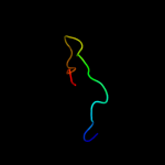
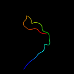
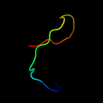
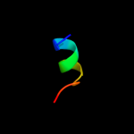
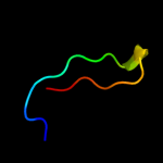
Image coloured by rainbow N → C terminus
Model (left) based on template c2j06B_
Top template information
PDB header:signal transduction
Chain: B: PDB Molecule:ras gtpase-activating protein 1;
PDBTitle: crystal structure of the rasgap sh3 domain at 1.8 angstrom2 resolution
Confidence and coverage
Confidence: 29.9% Coverage: 21%
22 residues ( 21% of your sequence) have been modelled with 29.9% confidence by the single highest scoring template.
You may wish to submit your sequence to Phyrealarm. This will automatically scan your sequence every week for new potential templates as they appear in the Phyre2 library.
Please note: You must be registered and logged in to use Phyrealarm.
3D viewing
Interactive 3D view in Jmol

Sequence analysis 

Secondary structure and disorder prediction 

   1........10.........20.........30.........40.........50.........60
Sequence  MIVQKELVAIYDYEVPVPEDPFSFRLEIHKCSELFTGSVYRLERFRLRPTFHQRDREDAD
Secondary structure 


SS confidence 


Disorder  ??????


?

????????
Disorder confidence 


   .........70.........80.........90.........100.....
Sequence  PLINDALIYIRDECIDERKLRGESPETVIAIFNRELQNIFNQEIE
Secondary structure 


SS confidence 


Disorder  ?


??????


?????
Disorder confidence 


Confidence Key
High(9)                    Low (0)
?Disordered
Alpha helix
Beta strand

Domain analysis 

Hover over an aligned region to see model and summary info

Please note, only up to the top 20 hits are modelled to reduce computer load

RankAligned region

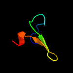
PDB 2j06 chain B

3D model

Region: 4 - 25
Aligned: 22
Modelled: 22
Confidence: 29.9%
Identity: 27%
PDB header:signal transduction
Chain: B: PDB Molecule:ras gtpase-activating protein 1;
PDBTitle: crystal structure of the rasgap sh3 domain at 1.8 angstrom2 resolution

Phyre2

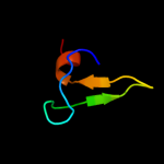
PDB 2ds5 chain A domain 1

3D model

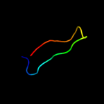
Region: 50 - 76
Aligned: 27
Modelled: 27
Confidence: 25.2%
Identity: 33%
Fold: Glucocorticoid receptor-like (DNA-binding domain)
Superfamily: Glucocorticoid receptor-like (DNA-binding domain)
Family: ClpX chaperone zinc binding domain

Phyre2

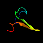
PDB 2ds8 chain A

3D model

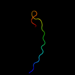
Region: 50 - 77
Aligned: 28
Modelled: 28
Confidence: 25.2%
Identity: 32%
PDB header:metal binding protein, protein binding
Chain: A: PDB Molecule:atp-dependent clp protease atp-binding subunit
PDBTitle: structure of the zbd-xb complex

Phyre2

PDB 1ovx chain B

3D model

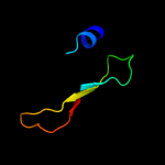
Region: 50 - 76
Aligned: 27
Modelled: 27
Confidence: 24.6%
Identity: 33%
PDB header:metal binding protein
Chain: B: PDB Molecule:atp-dependent clp protease atp-binding subunit clpx;
PDBTitle: nmr structure of the e. coli clpx chaperone zinc binding domain dimer

Phyre2

PDB 2egc chain A

3D model

Region: 3 - 25
Aligned: 21
Modelled: 23
Confidence: 24.6%
Identity: 33%
PDB header:signaling protein
Chain: A: PDB Molecule:sh3 and px domain-containing protein 2a;
PDBTitle: solution structure of the fifth sh3 domain from human2 kiaa0418 protein

Phyre2

PDB 3trk chain A

3D model

Region: 57 - 96
Aligned: 40
Modelled: 40
Confidence: 22.3%
Identity: 25%
PDB header:hydrolase
Chain: A: PDB Molecule:nonstructural polyprotein;
PDBTitle: structure of the chikungunya virus nsp2 protease

Phyre2

PDB 1t0j chain A

3D model

Region: 3 - 25
Aligned: 23
Modelled: 23
Confidence: 20.7%
Identity: 39%
PDB header:signaling protein
Chain: A: PDB Molecule:voltage-gated calcium channel subunit beta2a;
PDBTitle: crystal structure of a complex between voltage-gated calcium channel2 beta2a subunit and a peptide of the alpha1c subunit

Phyre2

PDB 2ege chain A

3D model

Region: 5 - 25
Aligned: 21
Modelled: 21
Confidence: 17.4%
Identity: 24%
PDB header:structural genomics, unknown function
Chain: A: PDB Molecule:uncharacterized protein kiaa1666;
PDBTitle: solution structure of the third sh3 domain from human2 kiaa1666 protein

Phyre2

PDB 1t0h chain A

3D model

Region: 3 - 25
Aligned: 23
Modelled: 23
Confidence: 16.2%
Identity: 39%
Fold: SH3-like barrel
Superfamily: SH3-domain
Family: SH3-domain

Phyre2

PDB 1k9a chain B

3D model

Region: 5 - 25
Aligned: 20
Modelled: 21
Confidence: 14.7%
Identity: 25%
PDB header:transferase
Chain: B: PDB Molecule:carboxyl-terminal src kinase;
PDBTitle: crystal structure analysis of full-length carboxyl-terminal2 src kinase at 2.5 a resolution

Phyre2

PDB 1gri chain A

3D model

Region: 6 - 25
Aligned: 19
Modelled: 20
Confidence: 13.6%
Identity: 32%
PDB header:signal transduction adaptor
Chain: A: PDB Molecule:growth factor bound protein 2;
PDBTitle: grb2

Phyre2

PDB 1vyv chain A domain 1

3D model

Region: 3 - 25
Aligned: 23
Modelled: 23
Confidence: 12.6%
Identity: 39%
Fold: SH3-like barrel
Superfamily: SH3-domain
Family: SH3-domain

Phyre2

PDB 3tbi chain A

3D model

Region: 70 - 81
Aligned: 12
Modelled: 12
Confidence: 12.0%
Identity: 58%
PDB header:transcription
Chain: A: PDB Molecule:rna polymerase-associated protein gp33;
PDBTitle: crystal structure of t4 gp33 bound to e. coli rnap beta-flap domain

Phyre2

PDB 2ke9 chain A

3D model

Region: 8 - 25
Aligned: 18
Modelled: 18
Confidence: 11.7%
Identity: 17%
PDB header:protein binding
Chain: A: PDB Molecule:caskin-2;
PDBTitle: nmr solution structure of the caskin sh3 domain

Phyre2

PDB 2lhf chain A

3D model

Region: 4 - 24
Aligned: 21
Modelled: 21
Confidence: 10.8%
Identity: 24%
PDB header:membrane protein
Chain: A: PDB Molecule:outer membrane protein h1;
PDBTitle: solution structure of outer membrane protein h (oprh) from p.2 aeruginosa in dhpc micelles

Phyre2

PDB 2nvu chain C domain 1

3D model

Region: 3 - 43
Aligned: 41
Modelled: 41
Confidence: 10.5%
Identity: 20%
Fold: UBC-like
Superfamily: UBC-like
Family: UBC-related

Phyre2

PDB 1jqq chain A

3D model

Region: 7 - 25
Aligned: 19
Modelled: 19
Confidence: 9.6%
Identity: 26%
Fold: SH3-like barrel
Superfamily: SH3-domain
Family: SH3-domain

Phyre2

PDB 2h8h chain A

3D model

Region: 8 - 25
Aligned: 17
Modelled: 18
Confidence: 9.5%
Identity: 53%
PDB header:transferase
Chain: A: PDB Molecule:proto-oncogene tyrosine-protein kinase src;
PDBTitle: src kinase in complex with a quinazoline inhibitor

Phyre2

PDB 1z9z chain A

3D model

Region: 4 - 25
Aligned: 21
Modelled: 22
Confidence: 8.7%
Identity: 14%
PDB header:structural protein
Chain: A: PDB Molecule:cytoskeleton assembly control protein sla1;
PDBTitle: crystal structure of yeast sla1 sh3 domain 3

Phyre2

PDB 2bnm chain A domain 1

3D model

Region: 80 - 95
Aligned: 16
Modelled: 16
Confidence: 8.4%
Identity: 63%
Fold: lambda repressor-like DNA-binding domains
Superfamily: lambda repressor-like DNA-binding domains
Family: SinR domain-like

Phyre2
1

c2j06B_
2

d2ds5a1
3

c2ds8A_
4

c1ovxB_
5

c2egcA_
6

c3trkA_
7

c1t0jA_
8

c2egeA_
9

d1t0ha_
10

c1k9aB_
11

c1griA_
12

d1vyva1
13

c3tbiA_
14

c2ke9A_
15

c2lhfA_
16

d2nvuc1
17

d1jqqa_
18

c2h8hA_
19

c1z9zA_
20

d2bnma1
21


22


23


24


25


26


27


28


29


30


31


32


33


34


35


36


37


38


39


40


41


42


43


44


45


46


47


Detailed template information 

#
Template Alignment Coverage3D Model Confidence
% i.d. Template Information
1c2j06B_


29.9 27 PDB header:signal transduction
Chain: B: PDB Molecule:ras gtpase-activating protein 1;
PDBTitle: crystal structure of the rasgap sh3 domain at 1.8 angstrom2 resolution
2d2ds5a1


25.2 33 Fold:Glucocorticoid receptor-like (DNA-binding domain)
Superfamily:Glucocorticoid receptor-like (DNA-binding domain)
Family:ClpX chaperone zinc binding domain
3c2ds8A_


25.2 32 PDB header:metal binding protein, protein binding
Chain: A: PDB Molecule:atp-dependent clp protease atp-binding subunit
PDBTitle: structure of the zbd-xb complex
4c1ovxB_


24.6 33 PDB header:metal binding protein
Chain: B: PDB Molecule:atp-dependent clp protease atp-binding subunit clpx;
PDBTitle: nmr structure of the e. coli clpx chaperone zinc binding domain dimer
5c2egcA_


24.6 33 PDB header:signaling protein
Chain: A: PDB Molecule:sh3 and px domain-containing protein 2a;
PDBTitle: solution structure of the fifth sh3 domain from human2 kiaa0418 protein
6c3trkA_


22.3 25 PDB header:hydrolase
Chain: A: PDB Molecule:nonstructural polyprotein;
PDBTitle: structure of the chikungunya virus nsp2 protease
7c1t0jA_


20.7 39 PDB header:signaling protein
Chain: A: PDB Molecule:voltage-gated calcium channel subunit beta2a;
PDBTitle: crystal structure of a complex between voltage-gated calcium channel2 beta2a subunit and a peptide of the alpha1c subunit
8c2egeA_


17.4 24 PDB header:structural genomics, unknown function
Chain: A: PDB Molecule:uncharacterized protein kiaa1666;
PDBTitle: solution structure of the third sh3 domain from human2 kiaa1666 protein
9d1t0ha_


16.2 39 Fold:SH3-like barrel
Superfamily:SH3-domain
Family:SH3-domain
10c1k9aB_


14.7 25 PDB header:transferase
Chain: B: PDB Molecule:carboxyl-terminal src kinase;
PDBTitle: crystal structure analysis of full-length carboxyl-terminal2 src kinase at 2.5 a resolution
11c1griA_


13.6 32 PDB header:signal transduction adaptor
Chain: A: PDB Molecule:growth factor bound protein 2;
PDBTitle: grb2
12d1vyva1


12.6 39 Fold:SH3-like barrel
Superfamily:SH3-domain
Family:SH3-domain
13c3tbiA_


12.0 58 PDB header:transcription
Chain: A: PDB Molecule:rna polymerase-associated protein gp33;
PDBTitle: crystal structure of t4 gp33 bound to e. coli rnap beta-flap domain
14c2ke9A_


11.7 17 PDB header:protein binding
Chain: A: PDB Molecule:caskin-2;
PDBTitle: nmr solution structure of the caskin sh3 domain
15c2lhfA_


10.8 24 PDB header:membrane protein
Chain: A: PDB Molecule:outer membrane protein h1;
PDBTitle: solution structure of outer membrane protein h (oprh) from p.2 aeruginosa in dhpc micelles
16d2nvuc1


10.5 20 Fold:UBC-like
Superfamily:UBC-like
Family:UBC-related
17d1jqqa_


9.6 26 Fold:SH3-like barrel
Superfamily:SH3-domain
Family:SH3-domain
18c2h8hA_


9.5 53 PDB header:transferase
Chain: A: PDB Molecule:proto-oncogene tyrosine-protein kinase src;
PDBTitle: src kinase in complex with a quinazoline inhibitor
19c1z9zA_


8.7 14 PDB header:structural protein
Chain: A: PDB Molecule:cytoskeleton assembly control protein sla1;
PDBTitle: crystal structure of yeast sla1 sh3 domain 3
20d2bnma1


8.4 63 Fold:lambda repressor-like DNA-binding domains
Superfamily:lambda repressor-like DNA-binding domains
Family:SinR domain-like
21c3u02C_


not modelled 8.3 21 PDB header:transcription
Chain: C: PDB Molecule:putative transcription-associated protein tfiis;
PDBTitle: crystal structure of the trna modifier tias from pyrococcus furiosus,2 northeast structural genomics consortium target pfr225
22d2jn9a1


not modelled 8.3 38 Fold:Split barrel-like
Superfamily:YkvR-like
Family:YkvR-like
23d1uj0a_


not modelled 8.3 30 Fold:SH3-like barrel
Superfamily:SH3-domain
Family:SH3-domain
24c1y57A_


not modelled 7.9 50 PDB header:transferase
Chain: A: PDB Molecule:proto-oncogene tyrosine-protein kinase src;
PDBTitle: structure of unphosphorylated c-src in complex with an inhibitor
25d1t3la1


not modelled 7.8 37 Fold:SH3-like barrel
Superfamily:SH3-domain
Family:SH3-domain
26c1s1nA_


not modelled 7.7 25 PDB header:cell adhesion
Chain: A: PDB Molecule:nephrocystin 1;
PDBTitle: sh3 domain of human nephrocystin
27c2c0iA_


not modelled 7.7 44 PDB header:transferase
Chain: A: PDB Molecule:tyrosine-protein kinase hck;
PDBTitle: src family kinase hck with bound inhibitor a-420983
28c1oplA_


not modelled 7.7 33 PDB header:transferase
Chain: A: PDB Molecule:proto-oncogene tyrosine-protein kinase;
PDBTitle: structural basis for the auto-inhibition of c-abl tyrosine2 kinase
29c2j6kE_


not modelled 7.4 32 PDB header:protein binding
Chain: E: PDB Molecule:cd2-associated protein;
PDBTitle: n-terminal sh3 domain of cms (cd2ap human homolog)
30c3rnjA_


not modelled 7.3 18 PDB header:protein binding
Chain: A: PDB Molecule:brain-specific angiogenesis inhibitor 1-associated protein
PDBTitle: crystal structure of the sh3 domain from irsp53 (baiap2)
31c2csqA_


not modelled 7.3 38 PDB header:endocytosis/exocytosis
Chain: A: PDB Molecule:rim binding protein 2;
PDBTitle: solution structure of the second sh3 domain of human rim-2 binding protein 2
32c2cucA_


not modelled 7.0 15 PDB header:signaling protein
Chain: A: PDB Molecule:sh3 domain containing ring finger 2;
PDBTitle: solution structure of the sh3 domain of the mouse2 hypothetical protein sh3rf2
33c2fo0A_


not modelled 6.6 33 PDB header:transferase
Chain: A: PDB Molecule:proto-oncogene tyrosine-protein kinase abl1 (1b isoform);
PDBTitle: organization of the sh3-sh2 unit in active and inactive forms of the2 c-abl tyrosine kinase
34c2pqhA_


not modelled 6.5 29 PDB header:structural protein
Chain: A: PDB Molecule:spectrin alpha chain, brain;
PDBTitle: structure of sh3 chimera with a type ii ligand linked to the chain c-2 terminal
35c2dm1A_


not modelled 6.3 25 PDB header:signaling protein
Chain: A: PDB Molecule:protein vav-2;
PDBTitle: solution structure of the second sh3 domain of human2 protein vav-2
36d1spka_


not modelled 6.2 14 Fold:SH3-like barrel
Superfamily:SH3-domain
Family:SH3-domain
37c1x27F_


not modelled 6.1 28 PDB header:signaling protein
Chain: F: PDB Molecule:proto-oncogene tyrosine-protein kinase lck;
PDBTitle: crystal structure of lck sh2-sh3 with sh2 binding site of2 p130cas
38d2v1ra1


not modelled 6.1 23 Fold:SH3-like barrel
Superfamily:SH3-domain
Family:SH3-domain
39c2hwkA_


not modelled 6.0 26 PDB header:hydrolase
Chain: A: PDB Molecule:helicase nsp2;
PDBTitle: crystal structure of venezuelan equine encephalitis2 alphavirus nsp2 protease domain
40d1awja_


not modelled 6.0 23 Fold:SH3-like barrel
Superfamily:SH3-domain
Family:SH3-domain
41c1g83A_


not modelled 5.8 50 PDB header:transferase
Chain: A: PDB Molecule:proto-oncogene tyrosine-protein kinase fyn;
PDBTitle: crystal structure of fyn sh3-sh2
42d1gria1


not modelled 5.7 32 Fold:SH3-like barrel
Superfamily:SH3-domain
Family:SH3-domain
43c2d8jA_


not modelled 5.6 35 PDB header:transferase
Chain: A: PDB Molecule:fyn-related kinase;
PDBTitle: solution structure of the sh3 domain of fyn-related kinase
44c1x2pA_


not modelled 5.4 45 PDB header:transferase
Chain: A: PDB Molecule:protein arginine n-methyltransferase 2;
PDBTitle: solution structure of the sh3 domain of the protein2 arginine n-methyltransferase 2
45c2dmoA_


not modelled 5.3 11 PDB header:signaling protein
Chain: A: PDB Molecule:neutrophil cytosol factor 2;
PDBTitle: refined solution structure of the 1st sh3 domain from human2 neutrophil cytosol factor 2 (ncf-2)
46c2dlpA_


not modelled 5.2 14 PDB header:structural protein
Chain: A: PDB Molecule:kiaa1783 protein;
PDBTitle: solution structure of the sh3 domain of human kiaa17832 protein
47d1lcka1


not modelled 5.1 24 Fold:SH3-like barrel
Superfamily:SH3-domain
Family:SH3-domain

Binding site prediction 

Due to computational demand, binding site predictions are not run for batch jobs

If you want to predict binding sites, please manually submit your model of choice to 3DLigandSite


Phyre is for academic use only

Please cite: Protein structure prediction on the web: a case study using the Phyre server
Kelley LA and Sternberg MJE. Nature Protocols 4, 363 - 371 (2009) [pdf] [Import into BibTeX]
 
If you use the binding site predictions from 3DLigandSite, please also cite:
3DLigandSite: predicting ligand-binding sites using similar structures.
Wass MN, Kelley LA and Sternberg MJ Nucleic Acids Research 38, W469-73 (2010) [PubMed]
 
© Structural Bioinformatics Group
Imperial College London
Lawrence Kelley, Benjamin Jefferys
Disclaimer
Terms and Conditions
Component software
Template detection: HHpred 1.51
Secondary structure prediction: Psi-pred 2.5
Disorder prediction: Disopred 2.4
Transmembrane prediction: Memsat_SVM
Multi-template modelling and ab initio: Poing 1.0