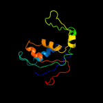
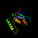
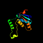
1 d2j01n1
100.0
56
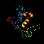
Fold: Ribosomal protein L13Superfamily: Ribosomal protein L13Family: Ribosomal protein L132 d2zjrg1
100.0
55
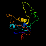
Fold: Ribosomal protein L13Superfamily: Ribosomal protein L13Family: Ribosomal protein L133 c3d5bN_
100.0
55
PDB header: ribosomeChain: N: PDB Molecule: 50s ribosomal protein l13;PDBTitle: structural basis for translation termination on the 70s ribosome. this2 file contains the 50s subunit of one 70s ribosome. the entire crystal3 structure contains two 70s ribosomes as described in remark 400.
4 c3cf5G_
100.0
55
PDB header: ribosome/antibioticChain: G: PDB Molecule: 50s ribosomal protein l13;PDBTitle: thiopeptide antibiotic thiostrepton bound to the large ribosomal2 subunit of deinococcus radiodurans
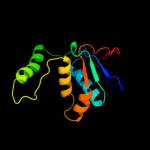
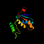
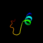
5 d2gych1
100.0
100
Fold: Ribosomal protein L13Superfamily: Ribosomal protein L13Family: Ribosomal protein L136 c2ftcH_
100.0
33
PDB header: ribosomeChain: H: PDB Molecule: 39s ribosomal protein l13, mitochondrial;PDBTitle: structural model for the large subunit of the mammalian mitochondrial2 ribosome
7 c3bboL_
100.0
59
PDB header: ribosomeChain: L: PDB Molecule: ribosomal protein l13;PDBTitle: homology model for the spinach chloroplast 50s subunit2 fitted to 9.4a cryo-em map of the 70s chlororibosome
8 c1s1iM_
100.0
27
PDB header: ribosomeChain: M: PDB Molecule: 60s ribosomal protein l16-a;PDBTitle: structure of the ribosomal 80s-eef2-sordarin complex from2 yeast obtained by docking atomic models for rna and protein3 components into a 11.7 a cryo-em map. this file, 1s1i,4 contains 60s subunit. the 40s ribosomal subunit is in file5 1s1h.
9 c3iz5K_
100.0
23
PDB header: ribosomeChain: K: PDB Molecule: 60s ribosomal protein l13a (l13p);PDBTitle: localization of the large subunit ribosomal proteins into a 5.5 a2 cryo-em map of triticum aestivum translating 80s ribosome
10 c2zkrj_
100.0
27
PDB header: ribosomal protein/rnaChain: J: PDB Molecule: rna expansion segment es15 part ii;PDBTitle: structure of a mammalian ribosomal 60s subunit within an2 80s complex obtained by docking homology models of the rna3 and proteins into an 8.7 a cryo-em map
11 c3izcK_
100.0
27
PDB header: ribosomeChain: K: PDB Molecule: 60s ribosomal protein rpl16 (l13p);PDBTitle: localization of the large subunit ribosomal proteins into a 6.1 a2 cryo-em map of saccharomyces cerevisiae translating 80s ribosome
12 c4a1aI_
100.0
29
PDB header: ribosomeChain: I: PDB Molecule: 60s ribosomal protein l13a;PDBTitle: t.thermophila 60s ribosomal subunit in complex with2 initiation factor 6. this file contains 5s rrna,3 5.8s rrna and proteins of molecule 3.
13 c3jywM_
100.0
28
PDB header: ribosomeChain: M: PDB Molecule: 60s ribosomal protein l16(a);PDBTitle: structure of the 60s proteins for eukaryotic ribosome based on cryo-em2 map of thermomyces lanuginosus ribosome at 8.9a resolution
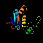
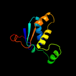
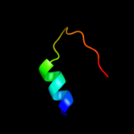
14 d1j3aa_
100.0
33
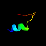
Fold: Ribosomal protein L13Superfamily: Ribosomal protein L13Family: Ribosomal protein L1315 d1vqoj1
100.0
26
Fold: Ribosomal protein L13Superfamily: Ribosomal protein L13Family: Ribosomal protein L1316 d1qvca_
34.0
30
Fold: OB-foldSuperfamily: Nucleic acid-binding proteinsFamily: Single strand DNA-binding domain, SSB17 c3kojA_
31.5
35
PDB header: dna binding proteinChain: A: PDB Molecule: uncharacterized protein ycf41;PDBTitle: crystal structure of the ssb domain of q5n255_synp6 protein2 from synechococcus sp. northeast structural genomics3 consortium target snr59a.
18 c1z9fA_
26.8
42
PDB header: dna binding proteinChain: A: PDB Molecule: single-strand binding protein;PDBTitle: crystal structure of single stranded dna-binding protein (tm0604) from2 thermotoga maritima at 2.60 a resolution
19 c3fmaD_
24.2
29
PDB header: protein bindingChain: D: PDB Molecule: protein smy2;PDBTitle: crystal structure of the gyf domain of smy2 in complex with a proline-2 rich peptide from bbp/scsf1
20 c2vw9B_
23.6
25
PDB header: dna-binding proteinChain: B: PDB Molecule: single-stranded dna binding protein;PDBTitle: single stranded dna binding protein complex from2 helicobacter pylori
21 c2ra9A_
not modelled
23.2
33
PDB header: unknown functionChain: A: PDB Molecule: uncharacterized protein duf1285;PDBTitle: crystal structure of a duf1285 family protein (sbal_2486) from2 shewanella baltica os155 at 1.40 a resolution
22 d2pstx1
not modelled
22.0
23
Fold: MbtH/L9 domain-likeSuperfamily: MbtH-likeFamily: MbtH-like23 c1eqqD_
not modelled
21.6
32
PDB header: replication/rnaChain: D: PDB Molecule: single stranded dna binding protein;PDBTitle: single stranded dna binding protein and ssdna complex
24 d1tzaa_
not modelled
18.1
50
Fold: Immunoglobulin-like beta-sandwichSuperfamily: ApaG-likeFamily: ApaG-like25 d1xq4a_
not modelled
17.8
21
Fold: Immunoglobulin-like beta-sandwichSuperfamily: ApaG-likeFamily: ApaG-like26 d2gpfa1
not modelled
17.4
23
Fold: MbtH/L9 domain-likeSuperfamily: MbtH-likeFamily: MbtH-like27 d2q07a1
not modelled
17.1
10
Fold: PUA domain-likeSuperfamily: PUA domain-likeFamily: PUA domain28 c2f1eA_
not modelled
17.0
43
PDB header: structural genomics, unknown functionChain: A: PDB Molecule: protein apag;PDBTitle: solution structure of apag protein
29 d1puja_
not modelled
17.0
27
Fold: P-loop containing nucleoside triphosphate hydrolasesSuperfamily: P-loop containing nucleoside triphosphate hydrolasesFamily: G proteins30 d1xvsa_
not modelled
16.9
43
Fold: Immunoglobulin-like beta-sandwichSuperfamily: ApaG-likeFamily: ApaG-like31 c2khrA_
not modelled
15.3
23
PDB header: biosynthetic proteinChain: A: PDB Molecule: protein mbth;PDBTitle: solution structure of rv2377c, a mbth-like protein from mycobacterium2 tuberculosis
32 d1eyga_
not modelled
15.0
30
Fold: OB-foldSuperfamily: Nucleic acid-binding proteinsFamily: Single strand DNA-binding domain, SSB33 d2bm8a1
not modelled
13.5
23
Fold: S-adenosyl-L-methionine-dependent methyltransferasesSuperfamily: S-adenosyl-L-methionine-dependent methyltransferasesFamily: CmcI-like34 c2p8tA_
not modelled
13.1
24
PDB header: structural genomics, unknown functionChain: A: PDB Molecule: hypothetical protein ph0730;PDBTitle: hypothetical protein ph0730 from pyrococcus horikoshii ot3
35 d1ue1a_
not modelled
13.0
35
Fold: OB-foldSuperfamily: Nucleic acid-binding proteinsFamily: Single strand DNA-binding domain, SSB36 d2d7na1
not modelled
12.7
19
Fold: Immunoglobulin-like beta-sandwichSuperfamily: E set domainsFamily: Filamin repeat (rod domain)37 c2q07A_
not modelled
12.2
10
PDB header: structural genomics, unknown functionChain: A: PDB Molecule: uncharacterized protein af0587;PDBTitle: crystal structure of af0587, a protein of unknown function
38 c3tqyA_
not modelled
12.1
35
PDB header: transferaseChain: A: PDB Molecule: single-stranded dna-binding protein;PDBTitle: structure of a single-stranded dna-binding protein (ssb), from2 coxiella burnetii
39 c2ihfA_
not modelled
11.2
37
PDB header: dna binding proteinChain: A: PDB Molecule: single-stranded dna-binding protein;PDBTitle: crystal structure of deletion mutant delta 228-252 r190a of the2 single-stranded dna binding protein from thermus aquaticus
40 c3eivB_
not modelled
11.1
32
PDB header: dna binding proteinChain: B: PDB Molecule: single-stranded dna-binding protein 2;PDBTitle: crystal structure of single-stranded dna-binding protein2 from streptomyces coelicolor
41 c2e6zA_
not modelled
10.3
17
PDB header: transcriptionChain: A: PDB Molecule: transcription elongation factor spt5;PDBTitle: solution structure of the second kow motif of human2 transcription elongation factor spt5
42 c3rhfB_
not modelled
10.2
44
PDB header: transferaseChain: B: PDB Molecule: putative polyphosphate kinase 2 family protein;PDBTitle: crystal structure of polyphosphate kinase 2 from arthrobacter2 aurescens tc1
43 c3pgzB_
not modelled
10.2
27
PDB header: dna binding proteinChain: B: PDB Molecule: single-stranded dna-binding protein;PDBTitle: crystal structure of a single strand binding protein (ssb) from2 bartonella henselae
44 c3czqA_
not modelled
9.9
24
PDB header: transferaseChain: A: PDB Molecule: putative polyphosphate kinase 2;PDBTitle: crystal structure of putative polyphosphate kinase 2 from2 sinorhizobium meliloti
45 c1ue7A_
not modelled
9.8
25
PDB header: dna binding proteinChain: A: PDB Molecule: single-strand binding protein;PDBTitle: crystal structure of the single-stranded dna-binding2 protein from mycobacterium tuberculosis
46 d1v1qa_
not modelled
9.0
17
Fold: OB-foldSuperfamily: Nucleic acid-binding proteinsFamily: Single strand DNA-binding domain, SSB47 c3czpA_
not modelled
8.8
31
PDB header: transferaseChain: A: PDB Molecule: putative polyphosphate kinase 2;PDBTitle: crystal structure of putative polyphosphate kinase 2 from pseudomonas2 aeruginosa pa01
48 c2iheA_
not modelled
8.6
36
PDB header: dna binding proteinChain: A: PDB Molecule: single-stranded dna-binding protein;PDBTitle: crystal structure of wild-type single-stranded dna binding protein2 from thermus aquaticus
49 c2l9dA_
not modelled
8.5
21
PDB header: structural genomics, unknown functionChain: A: PDB Molecule: uncharacterized protein;PDBTitle: solution structure of the protein yp_546394.1, the first structural2 representative of the pfam family pf12112
50 c2rndA_
not modelled
8.4
29
PDB header: endocytosisChain: A: PDB Molecule: myc box-dependent-interacting protein 1;PDBTitle: structure of the n-terminal barpeptide in dpc micelles
51 c1is7F_
not modelled
8.2
14
PDB header: hydrolase/protein bindingChain: F: PDB Molecule: gtp cyclohydrolase i;PDBTitle: crystal structure of rat gtpchi/gfrp stimulatory complex
52 d1a8ra_
not modelled
8.1
17
Fold: T-foldSuperfamily: Tetrahydrobiopterin biosynthesis enzymes-likeFamily: GTP cyclohydrolase I53 d2d7pa1
not modelled
8.1
22
Fold: Immunoglobulin-like beta-sandwichSuperfamily: E set domainsFamily: Filamin repeat (rod domain)54 d2d7ma1
not modelled
7.6
17
Fold: Immunoglobulin-like beta-sandwichSuperfamily: E set domainsFamily: Filamin repeat (rod domain)55 c3iswA_
not modelled
7.4
17
PDB header: structural proteinChain: A: PDB Molecule: filamin-a;PDBTitle: crystal structure of filamin-a immunoglobulin-like repeat 21 bound to2 an n-terminal peptide of cftr
56 c3mx7A_
not modelled
7.1
19
PDB header: apoptosisChain: A: PDB Molecule: fas apoptotic inhibitory molecule 1;PDBTitle: crystal structure analysis of human faim-ntd
57 d2w0pa1
not modelled
6.9
17
Fold: Immunoglobulin-like beta-sandwichSuperfamily: E set domainsFamily: Filamin repeat (rod domain)58 d2ntka1
not modelled
6.9
26
Fold: Ntn hydrolase-likeSuperfamily: Archaeal IMP cyclohydrolase PurOFamily: Archaeal IMP cyclohydrolase PurO59 d2do3a1
not modelled
6.7
25
Fold: SH3-like barrelSuperfamily: Translation proteins SH3-like domainFamily: SPT5 KOW domain-like60 c2l8kA_
not modelled
6.6
31
PDB header: viral proteinChain: A: PDB Molecule: non-structural protein 7;PDBTitle: nmr structure of the arterivirus nonstructural protein 7 alpha (nsp72 alpha)
61 d1txya_
not modelled
6.6
17
Fold: OB-foldSuperfamily: Nucleic acid-binding proteinsFamily: Single strand DNA-binding domain, SSB62 d2exna1
not modelled
6.4
31
Fold: MOSC N-terminal domain-likeSuperfamily: MOSC N-terminal domain-likeFamily: MOSC N-terminal domain-like63 c3ec1A_
not modelled
6.4
23
PDB header: hydrolase, signaling proteinChain: A: PDB Molecule: yqeh gtpase;PDBTitle: structure of yqeh gtpase from geobacillus stearothermophilus2 (an atnos1 / atnoa1 ortholog)
64 c2wojD_
not modelled
6.3
19
PDB header: hydrolaseChain: D: PDB Molecule: atpase get3;PDBTitle: adp-alf4 complex of s. cerevisiae get3
65 c3iswB_
not modelled
6.3
17
PDB header: structural proteinChain: B: PDB Molecule: filamin-a;PDBTitle: crystal structure of filamin-a immunoglobulin-like repeat 21 bound to2 an n-terminal peptide of cftr
66 c2brqA_
not modelled
6.3
17
PDB header: structural proteinChain: A: PDB Molecule: filamin a;PDBTitle: crystal structure of the filamin a repeat 21 complexed with2 the integrin beta7 cytoplasmic tail peptide
67 c2w0pB_
not modelled
6.3
17
PDB header: cell adhesionChain: B: PDB Molecule: filamin-a;PDBTitle: crystal structure of the filamin a repeat 21 complexed with2 the migfilin peptide
68 d1wpla_
not modelled
6.2
14
Fold: T-foldSuperfamily: Tetrahydrobiopterin biosynthesis enzymes-likeFamily: GTP cyclohydrolase I69 c3k6qB_
not modelled
6.2
11
PDB header: ligand binding proteinChain: B: PDB Molecule: putative ligand binding protein;PDBTitle: crystal structure of an antitoxin part of a putative toxin/antitoxin2 system (swol_0700) from syntrophomonas wolfei subsp. wolfei at 1.80 a3 resolution
70 d1a9xa3
not modelled
6.1
19
Fold: PreATP-grasp domainSuperfamily: PreATP-grasp domainFamily: BC N-terminal domain-like71 d1v5pa_
not modelled
6.1
21
Fold: PH domain-like barrelSuperfamily: PH domain-likeFamily: Pleckstrin-homology domain (PH domain)72 c3cf4A_
not modelled
5.8
19
PDB header: oxidoreductaseChain: A: PDB Molecule: acetyl-coa decarboxylase/synthase alpha subunit;PDBTitle: structure of the codh component of the m. barkeri acds complex
73 c3bjoA_
not modelled
5.8
19
PDB header: nucleotide binding proteinChain: A: PDB Molecule: uncharacterized atp-binding protein mj1010;PDBTitle: crystal structure of the c-terminal domain of a possible atp-binding2 protein from methanocaldococcus jannaschii dsm 2661
74 c2k7qA_
not modelled
5.6
14
PDB header: structural proteinChain: A: PDB Molecule: filamin-a;PDBTitle: filamin a ig-like domains 18-19
75 c2r37A_
not modelled
5.6
21
PDB header: oxidoreductaseChain: A: PDB Molecule: glutathione peroxidase 3;PDBTitle: crystal structure of human glutathione peroxidase 3 (selenocysteine to2 glycine mutant)
76 d2bp3a1
not modelled
5.5
31
Fold: Immunoglobulin-like beta-sandwichSuperfamily: E set domainsFamily: Filamin repeat (rod domain)77 d2h9fa2
not modelled
5.5
19
Fold: Diaminopimelate epimerase-likeSuperfamily: Diaminopimelate epimerase-likeFamily: PA0793-like