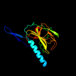
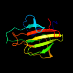
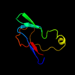
| 1 | c2gu1A_
|
|
 |
100.0 |
31 |
PDB header:hydrolase
Chain: A: PDB Molecule:zinc peptidase;
PDBTitle: crystal structure of a zinc containing peptidase from2 vibrio cholerae
|
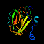
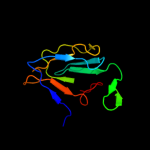
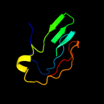
| 2 | c2hsiB_
|
|
 |
100.0 |
23 |
PDB header:structural genomics, unknown function
Chain: B: PDB Molecule:putative peptidase m23;
PDBTitle: crystal structure of putative peptidase m23 from2 pseudomonas aeruginosa, new york structural genomics3 consortium
|
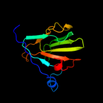
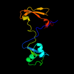
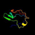
| 3 | d1qwya_
|
|
 |
100.0 |
26 |
Fold:Barrel-sandwich hybrid
Superfamily:Duplicated hybrid motif
Family:Peptidoglycan hydrolase LytM |
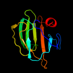
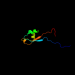
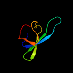
| 4 | c3nyyA_
|
|
 |
100.0 |
25 |
PDB header:hydrolase
Chain: A: PDB Molecule:putative glycyl-glycine endopeptidase lytm;
PDBTitle: crystal structure of a putative glycyl-glycine endopeptidase lytm2 (rumgna_02482) from ruminococcus gnavus atcc 29149 at 1.60 a3 resolution
|
| 5 | c2b44A_
|
|
 |
100.0 |
37 |
PDB header:hydrolase
Chain: A: PDB Molecule:glycyl-glycine endopeptidase lytm;
PDBTitle: truncated s. aureus lytm, p 32 2 1 crystal form
|
| 6 | c3it5B_
|
|
 |
99.9 |
22 |
PDB header:hydrolase
Chain: B: PDB Molecule:protease lasa;
PDBTitle: crystal structure of the lasa virulence factor from pseudomonas2 aeruginosa
|
| 7 | c3csqC_
|
|
 |
99.9 |
19 |
PDB header:hydrolase
Chain: C: PDB Molecule:morphogenesis protein 1;
PDBTitle: crystal and cryoem structural studies of a cell wall2 degrading enzyme in the bacteriophage phi29 tail
|
| 8 | c2l9yA_
|
|
 |
98.8 |
23 |
PDB header:sugar binding protein
Chain: A: PDB Molecule:cvnh-lysm lectin;
PDBTitle: solution structure of the mocvnh-lysm module from the rice blast2 fungus magnaporthe oryzae protein (mgg_03307)
|
| 9 | c2djpA_
|
|
 |
98.6 |
19 |
PDB header:structural genomics, unknown function
Chain: A: PDB Molecule:hypothetical protein sb145;
PDBTitle: the solution structure of the lysm domain of human2 hypothetical protein sb145
|
| 10 | d1e0ga_
|
|
 |
98.3 |
29 |
Fold:LysM domain
Superfamily:LysM domain
Family:LysM domain |
| 11 | d1y7ma2
|
|
 |
98.2 |
31 |
Fold:LysM domain
Superfamily:LysM domain
Family:LysM domain |
| 12 | c1y7mB_
|
|
 |
97.4 |
23 |
PDB header:structural genomics, unknown function
Chain: B: PDB Molecule:hypothetical protein bsu14040;
PDBTitle: crystal structure of the b. subtilis ykud protein at 2 a2 resolution
|
| 13 | d2f3ga_
|
|
 |
96.2 |
20 |
Fold:Barrel-sandwich hybrid
Superfamily:Duplicated hybrid motif
Family:Glucose permease-like |
| 14 | d1glaf_
|
|
 |
95.9 |
20 |
Fold:Barrel-sandwich hybrid
Superfamily:Duplicated hybrid motif
Family:Glucose permease-like |
| 15 | d1gpra_
|
|
 |
95.7 |
29 |
Fold:Barrel-sandwich hybrid
Superfamily:Duplicated hybrid motif
Family:Glucose permease-like |
| 16 | d2gpra_
|
|
 |
95.7 |
24 |
Fold:Barrel-sandwich hybrid
Superfamily:Duplicated hybrid motif
Family:Glucose permease-like |
| 17 | d1e2wa2
|
|
 |
95.6 |
25 |
Fold:Barrel-sandwich hybrid
Superfamily:Rudiment single hybrid motif
Family:Cytochrome f, small domain |
| 18 | d1ci3m2
|
|
 |
95.1 |
19 |
Fold:Barrel-sandwich hybrid
Superfamily:Rudiment single hybrid motif
Family:Cytochrome f, small domain |
| 19 | c2aukA_
|
|
 |
94.1 |
15 |
PDB header:transferase
Chain: A: PDB Molecule:dna-directed rna polymerase beta' chain;
PDBTitle: structure of e. coli rna polymerase beta' g/g' insert
|
| 20 | c1e2vB_
|
|
 |
93.8 |
25 |
PDB header:electron transport proteins
Chain: B: PDB Molecule:cytochrome f;
PDBTitle: n153q mutant of cytochrome f from chlamydomonas reinhardtii
|
| 21 | c1ctmA_ |
|
not modelled |
93.4 |
19 |
PDB header:electron transport(cytochrome)
Chain: A: PDB Molecule:cytochrome f;
PDBTitle: crystal structure of chloroplast cytochrome f reveals a2 novel cytochrome fold and unexpected heme ligation
|
| 22 | c1q90A_ |
|
not modelled |
93.3 |
25 |
PDB header:photosynthesis
Chain: A: PDB Molecule:apocytochrome f;
PDBTitle: structure of the cytochrome b6f (plastohydroquinone : plastocyanin2 oxidoreductase) from chlamydomonas reinhardtii
|
| 23 | d1dcza_ |
|
not modelled |
92.7 |
23 |
Fold:Barrel-sandwich hybrid
Superfamily:Single hybrid motif
Family:Biotinyl/lipoyl-carrier proteins and domains |
| 24 | c2ejgD_ |
|
not modelled |
91.8 |
31 |
PDB header:ligase
Chain: D: PDB Molecule:149aa long hypothetical methylmalonyl-coa decarboxylase
PDBTitle: crystal structure of the biotin protein ligase (mutation r48a) and2 biotin carboxyl carrier protein complex from pyrococcus horikoshii3 ot3
|
| 25 | d1bdoa_ |
|
not modelled |
91.3 |
18 |
Fold:Barrel-sandwich hybrid
Superfamily:Single hybrid motif
Family:Biotinyl/lipoyl-carrier proteins and domains |
| 26 | c2kccA_ |
|
not modelled |
90.6 |
18 |
PDB header:ligase
Chain: A: PDB Molecule:acetyl-coa carboxylase 2;
PDBTitle: solution structure of biotinoyl domain from human acetyl-2 coa carboxylase 2
|
| 27 | c3n6rK_ |
|
not modelled |
90.4 |
28 |
PDB header:ligase
Chain: K: PDB Molecule:propionyl-coa carboxylase, alpha subunit;
PDBTitle: crystal structure of the holoenzyme of propionyl-coa carboxylase (pcc)
|
| 28 | c2b8gA_ |
|
not modelled |
90.4 |
18 |
PDB header:biosynthetic protein
Chain: A: PDB Molecule:biotin/lipoyl attachment protein;
PDBTitle: solution structure of bacillus subtilis blap biotinylated-2 form (energy minimized mean structure)
|
| 29 | d1hcza2 |
|
not modelled |
90.3 |
19 |
Fold:Barrel-sandwich hybrid
Superfamily:Rudiment single hybrid motif
Family:Cytochrome f, small domain |
| 30 | c1t5eB_ |
|
not modelled |
90.1 |
30 |
PDB header:transport protein
Chain: B: PDB Molecule:multidrug resistance protein mexa;
PDBTitle: the structure of mexa
|
| 31 | c2jxmB_ |
|
not modelled |
89.9 |
23 |
PDB header:electron transport
Chain: B: PDB Molecule:cytochrome f;
PDBTitle: ensemble of twenty structures of the prochlorothrix2 hollandica plastocyanin- cytochrome f complex
|
| 32 | d1o78a_ |
|
not modelled |
89.7 |
35 |
Fold:Barrel-sandwich hybrid
Superfamily:Single hybrid motif
Family:Biotinyl/lipoyl-carrier proteins and domains |
| 33 | c3lnnB_ |
|
not modelled |
89.7 |
26 |
PDB header:metal transport
Chain: B: PDB Molecule:membrane fusion protein (mfp) heavy metal cation efflux
PDBTitle: crystal structure of zneb from cupriavidus metallidurans
|
| 34 | c2aujD_ |
|
not modelled |
89.4 |
17 |
PDB header:transferase
Chain: D: PDB Molecule:dna-directed rna polymerase beta' chain;
PDBTitle: structure of thermus aquaticus rna polymerase beta'-subunit2 insert
|
| 35 | c2f1mA_ |
|
not modelled |
89.0 |
13 |
PDB header:transport protein
Chain: A: PDB Molecule:acriflavine resistance protein a;
PDBTitle: conformational flexibility in the multidrug efflux system protein acra
|
| 36 | c1tu2B_ |
|
not modelled |
88.8 |
26 |
PDB header:electron transport
Chain: B: PDB Molecule:apocytochrome f;
PDBTitle: the complex of nostoc cytochrome f and plastocyanin determin with2 paramagnetic nmr. based on the structures of cytochrome f and3 plastocyanin, 10 structures
|
| 37 | c2k33A_ |
|
not modelled |
88.7 |
26 |
PDB header:membrane protein, transport protein
Chain: A: PDB Molecule:acra;
PDBTitle: solution structure of an n-glycosylated protein using in2 vitro glycosylation
|
| 38 | c3fppB_ |
|
not modelled |
88.0 |
26 |
PDB header:membrane protein
Chain: B: PDB Molecule:macrolide-specific efflux protein maca;
PDBTitle: crystal structure of e.coli maca
|
| 39 | d1vf7a_ |
|
not modelled |
87.9 |
30 |
Fold:HlyD-like secretion proteins
Superfamily:HlyD-like secretion proteins
Family:HlyD-like secretion proteins |
| 40 | c2ejmA_ |
|
not modelled |
87.5 |
30 |
PDB header:ligase
Chain: A: PDB Molecule:methylcrotonoyl-coa carboxylase subunit alpha;
PDBTitle: solution structure of ruh-072, an apo-biotnyl domain form2 human acetyl coenzyme a carboxylase
|
| 41 | c2dn8A_ |
|
not modelled |
87.3 |
30 |
PDB header:ligase
Chain: A: PDB Molecule:acetyl-coa carboxylase 2;
PDBTitle: solution structure of rsgi ruh-053, an apo-biotin carboxy2 carrier protein from human transcarboxylase
|
| 42 | d1brwa3 |
|
not modelled |
87.0 |
24 |
Fold:alpha/beta-Hammerhead
Superfamily:Pyrimidine nucleoside phosphorylase C-terminal domain
Family:Pyrimidine nucleoside phosphorylase C-terminal domain |
| 43 | d1laba_ |
|
not modelled |
86.9 |
13 |
Fold:Barrel-sandwich hybrid
Superfamily:Single hybrid motif
Family:Biotinyl/lipoyl-carrier proteins and domains |
| 44 | d1tu2b2 |
|
not modelled |
85.9 |
21 |
Fold:Barrel-sandwich hybrid
Superfamily:Rudiment single hybrid motif
Family:Cytochrome f, small domain |
| 45 | c2q8iB_ |
|
not modelled |
85.7 |
13 |
PDB header:transferase
Chain: B: PDB Molecule:dihydrolipoyllysine-residue acetyltransferase component of
PDBTitle: pyruvate dehydrogenase kinase isoform 3 in complex with antitumor drug2 radicicol
|
| 46 | c2l5tA_ |
|
not modelled |
84.5 |
17 |
PDB header:transferase
Chain: A: PDB Molecule:lipoamide acyltransferase;
PDBTitle: solution nmr structure of e2 lipoyl domain from thermoplasma2 acidophilum
|
| 47 | d1y8ob1 |
|
not modelled |
84.3 |
13 |
Fold:Barrel-sandwich hybrid
Superfamily:Single hybrid motif
Family:Biotinyl/lipoyl-carrier proteins and domains |
| 48 | d1qjoa_ |
|
not modelled |
83.7 |
26 |
Fold:Barrel-sandwich hybrid
Superfamily:Single hybrid motif
Family:Biotinyl/lipoyl-carrier proteins and domains |
| 49 | d2pnrc1 |
|
not modelled |
82.1 |
13 |
Fold:Barrel-sandwich hybrid
Superfamily:Single hybrid motif
Family:Biotinyl/lipoyl-carrier proteins and domains |
| 50 | c2dncA_ |
|
not modelled |
81.0 |
22 |
PDB header:transferase
Chain: A: PDB Molecule:pyruvate dehydrogenase protein x component;
PDBTitle: solution structure of rsgi ruh-054, a lipoyl domain from2 human 2-oxoacid dehydrogenase
|
| 51 | d1ghja_ |
|
not modelled |
80.8 |
16 |
Fold:Barrel-sandwich hybrid
Superfamily:Single hybrid motif
Family:Biotinyl/lipoyl-carrier proteins and domains |
| 52 | c2dneA_ |
|
not modelled |
80.0 |
17 |
PDB header:transferase
Chain: A: PDB Molecule:dihydrolipoyllysine-residue acetyltransferase
PDBTitle: solution structure of rsgi ruh-058, a lipoyl domain of2 human 2-oxoacid dehydrogenase
|
| 53 | c3h9iB_ |
|
not modelled |
78.2 |
17 |
PDB header:transport protein
Chain: B: PDB Molecule:cation efflux system protein cusb;
PDBTitle: crystal structure of the membrane fusion protein cusb from escherichia2 coli
|
| 54 | c2j0fC_ |
|
not modelled |
77.9 |
25 |
PDB header:transferase
Chain: C: PDB Molecule:thymidine phosphorylase;
PDBTitle: structural basis for non-competitive product inhibition in2 human thymidine phosphorylase: implication for drug design
|
| 55 | c1otpA_ |
|
not modelled |
77.7 |
24 |
PDB header:phosphorylase
Chain: A: PDB Molecule:thymidine phosphorylase;
PDBTitle: structural and theoretical studies suggest domain movement produces an2 active conformation of thymidine phosphorylase
|
| 56 | d1uoua3 |
|
not modelled |
77.2 |
26 |
Fold:alpha/beta-Hammerhead
Superfamily:Pyrimidine nucleoside phosphorylase C-terminal domain
Family:Pyrimidine nucleoside phosphorylase C-terminal domain |
| 57 | d1k8ma_ |
|
not modelled |
76.7 |
9 |
Fold:Barrel-sandwich hybrid
Superfamily:Single hybrid motif
Family:Biotinyl/lipoyl-carrier proteins and domains |
| 58 | c2dsjA_ |
|
not modelled |
76.6 |
27 |
PDB header:transferase
Chain: A: PDB Molecule:pyrimidine-nucleoside (thymidine) phosphorylase;
PDBTitle: crystal structure of project id tt0128 from thermus thermophilus hb8
|
| 59 | c2jkuA_ |
|
not modelled |
76.1 |
26 |
PDB header:ligase
Chain: A: PDB Molecule:propionyl-coa carboxylase alpha chain,
PDBTitle: crystal structure of the n-terminal region of the biotin2 acceptor domain of human propionyl-coa carboxylase
|
| 60 | c3fmcC_ |
|
not modelled |
75.7 |
20 |
PDB header:hydrolase
Chain: C: PDB Molecule:putative succinylglutamate desuccinylase / aspartoacylase;
PDBTitle: crystal structure of a putative succinylglutamate desuccinylase /2 aspartoacylase family protein (sama_0604) from shewanella amazonensis3 sb2b at 1.80 a resolution
|
| 61 | c2e75C_ |
|
not modelled |
75.6 |
21 |
PDB header:photosynthesis
Chain: C: PDB Molecule:apocytochrome f;
PDBTitle: crystal structure of the cytochrome b6f complex with 2-nonyl-4-2 hydroxyquinoline n-oxide (nqno) from m.laminosus
|
| 62 | c2qj8B_ |
|
not modelled |
72.6 |
17 |
PDB header:hydrolase
Chain: B: PDB Molecule:mlr6093 protein;
PDBTitle: crystal structure of an aspartoacylase family protein (mlr6093) from2 mesorhizobium loti maff303099 at 2.00 a resolution
|
| 63 | c2qf7A_ |
|
not modelled |
71.9 |
19 |
PDB header:ligase
Chain: A: PDB Molecule:pyruvate carboxylase protein;
PDBTitle: crystal structure of a complete multifunctional pyruvate carboxylase2 from rhizobium etli
|
| 64 | d1pmra_ |
|
not modelled |
71.3 |
19 |
Fold:Barrel-sandwich hybrid
Superfamily:Single hybrid motif
Family:Biotinyl/lipoyl-carrier proteins and domains |
| 65 | c3cdxB_ |
|
not modelled |
70.7 |
21 |
PDB header:hydrolase
Chain: B: PDB Molecule:succinylglutamatedesuccinylase/aspartoacylase;
PDBTitle: crystal structure of2 succinylglutamatedesuccinylase/aspartoacylase from3 rhodobacter sphaeroides
|
| 66 | d1gjxa_ |
|
not modelled |
70.7 |
17 |
Fold:Barrel-sandwich hybrid
Superfamily:Single hybrid motif
Family:Biotinyl/lipoyl-carrier proteins and domains |
| 67 | c3na6A_ |
|
not modelled |
70.1 |
28 |
PDB header:hydrolase
Chain: A: PDB Molecule:succinylglutamate desuccinylase/aspartoacylase;
PDBTitle: crystal structure of a succinylglutamate desuccinylase (tm1040_2694)2 from silicibacter sp. tm1040 at 2.00 a resolution
|
| 68 | d2tpta3 |
|
not modelled |
70.1 |
21 |
Fold:alpha/beta-Hammerhead
Superfamily:Pyrimidine nucleoside phosphorylase C-terminal domain
Family:Pyrimidine nucleoside phosphorylase C-terminal domain |
| 69 | d1mzya2 |
|
not modelled |
65.8 |
11 |
Fold:Cupredoxin-like
Superfamily:Cupredoxins
Family:Multidomain cupredoxins |
| 70 | d1iyua_ |
|
not modelled |
65.8 |
14 |
Fold:Barrel-sandwich hybrid
Superfamily:Single hybrid motif
Family:Biotinyl/lipoyl-carrier proteins and domains |
| 71 | c3ozxA_ |
|
not modelled |
65.4 |
14 |
PDB header:hydrolase, translation
Chain: A: PDB Molecule:rnase l inhibitor;
PDBTitle: crystal structure of abce1 of sulfolubus solfataricus (-fes domain)
|
| 72 | c2xhaB_ |
|
not modelled |
62.1 |
20 |
PDB header:transcription
Chain: B: PDB Molecule:transcription antitermination protein nusg;
PDBTitle: crystal structure of domain 2 of thermotoga maritima n-utilization2 substance g (nusg)
|
| 73 | c2dafA_
|
|
 |
54.6 |
30 |
PDB header:structural genomics, unknown function
Chain: A: PDB Molecule:flj35834 protein;
PDBTitle: solution structure of the novel identified ubiquitin-like2 domain in the human hypothetical protein flj35834
|
| 74 | c3h5qA_ |
|
not modelled |
54.0 |
21 |
PDB header:transferase
Chain: A: PDB Molecule:pyrimidine-nucleoside phosphorylase;
PDBTitle: crystal structure of a putative pyrimidine-nucleoside phosphorylase2 from staphylococcus aureus
|
| 75 | c1brwB_ |
|
not modelled |
53.7 |
26 |
PDB header:transferase
Chain: B: PDB Molecule:protein (pyrimidine nucleoside phosphorylase);
PDBTitle: the crystal structure of pyrimidine nucleoside2 phosphorylase in a closed conformation
|
| 76 | d1o4ua2 |
|
not modelled |
49.5 |
16 |
Fold:alpha/beta-Hammerhead
Superfamily:Nicotinate/Quinolinate PRTase N-terminal domain-like
Family:NadC N-terminal domain-like |
| 77 | d1qapa2 |
|
not modelled |
45.4 |
26 |
Fold:alpha/beta-Hammerhead
Superfamily:Nicotinate/Quinolinate PRTase N-terminal domain-like
Family:NadC N-terminal domain-like |
| 78 | d1k8ga2 |
|
not modelled |
41.4 |
19 |
Fold:OB-fold
Superfamily:Nucleic acid-binding proteins
Family:Single strand DNA-binding domain, SSB |
| 79 | d1qpoa2 |
|
not modelled |
39.5 |
26 |
Fold:alpha/beta-Hammerhead
Superfamily:Nicotinate/Quinolinate PRTase N-terminal domain-like
Family:NadC N-terminal domain-like |
| 80 | d1jb7a2 |
|
not modelled |
39.3 |
19 |
Fold:OB-fold
Superfamily:Nucleic acid-binding proteins
Family:Single strand DNA-binding domain, SSB |
| 81 | c2xhcA_ |
|
not modelled |
38.7 |
20 |
PDB header:transcription
Chain: A: PDB Molecule:transcription antitermination protein nusg;
PDBTitle: crystal structure of thermotoga maritima n-utilization substance g2 (nusg)
|
| 82 | c3camB_ |
|
not modelled |
36.3 |
26 |
PDB header:gene regulation
Chain: B: PDB Molecule:cold-shock domain family protein;
PDBTitle: crystal structure of the cold shock domain protein from neisseria2 meningitidis
|
| 83 | c3a5dB_ |
|
not modelled |
34.5 |
21 |
PDB header:hydrolase
Chain: B: PDB Molecule:v-type atp synthase alpha chain;
PDBTitle: inter-subunit interaction and quaternary rearrangement2 defined by the central stalk of prokaryotic v1-atpase
|
| 84 | d1ndsa2 |
|
not modelled |
33.0 |
17 |
Fold:Cupredoxin-like
Superfamily:Cupredoxins
Family:Multidomain cupredoxins |
| 85 | d2ns0a1 |
|
not modelled |
29.9 |
13 |
Fold:DNA/RNA-binding 3-helical bundle
Superfamily:"Winged helix" DNA-binding domain
Family:RHA1 ro06458-like |
| 86 | d2zjrt1 |
|
not modelled |
28.3 |
28 |
Fold:Barrel-sandwich hybrid
Superfamily:Ribosomal L27 protein-like
Family:Ribosomal L27 protein |
| 87 | d1v8qa_ |
|
not modelled |
28.1 |
19 |
Fold:Barrel-sandwich hybrid
Superfamily:Ribosomal L27 protein-like
Family:Ribosomal L27 protein |
| 88 | c3fin0_ |
|
not modelled |
25.6 |
20 |
PDB header:ribosome
Chain: 0: PDB Molecule:50s ribosomal protein l27;
PDBTitle: t. thermophilus 70s ribosome in complex with mrna, trnas2 and ef-tu.gdp.kirromycin ternary complex, fitted to a 6.43 a cryo-em map. this file contains the 50s subunit.
|
| 89 | c2boyC_ |
|
not modelled |
25.3 |
24 |
PDB header:oxidoreductase
Chain: C: PDB Molecule:3-chlorocatechol 1,2-dioxygenase;
PDBTitle: crystal structure of 3-chlorocatechol 1,2-dioxygenase from2 rhodococcus opacus 1cp
|
| 90 | d1vf5c2 |
|
not modelled |
24.3 |
44 |
Fold:Barrel-sandwich hybrid
Superfamily:Rudiment single hybrid motif
Family:Cytochrome f, small domain |
| 91 | d1snra2 |
|
not modelled |
24.2 |
16 |
Fold:Cupredoxin-like
Superfamily:Cupredoxins
Family:Multidomain cupredoxins |
| 92 | c2elmA_ |
|
not modelled |
24.0 |
27 |
PDB header:transcription
Chain: A: PDB Molecule:zinc finger protein 406;
PDBTitle: solution structure of the 10th c2h2 zinc finger of human2 zinc finger protein 406
|
| 93 | d3pcca_ |
|
not modelled |
23.5 |
9 |
Fold:Prealbumin-like
Superfamily:Aromatic compound dioxygenase
Family:Aromatic compound dioxygenase |
| 94 | c2kw8A_ |
|
not modelled |
23.5 |
3 |
PDB header:protein binding
Chain: A: PDB Molecule:lpxtg-site transpeptidase family protein;
PDBTitle: solution structure of bacillus anthracis sortase a (srta)2 transpeptidase
|
| 95 | d2awna2 |
|
not modelled |
23.1 |
12 |
Fold:P-loop containing nucleoside triphosphate hydrolases
Superfamily:P-loop containing nucleoside triphosphate hydrolases
Family:ABC transporter ATPase domain-like |
| 96 | d2bura1 |
|
not modelled |
22.7 |
18 |
Fold:Prealbumin-like
Superfamily:Aromatic compound dioxygenase
Family:Aromatic compound dioxygenase |
| 97 | d2es2a1 |
|
not modelled |
22.5 |
28 |
Fold:OB-fold
Superfamily:Nucleic acid-binding proteins
Family:Cold shock DNA-binding domain-like |
| 98 | c2yz2B_ |
|
not modelled |
22.2 |
18 |
PDB header:hydrolase
Chain: B: PDB Molecule:putative abc transporter atp-binding protein tm_0222;
PDBTitle: crystal structure of the abc transporter in the cobalt transport2 system
|
| 99 | d2burb1 |
|
not modelled |
21.6 |
18 |
Fold:Prealbumin-like
Superfamily:Aromatic compound dioxygenase
Family:Aromatic compound dioxygenase |