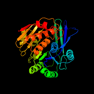
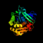
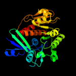
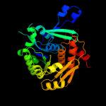
| 1 | c2jg1C_
|
|
 |
100.0 |
25 |
PDB header:transferase
Chain: C: PDB Molecule:tagatose-6-phosphate kinase;
PDBTitle: structure of staphylococcus aureus d-tagatose-6-phosphate2 kinase with cofactor and substrate
|
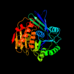
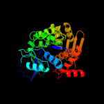
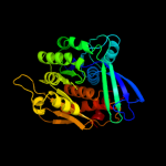
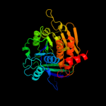
| 2 | c2jg5B_
|
|
 |
100.0 |
29 |
PDB header:transferase
Chain: B: PDB Molecule:fructose 1-phosphate kinase;
PDBTitle: crystal structure of a putative phosphofructokinase from2 staphylococcus aureus
|
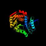
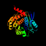
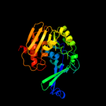
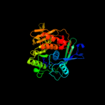
| 3 | d2f02a1
|
|
 |
100.0 |
25 |
Fold:Ribokinase-like
Superfamily:Ribokinase-like
Family:Ribokinase-like |
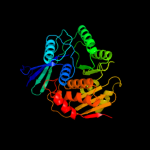
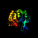
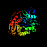
| 4 | c3cqdB_
|
|
 |
100.0 |
28 |
PDB header:transferase
Chain: B: PDB Molecule:6-phosphofructokinase isozyme 2;
PDBTitle: structure of the tetrameric inhibited form of2 phosphofructokinase-2 from escherichia coli
|
| 5 | d2abqa1
|
|
 |
100.0 |
28 |
Fold:Ribokinase-like
Superfamily:Ribokinase-like
Family:Ribokinase-like |
| 6 | d2ajra1
|
|
 |
100.0 |
24 |
Fold:Ribokinase-like
Superfamily:Ribokinase-like
Family:Ribokinase-like |
| 7 | c3julA_
|
|
 |
100.0 |
27 |
PDB header:transferase
Chain: A: PDB Molecule:lin2199 protein;
PDBTitle: crystal structure of listeria innocua d-tagatose-6-phosphate2 kinase bound with substrate
|
| 8 | d1rkda_
|
|
 |
100.0 |
21 |
Fold:Ribokinase-like
Superfamily:Ribokinase-like
Family:Ribokinase-like |
| 9 | d1vm7a_
|
|
 |
100.0 |
23 |
Fold:Ribokinase-like
Superfamily:Ribokinase-like
Family:Ribokinase-like |
| 10 | c3kzhA_
|
|
 |
100.0 |
21 |
PDB header:transferase
Chain: A: PDB Molecule:probable sugar kinase;
PDBTitle: crystal structure of a putative sugar kinase from2 clostridium perfringens
|
| 11 | c3pl2D_
|
|
 |
100.0 |
21 |
PDB header:transferase
Chain: D: PDB Molecule:sugar kinase, ribokinase family;
PDBTitle: crystal structure of a 5-keto-2-deoxygluconokinase (ncgl0155, cgl0158)2 from corynebacterium glutamicum atcc 13032 kitasato at 1.89 a3 resolution
|
| 12 | c2rbcA_
|
|
 |
100.0 |
19 |
PDB header:transferase
Chain: A: PDB Molecule:sugar kinase;
PDBTitle: crystal structure of a putative ribokinase from agrobacterium2 tumefaciens
|
| 13 | c2nwhA_
|
|
 |
100.0 |
18 |
PDB header:signaling protein,transferase
Chain: A: PDB Molecule:carbohydrate kinase;
PDBTitle: carbohydrate kinase from agrobacterium tumefaciens
|
| 14 | d1bx4a_
|
|
 |
100.0 |
17 |
Fold:Ribokinase-like
Superfamily:Ribokinase-like
Family:Ribokinase-like |
| 15 | c2pkkA_
|
|
 |
100.0 |
20 |
PDB header:transferase
Chain: A: PDB Molecule:adenosine kinase;
PDBTitle: crystal structure of m tuberculosis adenosine kinase complexed with 2-2 fluro adenosine
|
| 16 | c3go6B_
|
|
 |
100.0 |
24 |
PDB header:transferase
Chain: B: PDB Molecule:ribokinase rbsk;
PDBTitle: crystal structure of m. tuberculosis ribokinase (rv2436) in2 complex with ribose and amp-pnp
|
| 17 | c2xtbA_
|
|
 |
100.0 |
16 |
PDB header:transferase
Chain: A: PDB Molecule:adenosine kinase;
PDBTitle: crystal structure of trypanosoma brucei rhodesiense2 adenosine kinase complexed with activator
|
| 18 | c2qcvA_
|
|
 |
100.0 |
19 |
PDB header:transferase
Chain: A: PDB Molecule:putative 5-dehydro-2-deoxygluconokinase;
PDBTitle: crystal structure of a putative 5-dehydro-2-deoxygluconokinase (iolc)2 from bacillus halodurans c-125 at 1.90 a resolution
|
| 19 | c2c49A_
|
|
 |
100.0 |
14 |
PDB header:transferase
Chain: A: PDB Molecule:sugar kinase mj0406;
PDBTitle: crystal structure of methanocaldococcus jannaschii2 nucleoside kinase - an archaeal member of the ribokinase3 family
|
| 20 | d1v19a_
|
|
 |
100.0 |
24 |
Fold:Ribokinase-like
Superfamily:Ribokinase-like
Family:Ribokinase-like |
| 21 | c3looC_ |
|
not modelled |
100.0 |
15 |
PDB header:transferase
Chain: C: PDB Molecule:anopheles gambiae adenosine kinase;
PDBTitle: crystal structure of anopheles gambiae adenosine kinase in complex2 with p1,p4-di(adenosine-5) tetraphosphate
|
| 22 | c3iq0B_ |
|
not modelled |
100.0 |
19 |
PDB header:transferase
Chain: B: PDB Molecule:putative ribokinase ii;
PDBTitle: crystal structure of a putative ribokinase ii in complex2 with atp and mg+2 from e.coli
|
| 23 | d2fv7a1 |
|
not modelled |
100.0 |
21 |
Fold:Ribokinase-like
Superfamily:Ribokinase-like
Family:Ribokinase-like |
| 24 | c3in1A_ |
|
not modelled |
100.0 |
20 |
PDB header:transferase
Chain: A: PDB Molecule:uncharacterized sugar kinase ydjh;
PDBTitle: crystal structure of a putative ribokinase in complex with2 adp from e.coli
|
| 25 | c3b1qD_ |
|
not modelled |
100.0 |
19 |
PDB header:transferase
Chain: D: PDB Molecule:ribokinase, putative;
PDBTitle: structure of burkholderia thailandensis nucleoside kinase (bthnk) in2 complex with inosine
|
| 26 | d2afba1 |
|
not modelled |
100.0 |
17 |
Fold:Ribokinase-like
Superfamily:Ribokinase-like
Family:Ribokinase-like |
| 27 | c3i3yB_ |
|
not modelled |
100.0 |
20 |
PDB header:transferase
Chain: B: PDB Molecule:carbohydrate kinase;
PDBTitle: crystal structure of ribokinase in complex with d-ribose from2 klebsiella pneumoniae
|
| 28 | d2absa1 |
|
not modelled |
100.0 |
21 |
Fold:Ribokinase-like
Superfamily:Ribokinase-like
Family:Ribokinase-like |
| 29 | c2absA_ |
|
not modelled |
100.0 |
21 |
PDB header:signaling protein,transferase
Chain: A: PDB Molecule:adenosine kinase;
PDBTitle: crystal structure of t. gondii adenosine kinase complexed2 with amp-pcp
|
| 30 | c3ktnA_ |
|
not modelled |
100.0 |
16 |
PDB header:transferase
Chain: A: PDB Molecule:carbohydrate kinase, pfkb family;
PDBTitle: crystal structure of a putative 2-keto-3-deoxygluconate2 kinase from enterococcus faecalis
|
| 31 | c2varB_ |
|
not modelled |
100.0 |
15 |
PDB header:transferase
Chain: B: PDB Molecule:fructokinase;
PDBTitle: crystal structure of sulfolobus solfataricus 2-keto-3-2 deoxygluconate kinase complexed with 2-keto-3-3 deoxygluconate
|
| 32 | d2dcna1 |
|
not modelled |
100.0 |
16 |
Fold:Ribokinase-like
Superfamily:Ribokinase-like
Family:Ribokinase-like |
| 33 | c3bf5A_ |
|
not modelled |
100.0 |
17 |
PDB header:transferase
Chain: A: PDB Molecule:ribokinase related protein;
PDBTitle: crystal structure of putative ribokinase (10640157) from thermoplasma2 acidophilum at 1.91 a resolution
|
| 34 | c3b3lC_ |
|
not modelled |
100.0 |
16 |
PDB header:transferase
Chain: C: PDB Molecule:ketohexokinase;
PDBTitle: crystal structures of alternatively-spliced isoforms of human2 ketohexokinase
|
| 35 | d1tyya_ |
|
not modelled |
100.0 |
28 |
Fold:Ribokinase-like
Superfamily:Ribokinase-like
Family:Ribokinase-like |
| 36 | c3gbuD_ |
|
not modelled |
100.0 |
19 |
PDB header:transferase
Chain: D: PDB Molecule:uncharacterized sugar kinase ph1459;
PDBTitle: crystal structure of an uncharacterized sugar kinase ph1459 from2 pyrococcus horikoshii in complex with atp
|
| 37 | c3lhxA_ |
|
not modelled |
100.0 |
18 |
PDB header:transferase
Chain: A: PDB Molecule:ketodeoxygluconokinase;
PDBTitle: crystal structure of a ketodeoxygluconokinase (kdgk) from2 shigella flexneri
|
| 38 | c3lkiA_ |
|
not modelled |
100.0 |
17 |
PDB header:transferase
Chain: A: PDB Molecule:fructokinase;
PDBTitle: crystal structure of fructokinase with bound atp from2 xylella fastidiosa
|
| 39 | c2qhpA_ |
|
not modelled |
100.0 |
19 |
PDB header:transferase
Chain: A: PDB Molecule:fructokinase;
PDBTitle: crystal structure of fructokinase (np_810670.1) from bacteroides2 thetaiotaomicron vpi-5482 at 1.80 a resolution
|
| 40 | c1tz6B_ |
|
not modelled |
100.0 |
29 |
PDB header:transferase
Chain: B: PDB Molecule:putative sugar kinase;
PDBTitle: crystal structure of aminoimidazole riboside kinase from2 salmonella enterica complexed with aminoimidazole riboside3 and atp analog
|
| 41 | c3kd6B_ |
|
not modelled |
100.0 |
19 |
PDB header:transferase
Chain: B: PDB Molecule:carbohydrate kinase, pfkb family;
PDBTitle: crystal structure of nucleoside kinase from chlorobium tepidum in2 complex with amp
|
| 42 | d1vk4a_ |
|
not modelled |
100.0 |
17 |
Fold:Ribokinase-like
Superfamily:Ribokinase-like
Family:Ribokinase-like |
| 43 | c3hj6B_ |
|
not modelled |
100.0 |
21 |
PDB header:transferase
Chain: B: PDB Molecule:fructokinase;
PDBTitle: structure of halothermothrix orenii fructokinase (frk)
|
| 44 | c2ddmA_ |
|
not modelled |
99.9 |
20 |
PDB header:transferase
Chain: A: PDB Molecule:pyridoxine kinase;
PDBTitle: crystal structure of pyridoxal kinase from the escherichia2 coli pdxk gene at 2.1 a resolution
|
| 45 | d1vi9a_ |
|
not modelled |
99.8 |
24 |
Fold:Ribokinase-like
Superfamily:Ribokinase-like
Family:PfkB-like kinase |
| 46 | c3mbjA_ |
|
not modelled |
99.8 |
16 |
PDB header:transferase
Chain: A: PDB Molecule:putative phosphomethylpyrimidine kinase;
PDBTitle: crystal structure of a putative phosphomethylpyrimidine kinase2 (bt_4458) from bacteroides thetaiotaomicron vpi-5482 at 2.10 a3 resolution (rhombohedral form)
|
| 47 | d1lhpa_ |
|
not modelled |
99.8 |
17 |
Fold:Ribokinase-like
Superfamily:Ribokinase-like
Family:PfkB-like kinase |
| 48 | d1ub0a_ |
|
not modelled |
99.7 |
23 |
Fold:Ribokinase-like
Superfamily:Ribokinase-like
Family:Thiamin biosynthesis kinases |
| 49 | c2i5bC_ |
|
not modelled |
99.7 |
19 |
PDB header:transferase
Chain: C: PDB Molecule:phosphomethylpyrimidine kinase;
PDBTitle: the crystal structure of an adp complex of bacillus2 subtilis pyridoxal kinase provides evidence for the3 parralel emergence of enzyme activity during evolution
|
| 50 | c3ibqA_ |
|
not modelled |
99.7 |
18 |
PDB header:transferase
Chain: A: PDB Molecule:pyridoxal kinase;
PDBTitle: crystal structure of pyridoxal kinase from lactobacillus2 plantarum in complex with atp
|
| 51 | c3rm5B_ |
|
not modelled |
99.6 |
15 |
PDB header:transferase
Chain: B: PDB Molecule:hydroxymethylpyrimidine/phosphomethylpyrimidine kinase
PDBTitle: structure of trifunctional thi20 from yeast
|
| 52 | c3dzvB_ |
|
not modelled |
99.5 |
14 |
PDB header:transferase
Chain: B: PDB Molecule:4-methyl-5-(beta-hydroxyethyl)thiazole kinase;
PDBTitle: crystal structure of 4-methyl-5-(beta-hydroxyethyl)thiazole2 kinase (np_816404.1) from enterococcus faecalis v583 at3 2.57 a resolution
|
| 53 | d1v8aa_ |
|
not modelled |
99.4 |
10 |
Fold:Ribokinase-like
Superfamily:Ribokinase-like
Family:Thiamin biosynthesis kinases |
| 54 | d1jxha_ |
|
not modelled |
99.4 |
19 |
Fold:Ribokinase-like
Superfamily:Ribokinase-like
Family:Thiamin biosynthesis kinases |
| 55 | d1kyha_ |
|
not modelled |
99.2 |
16 |
Fold:Ribokinase-like
Superfamily:Ribokinase-like
Family:YjeF C-terminal domain-like |
| 56 | d2ax3a1 |
|
not modelled |
99.0 |
15 |
Fold:Ribokinase-like
Superfamily:Ribokinase-like
Family:YjeF C-terminal domain-like |
| 57 | d1ekqa_ |
|
not modelled |
99.0 |
16 |
Fold:Ribokinase-like
Superfamily:Ribokinase-like
Family:Thiamin biosynthesis kinases |
| 58 | c2ax3A_ |
|
not modelled |
98.9 |
14 |
PDB header:transferase
Chain: A: PDB Molecule:hypothetical protein tm0922;
PDBTitle: crystal structure of a putative carbohydrate kinase (tm0922) from2 thermotoga maritima msb8 at 2.25 a resolution
|
| 59 | d1gc5a_ |
|
not modelled |
98.8 |
18 |
Fold:Ribokinase-like
Superfamily:Ribokinase-like
Family:ADP-specific Phosphofructokinase/Glucokinase |
| 60 | c2r3bA_ |
|
not modelled |
98.8 |
13 |
PDB header:transferase
Chain: A: PDB Molecule:yjef-related protein;
PDBTitle: crystal structure of a ribokinase-like superfamily protein (ef1790)2 from enterococcus faecalis v583 at 1.80 a resolution
|
| 61 | c3bgkA_ |
|
not modelled |
98.7 |
14 |
PDB header:unknown function
Chain: A: PDB Molecule:putative uncharacterized protein;
PDBTitle: the crystal structure of hypothetic protein smu.573 from2 streptococcus mutans
|
| 62 | d1l2la_ |
|
not modelled |
98.6 |
14 |
Fold:Ribokinase-like
Superfamily:Ribokinase-like
Family:ADP-specific Phosphofructokinase/Glucokinase |
| 63 | d1ua4a_ |
|
not modelled |
98.6 |
18 |
Fold:Ribokinase-like
Superfamily:Ribokinase-like
Family:ADP-specific Phosphofructokinase/Glucokinase |
| 64 | c3nm3D_ |
|
not modelled |
98.6 |
14 |
PDB header:transferase
Chain: D: PDB Molecule:thiamine biosynthetic bifunctional enzyme;
PDBTitle: the crystal structure of candida glabrata thi6, a bifunctional enzyme2 involved in thiamin biosyhthesis of eukaryotes
|
| 65 | d1u2xa_ |
|
not modelled |
98.6 |
17 |
Fold:Ribokinase-like
Superfamily:Ribokinase-like
Family:ADP-specific Phosphofructokinase/Glucokinase |
| 66 | c3drwA_ |
|
not modelled |
98.5 |
16 |
PDB header:transferase
Chain: A: PDB Molecule:adp-specific phosphofructokinase;
PDBTitle: crystal structure of a phosphofructokinase from pyrococcus2 horikoshii ot3 with amp
|
| 67 | c3k5wA_ |
|
not modelled |
98.3 |
13 |
PDB header:transferase
Chain: A: PDB Molecule:carbohydrate kinase;
PDBTitle: crystal structure of a carbohydrate kinase (yjef family)from2 helicobacter pylori
|
| 68 | c3k96B_ |
|
not modelled |
58.2 |
14 |
PDB header:oxidoreductase
Chain: B: PDB Molecule:glycerol-3-phosphate dehydrogenase [nad(p)+];
PDBTitle: 2.1 angstrom resolution crystal structure of glycerol-3-phosphate2 dehydrogenase (gpsa) from coxiella burnetii
|
| 69 | c2vawA_ |
|
not modelled |
49.6 |
11 |
PDB header:cell cycle
Chain: A: PDB Molecule:cell division protein ftsz;
PDBTitle: ftsz pseudomonas aeruginosa gdp
|
| 70 | d1djqa2 |
|
not modelled |
44.4 |
15 |
Fold:FAD/NAD(P)-binding domain
Superfamily:FAD/NAD(P)-binding domain
Family:C-terminal domain of adrenodoxin reductase-like |
| 71 | c3d8xB_ |
|
not modelled |
43.1 |
16 |
PDB header:oxidoreductase
Chain: B: PDB Molecule:thioredoxin reductase 1;
PDBTitle: crystal structure of saccharomyces cerevisiae nadph dependent2 thioredoxin reductase 1
|
| 72 | d1ofua1 |
|
not modelled |
38.1 |
11 |
Fold:Tubulin nucleotide-binding domain-like
Superfamily:Tubulin nucleotide-binding domain-like
Family:Tubulin, GTPase domain |
| 73 | d2d5ba2 |
|
not modelled |
32.0 |
12 |
Fold:Adenine nucleotide alpha hydrolase-like
Superfamily:Nucleotidylyl transferase
Family:Class I aminoacyl-tRNA synthetases (RS), catalytic domain |
| 74 | c2dlnA_ |
|
not modelled |
31.9 |
20 |
PDB header:ligase(peptidoglycan synthesis)
Chain: A: PDB Molecule:d-alanine--d-alanine ligase;
PDBTitle: vancomycin resistance: structure of d-alanine:d-alanine2 ligase at 2.3 angstroms resolution
|
| 75 | d1d7ya1 |
|
not modelled |
31.8 |
18 |
Fold:FAD/NAD(P)-binding domain
Superfamily:FAD/NAD(P)-binding domain
Family:FAD/NAD-linked reductases, N-terminal and central domains |
| 76 | d1rq2a1 |
|
not modelled |
31.2 |
18 |
Fold:Tubulin nucleotide-binding domain-like
Superfamily:Tubulin nucleotide-binding domain-like
Family:Tubulin, GTPase domain |
| 77 | d2obba1 |
|
not modelled |
30.7 |
8 |
Fold:HAD-like
Superfamily:HAD-like
Family:BT0820-like |
| 78 | c1gqqA_ |
|
not modelled |
28.4 |
14 |
PDB header:cell wall biosynthesis
Chain: A: PDB Molecule:udp-n-acetylmuramate-l-alanine ligase;
PDBTitle: murc - crystal structure of the apo-enzyme from haemophilus2 influenzae
|
| 79 | c1u5tB_ |
|
not modelled |
27.4 |
14 |
PDB header:transport protein
Chain: B: PDB Molecule:defective in vacuolar protein sorting; vps36p;
PDBTitle: structure of the escrt-ii endosomal trafficking complex
|
| 80 | c1ofuB_ |
|
not modelled |
27.4 |
11 |
PDB header:bacterial cell division inhibitor
Chain: B: PDB Molecule:cell division protein ftsz;
PDBTitle: crystal structure of sula:ftsz from pseudomonas aeruginosa
|
| 81 | c1w7pD_ |
|
not modelled |
27.3 |
14 |
PDB header:protein transport
Chain: D: PDB Molecule:vps36p, ylr417w;
PDBTitle: the crystal structure of endosomal complex escrt-ii2 (vps22/vps25/vps36)
|
| 82 | c3sc6F_ |
|
not modelled |
25.7 |
23 |
PDB header:oxidoreductase
Chain: F: PDB Molecule:dtdp-4-dehydrorhamnose reductase;
PDBTitle: 2.65 angstrom resolution crystal structure of dtdp-4-dehydrorhamnose2 reductase (rfbd) from bacillus anthracis str. ames in complex with3 nadp
|
| 83 | d1nhpa2 |
|
not modelled |
23.6 |
28 |
Fold:FAD/NAD(P)-binding domain
Superfamily:FAD/NAD(P)-binding domain
Family:FAD/NAD-linked reductases, N-terminal and central domains |
| 84 | c1w59B_ |
|
not modelled |
22.5 |
14 |
PDB header:cell division
Chain: B: PDB Molecule:cell division protein ftsz homolog 1;
PDBTitle: ftsz dimer, empty (m. jannaschii)
|
| 85 | c2zj3A_ |
|
not modelled |
20.5 |
9 |
PDB header:transferase
Chain: A: PDB Molecule:glucosamine--fructose-6-phosphate
PDBTitle: isomerase domain of human glucose:fructose-6-phosphate2 amidotransferase
|
| 86 | c1v59B_ |
|
not modelled |
20.2 |
29 |
PDB header:oxidoreductase
Chain: B: PDB Molecule:dihydrolipoamide dehydrogenase;
PDBTitle: crystal structure of yeast lipoamide dehydrogenase2 complexed with nad+
|
| 87 | c3crcB_ |
|
not modelled |
19.7 |
19 |
PDB header:hydrolase
Chain: B: PDB Molecule:protein mazg;
PDBTitle: crystal structure of escherichia coli mazg, the regulator2 of nutritional stress response
|
| 88 | d2vapa1 |
|
not modelled |
19.2 |
16 |
Fold:Tubulin nucleotide-binding domain-like
Superfamily:Tubulin nucleotide-binding domain-like
Family:Tubulin, GTPase domain |
| 89 | d1y5ea1 |
|
not modelled |
18.7 |
18 |
Fold:Molybdenum cofactor biosynthesis proteins
Superfamily:Molybdenum cofactor biosynthesis proteins
Family:MogA-like |
| 90 | c2puwA_ |
|
not modelled |
18.2 |
13 |
PDB header:transferase
Chain: A: PDB Molecule:isomerase domain of glutamine-fructose-6-phosphate
PDBTitle: the crystal structure of isomerase domain of glucosamine-6-phosphate2 synthase from candida albicans
|
| 91 | c2f00A_ |
|
not modelled |
17.2 |
12 |
PDB header:ligase
Chain: A: PDB Molecule:udp-n-acetylmuramate--l-alanine ligase;
PDBTitle: escherichia coli murc
|
| 92 | d1ztda1 |
|
not modelled |
16.7 |
20 |
Fold:RNase III domain-like
Superfamily:RNase III domain-like
Family:PF0609-like |
| 93 | d1w5fa1 |
|
not modelled |
16.3 |
14 |
Fold:Tubulin nucleotide-binding domain-like
Superfamily:Tubulin nucleotide-binding domain-like
Family:Tubulin, GTPase domain |
| 94 | d1p3da1 |
|
not modelled |
15.9 |
17 |
Fold:MurCD N-terminal domain
Superfamily:MurCD N-terminal domain
Family:MurCD N-terminal domain |
| 95 | c2nm0B_ |
|
not modelled |
15.9 |
35 |
PDB header:oxidoreductase
Chain: B: PDB Molecule:probable 3-oxacyl-(acyl-carrier-protein) reductase;
PDBTitle: crystal structure of sco1815: a beta-ketoacyl-acyl carrier protein2 reductase from streptomyces coelicolor a3(2)
|
| 96 | d2gtad1 |
|
not modelled |
15.4 |
15 |
Fold:all-alpha NTP pyrophosphatases
Superfamily:all-alpha NTP pyrophosphatases
Family:MazG-like |
| 97 | c2gr2A_ |
|
not modelled |
13.9 |
25 |
PDB header:oxidoreductase
Chain: A: PDB Molecule:ferredoxin reductase;
PDBTitle: crystal structure of ferredoxin reductase, bpha4 (oxidized form)
|
| 98 | d1ml4a2 |
|
not modelled |
13.9 |
9 |
Fold:ATC-like
Superfamily:Aspartate/ornithine carbamoyltransferase
Family:Aspartate/ornithine carbamoyltransferase |
| 99 | c2is8A_ |
|
not modelled |
13.8 |
9 |
PDB header:structural protein
Chain: A: PDB Molecule:molybdopterin biosynthesis enzyme, moab;
PDBTitle: crystal structure of the molybdopterin biosynthesis enzyme moab2 (ttha0341) from thermus theromophilus hb8
|