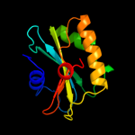
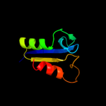
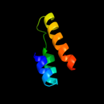
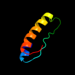
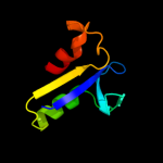
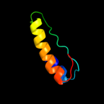
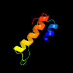
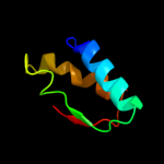
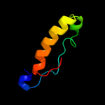
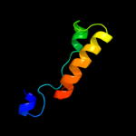
1 c2qlcC_
100.0
39
PDB header: dna binding proteinChain: C: PDB Molecule: dna repair protein radc homolog;PDBTitle: the crystal structure of dna repair protein radc from chlorobium2 tepidum tls
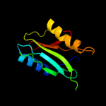
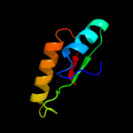
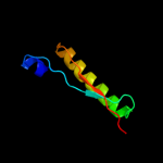
2 d1oi0a_
96.7
21
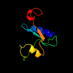
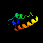
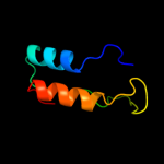
Fold: Cytidine deaminase-likeSuperfamily: JAB1/MPN domainFamily: JAB1/MPN domain3 c2kcqA_
95.3
15
PDB header: structural genomics, unknown functionChain: A: PDB Molecule: mov34/mpn/pad-1 family;PDBTitle: solution structure of protein sru_2040 from salinibacter2 ruber (strain dsm 13855) . northeast structural genomics3 consortium target srr106
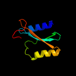
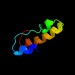
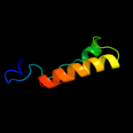
4 c2kksA_
94.9
18
PDB header: structural genomics, unknown functionChain: A: PDB Molecule: uncharacterized protein;PDBTitle: solution structure of protein dsy2949 from desulfitobacterium2 hafniense. northeast structural genomics consortium target dhr27
5 c2w6rA_
80.2
11
PDB header: lyaseChain: A: PDB Molecule: imidazole glycerol phosphate synthase subunitPDBTitle: crystal structure of an artificial (ba)8-barrel protein2 designed from identical half barrels
6 c3q94B_
60.3
18
PDB header: lyaseChain: B: PDB Molecule: fructose-bisphosphate aldolase, class ii;PDBTitle: the crystal structure of fructose 1,6-bisphosphate aldolase from2 bacillus anthracis str. 'ames ancestor'
7 d1gvfa_
58.2
16
Fold: TIM beta/alpha-barrelSuperfamily: AldolaseFamily: Class II FBP aldolase8 c3tdmD_
57.0
10
PDB header: de novo proteinChain: D: PDB Molecule: computationally designed two-fold symmetric tim-barrelPDBTitle: computationally designed tim-barrel protein, halfflr
9 d2csua1
56.8
13
Fold: NAD(P)-binding Rossmann-fold domainsSuperfamily: NAD(P)-binding Rossmann-fold domainsFamily: CoA-binding domain10 c3c52B_
55.4
14
PDB header: lyaseChain: B: PDB Molecule: fructose-bisphosphate aldolase;PDBTitle: class ii fructose-1,6-bisphosphate aldolase from2 helicobacter pylori in complex with3 phosphoglycolohydroxamic acid, a competitive inhibitor
11 c2yciX_
54.5
17
PDB header: transferaseChain: X: PDB Molecule: 5-methyltetrahydrofolate corrinoid/iron sulfur proteinPDBTitle: methyltransferase native
12 c3pm6B_
54.3
20
PDB header: lyaseChain: B: PDB Molecule: putative fructose-bisphosphate aldolase;PDBTitle: crystal structure of a putative fructose-1,6-biphosphate aldolase from2 coccidioides immitis solved by combined sad mr
13 c2iswB_
54.2
20
PDB header: lyaseChain: B: PDB Molecule: putative fructose-1,6-bisphosphate aldolase;PDBTitle: structure of giardia fructose-1,6-biphosphate aldolase in2 complex with phosphoglycolohydroxamate
14 d1rvga_
51.2
25
Fold: TIM beta/alpha-barrelSuperfamily: AldolaseFamily: Class II FBP aldolase15 c3jrkG_
43.5
19
PDB header: lyaseChain: G: PDB Molecule: tagatose 1,6-diphosphate aldolase 2;PDBTitle: a putative tagatose 1,6-diphosphate aldolase from streptococcus2 pyogenes
16 d1hl9a2
40.2
19
Fold: TIM beta/alpha-barrelSuperfamily: (Trans)glycosidasesFamily: Putative alpha-L-fucosidase, catalytic domain17 c3elfA_
35.3
14
PDB header: lyaseChain: A: PDB Molecule: fructose-bisphosphate aldolase;PDBTitle: structural characterization of tetrameric mycobacterium tuberculosis2 fructose 1,6-bisphosphate aldolase - substrate binding and catalysis3 mechanism of a class iia bacterial aldolase
18 c3e49A_
34.8
25
PDB header: metal binding proteinChain: A: PDB Molecule: uncharacterized protein duf849 with a tim barrel fold;PDBTitle: crystal structure of a prokaryotic domain of unknown function (duf849)2 with a tim barrel fold (bxe_c0966) from burkholderia xenovorans lb4003 at 1.75 a resolution
19 d1dosa_
30.9
2
Fold: TIM beta/alpha-barrelSuperfamily: AldolaseFamily: Class II FBP aldolase20 c3qm3C_
30.8
11
PDB header: lyaseChain: C: PDB Molecule: fructose-bisphosphate aldolase;PDBTitle: 1.85 angstrom resolution crystal structure of fructose-bisphosphate2 aldolase (fba) from campylobacter jejuni
21 d3bzka5
not modelled
29.9
11
Fold: Ribonuclease H-like motifSuperfamily: Ribonuclease H-likeFamily: Tex RuvX-like domain-like22 c2xrfA_
not modelled
29.9
8
PDB header: transferaseChain: A: PDB Molecule: uridine phosphorylase 2;PDBTitle: crystal structure of human uridine phosphorylase 2
23 d1je0a_
not modelled
29.5
12
Fold: Phosphorylase/hydrolase-likeSuperfamily: Purine and uridine phosphorylasesFamily: Purine and uridine phosphorylases24 d1ybfa_
not modelled
27.9
11
Fold: Phosphorylase/hydrolase-likeSuperfamily: Purine and uridine phosphorylasesFamily: Purine and uridine phosphorylases25 c3stgA_
not modelled
26.9
9
PDB header: transferaseChain: A: PDB Molecule: 2-dehydro-3-deoxyphosphooctonate aldolase;PDBTitle: crystal structure of a58p, del(n59), and loop 7 truncated mutant of 3-2 deoxy-d-manno-octulosonate 8-phosphate synthase (kdo8ps) from3 neisseria meningitidis
26 d2isya2
not modelled
26.2
25
Fold: Iron-dependent repressor protein, dimerization domainSuperfamily: Iron-dependent repressor protein, dimerization domainFamily: Iron-dependent repressor protein, dimerization domain27 c3lotC_
not modelled
22.3
19
PDB header: structure genomics, unknown functionChain: C: PDB Molecule: uncharacterized protein;PDBTitle: crystal structure of protein of unknown function (np_070038.1) from2 archaeoglobus fulgidus at 1.89 a resolution
28 c3t18D_
not modelled
22.0
17
PDB header: transferaseChain: D: PDB Molecule: aminotransferase class i and ii;PDBTitle: crystal structure of aminotransferase from anaerococcus prevotii dsm2 20548.
29 c3zqoK_
not modelled
19.9
19
PDB header: dna-binding proteinChain: K: PDB Molecule: terminase small subunit;PDBTitle: crystal structure of the small terminase oligomerization2 core domain from a spp1-like bacteriophage (crystal form 3)
30 c3c6cA_
not modelled
19.2
17
PDB header: hydrolaseChain: A: PDB Molecule: 3-keto-5-aminohexanoate cleavage enzyme;PDBTitle: crystal structure of a putative 3-keto-5-aminohexanoate cleavage2 enzyme (reut_c6226) from ralstonia eutropha jmp134 at 1.72 a3 resolution
31 c3op1A_
not modelled
18.5
15
PDB header: transferaseChain: A: PDB Molecule: macrolide-efflux protein;PDBTitle: crystal structure of macrolide-efflux protein sp_1110 from2 streptococcus pneumoniae
32 c3eypB_
not modelled
18.2
25
PDB header: structural genomics, unknown functionChain: B: PDB Molecule: putative alpha-l-fucosidase;PDBTitle: crystal structure of putative alpha-l-fucosidase from bacteroides2 thetaiotaomicron
33 d1k9sa_
not modelled
18.2
14
Fold: Phosphorylase/hydrolase-likeSuperfamily: Purine and uridine phosphorylasesFamily: Purine and uridine phosphorylases34 c3k13A_
not modelled
17.8
19
PDB header: transferaseChain: A: PDB Molecule: 5-methyltetrahydrofolate-homocysteine methyltransferase;PDBTitle: structure of the pterin-binding domain metr of 5-2 methyltetrahydrofolate-homocysteine methyltransferase from3 bacteroides thetaiotaomicron
35 d1ywxa1
not modelled
17.5
22
Fold: Ribosomal proteins S24e, L23 and L15eSuperfamily: Ribosomal proteins S24e, L23 and L15eFamily: Ribosomal protein S24e36 d1rw0a_
not modelled
17.3
25
Fold: CNF1/YfiH-like putative cysteine hydrolasesSuperfamily: CNF1/YfiH-like putative cysteine hydrolasesFamily: YfiH-like37 d1rv9a_
not modelled
17.2
19
Fold: CNF1/YfiH-like putative cysteine hydrolasesSuperfamily: CNF1/YfiH-like putative cysteine hydrolasesFamily: YfiH-like38 c1k97A_
not modelled
16.1
18
PDB header: ligaseChain: A: PDB Molecule: argininosuccinate synthase;PDBTitle: crystal structure of e. coli argininosuccinate synthetase in complex2 with aspartate and citrulline
39 c2wvsD_
not modelled
16.0
28
PDB header: hydrolaseChain: D: PDB Molecule: alpha-l-fucosidase;PDBTitle: crystal structure of an alpha-l-fucosidase gh29 trapped2 covalent intermediate from bacteroides thetaiotaomicron in3 complex with 2-fluoro-fucosyl fluoride using an e288q4 mutant
40 c3gndC_
not modelled
16.0
20
PDB header: lyaseChain: C: PDB Molecule: aldolase lsrf;PDBTitle: crystal structure of e. coli lsrf in complex with ribulose-5-phosphate
41 c3fiuD_
not modelled
15.8
16
PDB header: ligaseChain: D: PDB Molecule: nh(3)-dependent nad(+) synthetase;PDBTitle: structure of nmn synthetase from francisella tularensis
42 c3av0A_
not modelled
15.0
23
PDB header: recombinationChain: A: PDB Molecule: dna double-strand break repair protein mre11;PDBTitle: crystal structure of mre11-rad50 bound to atp s
43 d1xafa_
not modelled
15.0
25
Fold: CNF1/YfiH-like putative cysteine hydrolasesSuperfamily: CNF1/YfiH-like putative cysteine hydrolasesFamily: YfiH-like44 d1ii7a_
not modelled
14.7
20
Fold: Metallo-dependent phosphatasesSuperfamily: Metallo-dependent phosphatasesFamily: DNA double-strand break repair nuclease45 c3mo4B_
not modelled
14.5
19
PDB header: hydrolaseChain: B: PDB Molecule: alpha-1,3/4-fucosidase;PDBTitle: the crystal structure of an alpha-(1-3,4)-fucosidase from2 bifidobacterium longum subsp. infantis atcc 15697
46 d1xn9a_
not modelled
14.3
20
Fold: Ribosomal proteins S24e, L23 and L15eSuperfamily: Ribosomal proteins S24e, L23 and L15eFamily: Ribosomal protein S24e47 d1g3wa2
not modelled
14.1
25
Fold: Iron-dependent repressor protein, dimerization domainSuperfamily: Iron-dependent repressor protein, dimerization domainFamily: Iron-dependent repressor protein, dimerization domain48 d1ka9f_
not modelled
13.5
18
Fold: TIM beta/alpha-barrelSuperfamily: Ribulose-phoshate binding barrelFamily: Histidine biosynthesis enzymes49 d1e0fi_
not modelled
13.3
44
Fold: Knottins (small inhibitors, toxins, lectins)Superfamily: Leech antihemostatic proteinsFamily: Hirudin-like50 c1e0fI_
not modelled
13.3
44
PDB header: coagulation/crystal structure/heparin-bChain: I: PDB Molecule: haemadin;PDBTitle: crystal structure of the human alpha-thrombin-haemadin2 complex: an exosite ii-binding inhibitor
51 d1f6ya_
not modelled
13.3
20
Fold: TIM beta/alpha-barrelSuperfamily: Dihydropteroate synthetase-likeFamily: Methyltetrahydrofolate-utiluzing methyltransferases52 c2it0A_
not modelled
12.7
25
PDB header: transcription/dnaChain: A: PDB Molecule: iron-dependent repressor ider;PDBTitle: crystal structure of a two-domain ider-dna complex crystal2 form ii
53 c1e0fJ_
not modelled
12.2
44
PDB header: coagulation/crystal structure/heparin-bChain: J: PDB Molecule: haemadin;PDBTitle: crystal structure of the human alpha-thrombin-haemadin2 complex: an exosite ii-binding inhibitor
54 c3no5C_
not modelled
12.1
19
PDB header: structural genomics, unknown functionChain: C: PDB Molecule: uncharacterized protein;PDBTitle: crystal structure of a pfam duf849 domain containing protein2 (reut_a1631) from ralstonia eutropha jmp134 at 1.90 a resolution
55 d1xi3a_
not modelled
12.0
21
Fold: TIM beta/alpha-barrelSuperfamily: Thiamin phosphate synthaseFamily: Thiamin phosphate synthase56 d1w5da1
not modelled
11.8
16
Fold: beta-lactamase/transpeptidase-likeSuperfamily: beta-lactamase/transpeptidase-likeFamily: Dac-like57 c3iz5w_
not modelled
11.8
42
PDB header: ribosomeChain: W: PDB Molecule: 60s ribosomal protein l22 (l22e);PDBTitle: localization of the large subunit ribosomal proteins into a 5.5 a2 cryo-em map of triticum aestivum translating 80s ribosome
58 c2y85D_
not modelled
11.6
13
PDB header: isomeraseChain: D: PDB Molecule: phosphoribosyl isomerase a;PDBTitle: crystal structure of mycobacterium tuberculosis phosphoribosyl2 isomerase with bound rcdrp
59 c3eufC_
not modelled
11.6
2
PDB header: transferaseChain: C: PDB Molecule: uridine phosphorylase 1;PDBTitle: crystal structure of bau-bound human uridine phosphorylase 1
60 c2j8qB_
not modelled
11.3
19
PDB header: nuclear proteinChain: B: PDB Molecule: cleavage and polyadenylation specificity factor 5;PDBTitle: crystal structure of human cleavage and polyadenylation2 specificity factor 5 (cpsf5) in complex with a sulphate3 ion.
61 c1jvnB_
not modelled
11.3
17
PDB header: transferaseChain: B: PDB Molecule: bifunctional histidine biosynthesis protein hishf;PDBTitle: crystal structure of imidazole glycerol phosphate synthase: a tunnel2 through a (beta/alpha)8 barrel joins two active sites
62 c1t3tA_
not modelled
10.9
67
PDB header: ligaseChain: A: PDB Molecule: phosphoribosylformylglycinamidine synthase;PDBTitle: structure of formylglycinamide synthetase
63 c2x0kB_
not modelled
10.6
16
PDB header: transferaseChain: B: PDB Molecule: riboflavin biosynthesis protein ribf;PDBTitle: crystal structure of modular fad synthetase from2 corynebacterium ammoniagenes
64 c1e0fK_
not modelled
10.4
44
PDB header: coagulation/crystal structure/heparin-bChain: K: PDB Molecule: haemadin;PDBTitle: crystal structure of the human alpha-thrombin-haemadin2 complex: an exosite ii-binding inhibitor
65 d2ex2a1
not modelled
10.2
19
Fold: beta-lactamase/transpeptidase-likeSuperfamily: beta-lactamase/transpeptidase-likeFamily: Dac-like66 d1odka_
not modelled
10.1
10
Fold: Phosphorylase/hydrolase-likeSuperfamily: Purine and uridine phosphorylasesFamily: Purine and uridine phosphorylases67 c3chvA_
not modelled
10.1
24
PDB header: metal binding proteinChain: A: PDB Molecule: prokaryotic domain of unknown function (duf849) with a timPDBTitle: crystal structure of a prokaryotic domain of unknown function (duf849)2 member (spoa0042) from silicibacter pomeroyi dss-3 at 1.45 a3 resolution
68 d1t3ta4
not modelled
10.0
67
Fold: Bacillus chorismate mutase-likeSuperfamily: PurM N-terminal domain-likeFamily: PurM N-terminal domain-like69 d1h5ya_
not modelled
9.8
14
Fold: TIM beta/alpha-barrelSuperfamily: Ribulose-phoshate binding barrelFamily: Histidine biosynthesis enzymes70 c3e02A_
not modelled
9.6
24
PDB header: metal binding proteinChain: A: PDB Molecule: uncharacterized protein duf849;PDBTitle: crystal structure of a duf849 family protein (bxe_c0271) from2 burkholderia xenovorans lb400 at 1.90 a resolution
71 c2w1oA_
not modelled
9.5
33
PDB header: translationChain: A: PDB Molecule: 60s acidic ribosomal protein p2;PDBTitle: nmr structure of dimerization domain of human ribosomal2 protein p2
72 c1o98A_
not modelled
9.4
16
PDB header: isomeraseChain: A: PDB Molecule: 2,3-bisphosphoglycerate-independentPDBTitle: 1.4a crystal structure of phosphoglycerate mutase from2 bacillus stearothermophilus complexed with3 2-phosphoglycerate
73 c3izcw_
not modelled
9.1
29
PDB header: ribosomeChain: W: PDB Molecule: 60s ribosomal protein rpl22 (l22e);PDBTitle: localization of the large subunit ribosomal proteins into a 6.1 a2 cryo-em map of saccharomyces cerevisiae translating 80s ribosome
74 d2qi2a3
not modelled
9.1
12
Fold: Bacillus chorismate mutase-likeSuperfamily: L30e-likeFamily: ERF1/Dom34 C-terminal domain-like75 d1yj5a1
not modelled
8.9
13
Fold: HAD-likeSuperfamily: HAD-likeFamily: phosphatase domain of polynucleotide kinase76 c3a3eB_
not modelled
8.8
22
PDB header: hydrolaseChain: B: PDB Molecule: penicillin-binding protein 4;PDBTitle: crystal structure of penicillin binding protein 4 (dacb)2 from haemophilus influenzae, complexed with novel beta-3 lactam (cmv)
77 c3f0hA_
not modelled
8.8
9
PDB header: transferaseChain: A: PDB Molecule: aminotransferase;PDBTitle: crystal structure of aminotransferase (rer070207000802) from2 eubacterium rectale at 1.70 a resolution
78 c3auzA_
not modelled
8.6
20
PDB header: recombinationChain: A: PDB Molecule: dna double-strand break repair protein mre11;PDBTitle: crystal structure of mre11 with manganese
79 c2y7eA_
not modelled
8.4
26
PDB header: lyaseChain: A: PDB Molecule: 3-keto-5-aminohexanoate cleavage enzyme;PDBTitle: crystal structure of the 3-keto-5-aminohexanoate cleavage enzyme2 (kce) from candidatus cloacamonas acidaminovorans (tetragonal form)
80 c3izbU_
not modelled
8.0
18
PDB header: ribosomeChain: U: PDB Molecule: 40s ribosomal protein s24;PDBTitle: localization of the small subunit ribosomal proteins into a 6.1 a2 cryo-em map of saccharomyces cerevisiae translating 80s ribosome
81 c3iz6U_
not modelled
8.0
45
PDB header: ribosomeChain: U: PDB Molecule: 40s ribosomal protein s24 (s24e);PDBTitle: localization of the small subunit ribosomal proteins into a 5.5 a2 cryo-em map of triticum aestivum translating 80s ribosome
82 d1s7ia_
not modelled
7.9
12
Fold: Ferredoxin-likeSuperfamily: Dimeric alpha+beta barrelFamily: DGPF domain (Pfam 04946)83 d1vhwa_
not modelled
7.8
13
Fold: Phosphorylase/hydrolase-likeSuperfamily: Purine and uridine phosphorylasesFamily: Purine and uridine phosphorylases84 d1dfoa_
not modelled
7.7
5
Fold: PLP-dependent transferase-likeSuperfamily: PLP-dependent transferasesFamily: GABA-aminotransferase-like85 c2bdqA_
not modelled
7.7
23
PDB header: metal transportChain: A: PDB Molecule: copper homeostasis protein cutc;PDBTitle: crystal structure of the putative copper homeostasis2 protein cutc from streptococcus agalactiae, northeast3 strucural genomics target sar15.
86 d1gpma1
not modelled
7.6
9
Fold: Adenine nucleotide alpha hydrolase-likeSuperfamily: Adenine nucleotide alpha hydrolases-likeFamily: N-type ATP pyrophosphatases87 c3guzB_
not modelled
7.5
9
PDB header: ligaseChain: B: PDB Molecule: pantothenate synthetase;PDBTitle: structural and substrate-binding studies of pantothenate2 synthenate (ps)provide insights into homotropic inhibition3 by pantoate in ps's
88 c3pj0D_
not modelled
7.2
15
PDB header: lyaseChain: D: PDB Molecule: lmo0305 protein;PDBTitle: crystal structure of a putative l-allo-threonine aldolase (lmo0305)2 from listeria monocytogenes egd-e at 1.80 a resolution
89 c2deoA_
not modelled
7.0
21
PDB header: hydrolaseChain: A: PDB Molecule: 441aa long hypothetical nfed protein;PDBTitle: 1510-n membrane protease specific for a stomatin homolog from2 pyrococcus horikoshii
90 c2csuB_
not modelled
7.0
13
PDB header: structural genomics, unknown functionChain: B: PDB Molecule: 457aa long hypothetical protein;PDBTitle: crystal structure of ph0766 from pyrococcus horikoshii ot3
91 c3gzaB_
not modelled
7.0
18
PDB header: hydrolaseChain: B: PDB Molecule: putative alpha-l-fucosidase;PDBTitle: crystal structure of putative alpha-l-fucosidase (np_812709.1) from2 bacteroides thetaiotaomicron vpi-5482 at 1.60 a resolution
92 d1q1ga_
not modelled
6.9
14
Fold: Phosphorylase/hydrolase-likeSuperfamily: Purine and uridine phosphorylasesFamily: Purine and uridine phosphorylases93 c1nw4C_
not modelled
6.9
14
PDB header: transferaseChain: C: PDB Molecule: uridine phosphorylase, putative;PDBTitle: crystal structure of plasmodium falciparum purine nucleoside2 phosphorylase in complex with immh and sulfate
94 c2xzmP_
not modelled
6.7
32
PDB header: ribosomeChain: P: PDB Molecule: rps24e;PDBTitle: crystal structure of the eukaryotic 40s ribosomal2 subunit in complex with initiation factor 1. this file3 contains the 40s subunit and initiation factor for4 molecule 1
95 c2x5fB_
not modelled
6.6
11
PDB header: transferaseChain: B: PDB Molecule: aspartate_tyrosine_phenylalanine pyridoxal-5'PDBTitle: crystal structure of the methicillin-resistant2 staphylococcus aureus sar2028, an3 aspartate_tyrosine_phenylalanine pyridoxal-5'-phosphate4 dependent aminotransferase
96 c1hl8B_
not modelled
6.6
19
PDB header: hydrolaseChain: B: PDB Molecule: putative alpha-l-fucosidase;PDBTitle: crystal structure of thermotoga maritima alpha-fucosidase
97 d1x6va2
not modelled
6.4
23
Fold: Adenine nucleotide alpha hydrolase-likeSuperfamily: Nucleotidylyl transferaseFamily: ATP sulfurylase catalytic domain98 c2vxoB_
not modelled
6.3
23
PDB header: ligaseChain: B: PDB Molecule: gmp synthase [glutamine-hydrolyzing];PDBTitle: human gmp synthetase in complex with xmp
99 c1z34A_
not modelled
6.3
14
PDB header: transferaseChain: A: PDB Molecule: purine nucleoside phosphorylase;PDBTitle: crystal structure of trichomonas vaginalis purine nucleoside2 phosphorylase complexed with 2-fluoro-2'-deoxyadenosine


