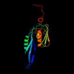
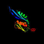
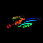
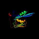
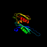
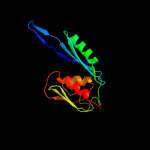
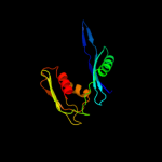
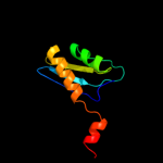
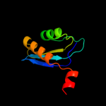
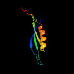
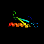
1 c3bbnE_
100.0
47
PDB header: ribosomeChain: E: PDB Molecule: ribosomal protein s5;PDBTitle: homology model for the spinach chloroplast 30s subunit2 fitted to 9.4a cryo-em map of the 70s chlororibosome.
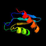
2 c1p6gE_
100.0
100
PDB header: ribosomeChain: E: PDB Molecule: 30s ribosomal protein s5;PDBTitle: real space refined coordinates of the 30s subunit fitted2 into the low resolution cryo-em map of the ef-g.gtp state3 of e. coli 70s ribosome
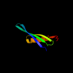
3 c2ow8f_
100.0
47
PDB header: ribosomeChain: F: PDB Molecule: PDBTitle: crystal structure of a 70s ribosome-trna complex reveals functional2 interactions and rearrangements. this file, 2ow8, contains the 30s3 ribosome subunit, two trna, and mrna molecules. 50s ribosome subunit4 is in the file 1vsa.
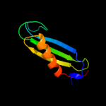
4 c2xzmE_
100.0
29
PDB header: ribosomeChain: E: PDB Molecule: ribosomal protein s5 containing protein;PDBTitle: crystal structure of the eukaryotic 40s ribosomal2 subunit in complex with initiation factor 1. this file3 contains the 40s subunit and initiation factor for4 molecule 1
5 c1eg0B_
100.0
54
PDB header: ribosomePDB COMPND: 6 c2zkqe_
100.0
29
PDB header: ribosomal protein/rnaChain: E: PDB Molecule: rna expansion segment es6 part ii;PDBTitle: structure of a mammalian ribosomal 40s subunit within an2 80s complex obtained by docking homology models of the rna3 and proteins into an 8.7 a cryo-em map
7 c1s1hE_
100.0
31
PDB header: ribosomeChain: E: PDB Molecule: 40s ribosomal protein s2;PDBTitle: structure of the ribosomal 80s-eef2-sordarin complex from2 yeast obtained by docking atomic models for rna and protein3 components into a 11.7 a cryo-em map. this file, 1s1h,4 contains 40s subunit. the 60s ribosomal subunit is in file5 1s1i.
8 c3izbE_
100.0
30
PDB header: ribosomeChain: E: PDB Molecule: 40s ribosomal protein rps2 (s5p);PDBTitle: localization of the small subunit ribosomal proteins into a 6.1 a2 cryo-em map of saccharomyces cerevisiae translating 80s ribosome
9 c3iz6E_
100.0
33
PDB header: ribosomeChain: E: PDB Molecule: 40s ribosomal protein s2 (s5p);PDBTitle: localization of the small subunit ribosomal proteins into a 5.5 a2 cryo-em map of triticum aestivum translating 80s ribosome
10 d2qale1
100.0
100
Fold: Ribosomal protein S5 domain 2-likeSuperfamily: Ribosomal protein S5 domain 2-likeFamily: Translational machinery components11 d2uube1
100.0
41
Fold: Ribosomal protein S5 domain 2-likeSuperfamily: Ribosomal protein S5 domain 2-likeFamily: Translational machinery components12 d1pkpa1
99.9
51
Fold: Ribosomal protein S5 domain 2-likeSuperfamily: Ribosomal protein S5 domain 2-likeFamily: Translational machinery components13 d2uube2
99.9
55
Fold: dsRBD-likeSuperfamily: dsRNA-binding domain-likeFamily: Ribosomal S5 protein, N-terminal domain14 d1pkpa2
99.9
59
Fold: dsRBD-likeSuperfamily: dsRNA-binding domain-likeFamily: Ribosomal S5 protein, N-terminal domain15 d2qale2
99.9
100
Fold: dsRBD-likeSuperfamily: dsRNA-binding domain-likeFamily: Ribosomal S5 protein, N-terminal domain16 d1p42a1
58.3
47
Fold: Ribosomal protein S5 domain 2-likeSuperfamily: Ribosomal protein S5 domain 2-likeFamily: UDP-3-O-[3-hydroxymyristoyl] N-acetylglucosamine deacetylase LpxC17 c2go4A_
54.3
47
PDB header: hydrolaseChain: A: PDB Molecule: udp-3-o-[3-hydroxymyristoyl] n-acetylglucosaminePDBTitle: crystal structure of aquifex aeolicus lpxc complexed with tu-514
18 c3nzkB_
51.9
40
PDB header: hydrolaseChain: B: PDB Molecule: udp-3-o-[3-hydroxymyristoyl] n-acetylglucosaminePDBTitle: structure of lpxc from yersinia enterocolitica complexed with chir0902 inhibitor
19 c2vesA_
50.3
33
PDB header: hydrolaseChain: A: PDB Molecule: udp-3-o-[3-hydroxymyristoyl] n-acetylglucosaminePDBTitle: crystal structure of lpxc from pseudomonas aeruginosa2 complexed with the potent bb-78485 inhibitor
20 d2b7ta1
49.0
22
Fold: dsRBD-likeSuperfamily: dsRNA-binding domain-likeFamily: Double-stranded RNA-binding domain (dsRBD)21 d2nuga2
not modelled
46.6
15
Fold: dsRBD-likeSuperfamily: dsRNA-binding domain-likeFamily: Double-stranded RNA-binding domain (dsRBD)22 d2pifa1
not modelled
46.5
22
Fold: PSTPO5379-likeSuperfamily: PSTPO5379-likeFamily: PSTPO5379-like23 c3adjA_
not modelled
43.1
21
PDB header: gene regulationChain: A: PDB Molecule: f21m12.9 protein;PDBTitle: structure of arabidopsis hyl1 and its molecular implications for mirna2 processing
24 d1x47a1
not modelled
42.1
24
Fold: dsRBD-likeSuperfamily: dsRNA-binding domain-likeFamily: Double-stranded RNA-binding domain (dsRBD)25 d2vapa2
not modelled
41.7
20
Fold: Bacillus chorismate mutase-likeSuperfamily: Tubulin C-terminal domain-likeFamily: Tubulin, C-terminal domain26 d1stua_
not modelled
40.5
18
Fold: dsRBD-likeSuperfamily: dsRNA-binding domain-likeFamily: Double-stranded RNA-binding domain (dsRBD)27 d1qu6a2
not modelled
40.1
19
Fold: dsRBD-likeSuperfamily: dsRNA-binding domain-likeFamily: Double-stranded RNA-binding domain (dsRBD)28 d1w5fa2
not modelled
39.2
36
Fold: Bacillus chorismate mutase-likeSuperfamily: Tubulin C-terminal domain-likeFamily: Tubulin, C-terminal domain29 d2b7va1
not modelled
38.1
28
Fold: dsRBD-likeSuperfamily: dsRNA-binding domain-likeFamily: Double-stranded RNA-binding domain (dsRBD)30 c2ljhA_
not modelled
37.8
21
PDB header: hydrolaseChain: A: PDB Molecule: double-stranded rna-specific editase adar;PDBTitle: nmr structure of double-stranded rna-specific editase adar
31 d1rq2a2
not modelled
37.4
24
Fold: Bacillus chorismate mutase-likeSuperfamily: Tubulin C-terminal domain-likeFamily: Tubulin, C-terminal domain32 d1ofua2
not modelled
34.2
20
Fold: Bacillus chorismate mutase-likeSuperfamily: Tubulin C-terminal domain-likeFamily: Tubulin, C-terminal domain33 d1o0wa2
not modelled
33.9
25
Fold: dsRBD-likeSuperfamily: dsRNA-binding domain-likeFamily: Double-stranded RNA-binding domain (dsRBD)34 c3adiC_
not modelled
30.5
25
PDB header: gene regulation/rnaChain: C: PDB Molecule: f21m12.9 protein;PDBTitle: structure of arabidopsis hyl1 and its molecular implications for mirna2 processing
35 c2zqeA_
not modelled
25.8
33
PDB header: dna binding proteinChain: A: PDB Molecule: muts2 protein;PDBTitle: crystal structure of the smr domain of thermus thermophilus muts2
36 d1bf4a_
not modelled
24.2
26
Fold: SH3-like barrelSuperfamily: Chromo domain-likeFamily: "Histone-like" proteins from archaea37 d1x48a1
not modelled
23.4
24
Fold: dsRBD-likeSuperfamily: dsRNA-binding domain-likeFamily: Double-stranded RNA-binding domain (dsRBD)38 d2dixa1
not modelled
22.4
20
Fold: dsRBD-likeSuperfamily: dsRNA-binding domain-likeFamily: Double-stranded RNA-binding domain (dsRBD)39 d1tzza2
not modelled
21.5
26
Fold: Enolase N-terminal domain-likeSuperfamily: Enolase N-terminal domain-likeFamily: Enolase N-terminal domain-like40 c1t4oA_
not modelled
21.4
30
PDB header: hydrolaseChain: A: PDB Molecule: ribonuclease iii;PDBTitle: crystal structure of rnt1p dsrbd
41 d1t4oa_
not modelled
21.4
30
Fold: dsRBD-likeSuperfamily: dsRNA-binding domain-likeFamily: Double-stranded RNA-binding domain (dsRBD)42 c3c4tA_
not modelled
20.6
23
PDB header: hydrolaseChain: A: PDB Molecule: endoribonuclease dicer;PDBTitle: structure of rnaseiiib and dsrna binding domains of mouse dicer
43 d1sr9a3
not modelled
20.3
15
Fold: 2-isopropylmalate synthase LeuA, allosteric (dimerisation) domainSuperfamily: 2-isopropylmalate synthase LeuA, allosteric (dimerisation) domainFamily: 2-isopropylmalate synthase LeuA, allosteric (dimerisation) domain44 c2l3jA_
not modelled
17.9
27
PDB header: hydrolase/rnaChain: A: PDB Molecule: double-stranded rna-specific editase 1;PDBTitle: the solution structure of the adar2 dsrbm-rna complex reveals a2 sequence-specific read out of the minor groove
45 d1r4ca_
not modelled
17.3
11
Fold: Cystatin-likeSuperfamily: Cystatin/monellinFamily: Cystatins46 c2ch9A_
not modelled
16.7
8
PDB header: inhibitorChain: A: PDB Molecule: cystatin f;PDBTitle: crystal structure of dimeric human cystatin f
47 d2dmya1
not modelled
15.8
17
Fold: dsRBD-likeSuperfamily: dsRNA-binding domain-likeFamily: Double-stranded RNA-binding domain (dsRBD)48 d1bqga2
not modelled
14.9
20
Fold: Enolase N-terminal domain-likeSuperfamily: Enolase N-terminal domain-likeFamily: Enolase N-terminal domain-like49 c2fkpC_
not modelled
14.6
13
PDB header: isomeraseChain: C: PDB Molecule: n-acylamino acid racemase;PDBTitle: the mutant g127c-t313c of deinococcus radiodurans n-2 acylamino acid racemase
50 d1uila_
not modelled
14.5
16
Fold: dsRBD-likeSuperfamily: dsRNA-binding domain-likeFamily: Double-stranded RNA-binding domain (dsRBD)51 d1wi9a_
not modelled
14.4
15
Fold: DNA/RNA-binding 3-helical bundleSuperfamily: "Winged helix" DNA-binding domainFamily: PCI domain (PINT motif)52 c1yywB_
not modelled
14.2
19
PDB header: hydrolase/rnaChain: B: PDB Molecule: ribonuclease iii;PDBTitle: crystal structure of rnase iii from aquifex aeolicus2 complexed with double stranded rna at 2.8-angstrom3 resolution
53 c3kk4B_
not modelled
14.1
21
PDB header: structural genomics, unknown functionChain: B: PDB Molecule: uncharacterized protein bp1543;PDBTitle: uncharacterized protein bp1543 from bordetella pertussis tohama i
54 c3adlA_
not modelled
14.0
19
PDB header: gene regulation/rnaChain: A: PDB Molecule: risc-loading complex subunit tarbp2;PDBTitle: structure of trbp2 and its molecule implications for mirna processing
55 d1r0ma2
not modelled
13.7
12
Fold: Enolase N-terminal domain-likeSuperfamily: Enolase N-terminal domain-likeFamily: Enolase N-terminal domain-like56 d1jpdx2
not modelled
13.6
9
Fold: Enolase N-terminal domain-likeSuperfamily: Enolase N-terminal domain-likeFamily: Enolase N-terminal domain-like57 d1jdfa2
not modelled
13.2
15
Fold: Enolase N-terminal domain-likeSuperfamily: Enolase N-terminal domain-likeFamily: Enolase N-terminal domain-like58 d1cewi_
not modelled
13.2
5
Fold: Cystatin-likeSuperfamily: Cystatin/monellinFamily: Cystatins59 d1nu5a2
not modelled
12.9
15
Fold: Enolase N-terminal domain-likeSuperfamily: Enolase N-terminal domain-likeFamily: Enolase N-terminal domain-like60 c3llhB_
not modelled
12.8
31
PDB header: rna binding proteinChain: B: PDB Molecule: risc-loading complex subunit tarbp2;PDBTitle: crystal structure of the first dsrbd of tar rna-binding protein 2
61 d1kn0a_
not modelled
12.7
20
Fold: dsRBD-likeSuperfamily: dsRNA-binding domain-likeFamily: The homologous-pairing domain of Rad52 recombinase62 c1znnF_
not modelled
12.2
19
PDB header: biosynthetic proteinChain: F: PDB Molecule: plp synthase;PDBTitle: structure of the synthase subunit of plp synthase
63 d1znna1
not modelled
12.2
19
Fold: TIM beta/alpha-barrelSuperfamily: Ribulose-phoshate binding barrelFamily: PdxS-like64 d1t4lb_
not modelled
12.2
33
Fold: dsRBD-likeSuperfamily: dsRNA-binding domain-likeFamily: Double-stranded RNA-binding domain (dsRBD)65 d1di2a_
not modelled
11.8
23
Fold: dsRBD-likeSuperfamily: dsRNA-binding domain-likeFamily: Double-stranded RNA-binding domain (dsRBD)66 c1w5fA_
not modelled
11.8
36
PDB header: cell divisionChain: A: PDB Molecule: cell division protein ftsz;PDBTitle: ftsz, t7 mutated, domain swapped (t. maritima)
67 d1uhza_
not modelled
10.7
17
Fold: dsRBD-likeSuperfamily: dsRNA-binding domain-likeFamily: Double-stranded RNA-binding domain (dsRBD)68 c1h2iG_
not modelled
9.9
20
PDB header: dna-binding proteinChain: G: PDB Molecule: dna repair protein rad52 homolog;PDBTitle: human rad52 protein, n-terminal domain
69 d2cpna1
not modelled
9.8
20
Fold: dsRBD-likeSuperfamily: dsRNA-binding domain-likeFamily: Double-stranded RNA-binding domain (dsRBD)70 d2mnra2
not modelled
9.6
21
Fold: Enolase N-terminal domain-likeSuperfamily: Enolase N-terminal domain-likeFamily: Enolase N-terminal domain-like71 d1iowa1
not modelled
9.6
21
Fold: PreATP-grasp domainSuperfamily: PreATP-grasp domainFamily: D-Alanine ligase N-terminal domain72 c1w59B_
not modelled
9.4
20
PDB header: cell divisionChain: B: PDB Molecule: cell division protein ftsz homolog 1;PDBTitle: ftsz dimer, empty (m. jannaschii)
73 d1x49a1
not modelled
9.4
22
Fold: dsRBD-likeSuperfamily: dsRNA-binding domain-likeFamily: Double-stranded RNA-binding domain (dsRBD)74 c2khxA_
not modelled
9.4
26
PDB header: gene regulation,nuclear proteinChain: A: PDB Molecule: ribonuclease 3;PDBTitle: drosha double-stranded rna binding motif
75 d1t4na_
not modelled
9.3
37
Fold: dsRBD-likeSuperfamily: dsRNA-binding domain-likeFamily: Double-stranded RNA-binding domain (dsRBD)76 c3mwzA_
not modelled
9.1
11
PDB header: hydrolase inhibitorChain: A: PDB Molecule: sialostatin l2;PDBTitle: crystal structure of the selenomethionine derivative of the l 22,47,2 100 m mutant of sialostatin l2
77 d2ux9a1
not modelled
9.0
26
Fold: Dodecin subunit-likeSuperfamily: Dodecin-likeFamily: Dodecin-like78 c3onrI_
not modelled
9.0
15
PDB header: metal binding proteinChain: I: PDB Molecule: protein transport protein sece2;PDBTitle: crystal structure of the calcium chelating immunodominant antigen,2 calcium dodecin (rv0379),from mycobacterium tuberculosis with a novel3 calcium-binding site
79 c2l2nA_
not modelled
9.0
19
PDB header: rna binding protein, plant proteinChain: A: PDB Molecule: hyponastic leave 1;PDBTitle: backbone 1h, 13c, and 15n chemical shift assignments for the first2 dsrbd of protein hyl1
80 c3oqtP_
not modelled
8.9
26
PDB header: flavoproteinChain: P: PDB Molecule: rv1498a protein;PDBTitle: crystal structure of rv1498a protein from mycobacterium tuberculosis
81 c3dfhC_
not modelled
8.7
20
PDB header: isomeraseChain: C: PDB Molecule: mandelate racemase;PDBTitle: crystal structure of putative mandelate racemase / muconate2 lactonizing enzyme from vibrionales bacterium swat-3
82 c2l33A_
not modelled
8.7
19
PDB header: transcription regulatorChain: A: PDB Molecule: interleukin enhancer-binding factor 3;PDBTitle: solution nmr structure of drbm 2 domain of interleukin enhancer-2 binding factor 3 from homo sapiens, northeast structural genomics3 consortium target hr4527e
83 c2rhoB_
not modelled
8.5
22
PDB header: cell cycleChain: B: PDB Molecule: cell division protein ftsz;PDBTitle: synthetic gene encoded bacillus subtilis ftsz ncs dimer with2 bound gdp and gtp-gamma-s
84 c3l0rA_
not modelled
8.5
17
PDB header: hydrolase inhibitorChain: A: PDB Molecule: cystatin-2;PDBTitle: crystal structure of salivary cystatin from the soft tick ornithodoros2 moubata
85 c1vz0B_
not modelled
8.1
25
PDB header: nuclear proteinChain: B: PDB Molecule: chromosome partitioning protein parb;PDBTitle: chromosome segregation protein spo0j from thermus2 thermophilus
86 d2bv3a3
not modelled
7.9
23
Fold: Ribosomal protein S5 domain 2-likeSuperfamily: Ribosomal protein S5 domain 2-likeFamily: Translational machinery components87 d1azpa_
not modelled
7.8
28
Fold: SH3-like barrelSuperfamily: Chromo domain-likeFamily: "Histone-like" proteins from archaea88 d1vz0a1
not modelled
7.7
26
Fold: DNA/RNA-binding 3-helical bundleSuperfamily: KorB DNA-binding domain-likeFamily: KorB DNA-binding domain-like89 c2vxyA_
not modelled
7.6
24
PDB header: cell cycleChain: A: PDB Molecule: cell division protein ftsz;PDBTitle: the structure of ftsz from bacillus subtilis at 1.7a2 resolution
90 c2vxaL_
not modelled
7.5
12
PDB header: flavoproteinChain: L: PDB Molecule: dodecin;PDBTitle: h.halophila dodecin in complex with riboflavin
91 c3n4fD_
not modelled
7.3
18
PDB header: isomeraseChain: D: PDB Molecule: mandelate racemase/muconate lactonizing protein;PDBTitle: crystal structure of mandelate racemase/muconate lactonizing protein2 from geobacillus sp. y412mc10
92 c2vawA_
not modelled
7.3
20
PDB header: cell cycleChain: A: PDB Molecule: cell division protein ftsz;PDBTitle: ftsz pseudomonas aeruginosa gdp
93 c1l0oC_
not modelled
7.3
13
PDB header: protein bindingChain: C: PDB Molecule: sigma factor;PDBTitle: crystal structure of the bacillus stearothermophilus anti-2 sigma factor spoiiab with the sporulation sigma factor3 sigmaf
94 d1l0oc_
not modelled
7.3
13
Fold: DNA/RNA-binding 3-helical bundleSuperfamily: Sigma3 and sigma4 domains of RNA polymerase sigma factorsFamily: Sigma4 domain95 d1roaa_
not modelled
7.1
15
Fold: Cystatin-likeSuperfamily: Cystatin/monellinFamily: Cystatins96 d1ekza_
not modelled
7.0
21
Fold: dsRBD-likeSuperfamily: dsRNA-binding domain-likeFamily: Double-stranded RNA-binding domain (dsRBD)97 d1yeya2
not modelled
6.8
12
Fold: Enolase N-terminal domain-likeSuperfamily: Enolase N-terminal domain-likeFamily: Enolase N-terminal domain-like98 c2ppgB_
not modelled
6.7
21
PDB header: isomeraseChain: B: PDB Molecule: putative isomerase;PDBTitle: crystal structure of putative isomerase from sinorhizobium meliloti
99 c2p88E_
not modelled
6.7
15
PDB header: lyaseChain: E: PDB Molecule: mandelate racemase/muconate lactonizing enzymePDBTitle: crystal structure of n-succinyl arg/lys racemase from2 bacillus cereus atcc 14579


