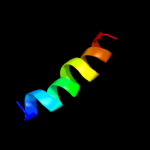
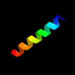
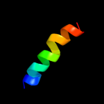
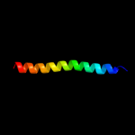
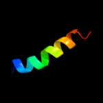
1 c2e76H_
46.4
30
PDB header: photosynthesisChain: H: PDB Molecule: cytochrome b6-f complex subunit 8;PDBTitle: crystal structure of the cytochrome b6f complex with tridecyl-2 stigmatellin (tds) from m.laminosus
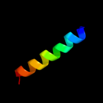
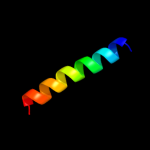
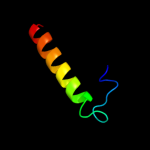
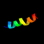
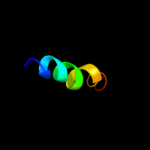
2 c2e74H_
46.4
30
PDB header: photosynthesisChain: H: PDB Molecule: cytochrome b6-f complex subunit 8;PDBTitle: crystal structure of the cytochrome b6f complex from m.laminosus
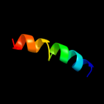
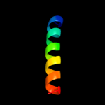
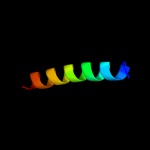
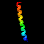
3 c2e75H_
46.4
30
PDB header: photosynthesisChain: H: PDB Molecule: cytochrome b6-f complex subunit 8;PDBTitle: crystal structure of the cytochrome b6f complex with 2-nonyl-4-2 hydroxyquinoline n-oxide (nqno) from m.laminosus
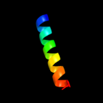
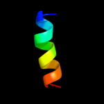
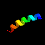
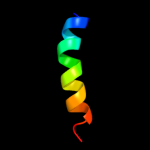
4 c1u0iA_
42.4
42
PDB header: de novo proteinChain: A: PDB Molecule: iaal-e3;PDBTitle: iaal-e3/k3 heterodimer
5 d2e74h1
41.9
30
Fold: Single transmembrane helixSuperfamily: PetN subunit of the cytochrome b6f complexFamily: PetN subunit of the cytochrome b6f complex6 c1ce0B_
37.8
29
PDB header: hiv-1 envelope proteinChain: B: PDB Molecule: protein (leucine zipper model h38-p1);PDBTitle: trimerization specificity in hiv-1 gp41: analysis with a2 gcn4 leucine zipper model
7 c3he5A_
37.6
26
PDB header: de novo proteinChain: A: PDB Molecule: synzip1;PDBTitle: heterospecific coiled-coil pair synzip2:synzip1
8 c3g9rF_
37.5
33
PDB header: viral proteinChain: F: PDB Molecule: fusion complex of hiv-1 envelope glycoproteinPDBTitle: structure of the hiv-1 gp41 membrane-proximal ectodomain2 region in a putative prefusion conformation
9 c3lt7D_
36.7
32
PDB header: cell adhesionChain: D: PDB Molecule: adhesin yada;PDBTitle: a transition from strong right-handed to canonical left-handed2 supercoiling in a conserved coiled coil segment of trimeric3 autotransporter adhesins - the m3 mutant structure
10 c1u2uA_
36.3
33
PDB header: transcriptionChain: A: PDB Molecule: general control protein gcn4;PDBTitle: nmr solution structure of a designed heterodimeric leucine2 zipper
11 c1vf5H_
33.9
30
PDB header: photosynthesisChain: H: PDB Molecule: protein pet n;PDBTitle: crystal structure of cytochrome b6f complex from m.laminosus
12 c2lf0A_
32.8
15
PDB header: structural genomics, unknown functionChain: A: PDB Molecule: uncharacterized protein yibl;PDBTitle: solution structure of sf3636, a two-domain unknown function protein2 from shigella flexneri 2a, determined by joint refinement of nmr,3 residual dipolar couplings and small-angle x-ray scatting, nesg4 target sfr339/ocsp target sf3636
13 c3ci9B_
32.5
30
PDB header: transcriptionChain: B: PDB Molecule: heat shock factor-binding protein 1;PDBTitle: crystal structure of the human hsbp1
14 c2zt9H_
32.2
43
PDB header: photosynthesisChain: H: PDB Molecule: cytochrome b6-f complex subunit 8;PDBTitle: crystal structure of the cytochrome b6f complex from nostoc sp. pcc2 7120
15 c1hf9B_
31.3
19
PDB header: atpase inhibitorChain: B: PDB Molecule: atpase inhibitor (mitochondrial);PDBTitle: c-terminal coiled-coil domain from bovine if1
16 c1vf5U_
30.7
30
PDB header: photosynthesisChain: U: PDB Molecule: protein pet n;PDBTitle: crystal structure of cytochrome b6f complex from m.laminosus
17 c1aq5C_
28.8
23
PDB header: coiled-coilChain: C: PDB Molecule: cartilage matrix protein;PDBTitle: high-resolution solution nmr structure of the trimeric coiled-coil2 domain of chicken cartilage matrix protein, 20 structures
18 d1v54m_
27.9
25
Fold: Single transmembrane helixSuperfamily: Mitochondrial cytochrome c oxidase subunit VIIIb (aka IX)Family: Mitochondrial cytochrome c oxidase subunit VIIIb (aka IX)19 c2y69Z_
26.9
25
PDB header: electron transportChain: Z: PDB Molecule: cytochrome c oxidase polypeptide 8h;PDBTitle: bovine heart cytochrome c oxidase re-refined with molecular2 oxygen
20 c2d2cH_
26.5
30
PDB header: photosynthesisChain: H: PDB Molecule: cytochrome b6-f complex subunit viii;PDBTitle: crystal structure of cytochrome b6f complex with dbmib from2 m. laminosus
21 c2d2cU_
not modelled
26.5
30
PDB header: photosynthesisChain: U: PDB Molecule: cytochrome b6-f complex subunit viii;PDBTitle: crystal structure of cytochrome b6f complex with dbmib from2 m. laminosus
22 c2ke4A_
not modelled
25.7
12
PDB header: membrane proteinChain: A: PDB Molecule: cdc42-interacting protein 4;PDBTitle: the nmr structure of the tc10 and cdc42 interacting domain2 of cip4
23 c3m9hB_
not modelled
23.9
24
PDB header: chaperoneChain: B: PDB Molecule: proteasome-associated atpase;PDBTitle: crystal structure of the amino terminal coiled coil domain of the2 mycobacterium tuberculosis proteasomal atpase mpa
24 c2qa7B_
not modelled
23.6
27
PDB header: actin bindingChain: B: PDB Molecule: huntingtin-interacting protein 1;PDBTitle: crystal structure of huntingtin-interacting protein 12 (hip1) coiled-coil domain with a basic surface suitable3 for hip-protein interactor (hippi)
25 c3rylB_
not modelled
22.3
42
PDB header: protein bindingChain: B: PDB Molecule: protein vpa1370;PDBTitle: dimerization domain of vibrio parahemolyticus vopl
26 d2gr7a1
not modelled
19.7
20
Fold: Pili subunitsSuperfamily: Pili subunitsFamily: YadA C-terminal domain-like27 c2gr7C_
not modelled
19.7
20
PDB header: membrane proteinChain: C: PDB Molecule: adhesin;PDBTitle: hia 992-1098
28 c3p8cE_
not modelled
19.5
21
PDB header: protein bindingChain: E: PDB Molecule: probable protein brick1;PDBTitle: structure and control of the actin regulatory wave complex
29 c1hwtC_
not modelled
18.4
26
PDB header: gene regulation/dnaChain: C: PDB Molecule: protein (heme activator protein);PDBTitle: structure of a hap1/dna complex reveals dramatically2 asymmetric dna binding by a homodimeric protein
30 c1ij2C_
not modelled
17.7
27
PDB header: transcriptionChain: C: PDB Molecule: general control protein gcn4;PDBTitle: gcn4-pvtl coiled-coil trimer with threonine at the a(16)2 position
31 c3k7zA_
not modelled
17.4
27
PDB header: dna binding proteinChain: A: PDB Molecule: general control protein gcn4;PDBTitle: gcn4-leucine zipper core mutant as n16a trigonal automatic2 solution
32 c1rb6C_
not modelled
17.4
27
PDB header: dna binding proteinChain: C: PDB Molecule: general control protein gcn4;PDBTitle: antiparallel trimer of gcn4-leucine zipper core mutant as2 n16a tetragonal form
33 c1swiA_
not modelled
17.4
27
PDB header: leucine zipperChain: A: PDB Molecule: gcn4p1;PDBTitle: gcn4-leucine zipper core mutant as n16a complexed with2 benzene
34 c1rb1A_
not modelled
17.4
27
PDB header: dna binding proteinChain: A: PDB Molecule: general control protein gcn4;PDBTitle: gcn4-leucine zipper core mutant as n16a trigonal automatic2 solution
35 c1rb1B_
not modelled
17.4
27
PDB header: dna binding proteinChain: B: PDB Molecule: general control protein gcn4;PDBTitle: gcn4-leucine zipper core mutant as n16a trigonal automatic2 solution
36 c3k7zB_
not modelled
17.4
27
PDB header: dna binding proteinChain: B: PDB Molecule: general control protein gcn4;PDBTitle: gcn4-leucine zipper core mutant as n16a trigonal automatic2 solution
37 d1vl2a2
not modelled
16.8
21
Fold: Argininosuccinate synthetase, C-terminal domainSuperfamily: Argininosuccinate synthetase, C-terminal domainFamily: Argininosuccinate synthetase, C-terminal domain38 c1ij3C_
not modelled
16.6
27
PDB header: transcriptionChain: C: PDB Molecule: general control protein gcn4;PDBTitle: gcn4-pvsl coiled-coil trimer with serine at the a(16)2 position
39 c1ij3B_
not modelled
16.6
27
PDB header: transcriptionChain: B: PDB Molecule: general control protein gcn4;PDBTitle: gcn4-pvsl coiled-coil trimer with serine at the a(16)2 position
40 d1slqa_
not modelled
16.2
8
Fold: VP4 membrane interaction domainSuperfamily: VP4 membrane interaction domainFamily: VP4 membrane interaction domain41 d1ioka2
not modelled
15.9
32
Fold: The "swivelling" beta/beta/alpha domainSuperfamily: GroEL apical domain-likeFamily: GroEL-like chaperone, apical domain42 d1ta8a_
not modelled
15.9
18
Fold: ATP-graspSuperfamily: DNA ligase/mRNA capping enzyme, catalytic domainFamily: Adenylation domain of NAD+-dependent DNA ligase43 c1ztaA_
not modelled
15.8
29
PDB header: dna-binding motifChain: A: PDB Molecule: leucine zipper monomer;PDBTitle: the solution structure of a leucine-zipper motif peptide
44 c1ij2B_
not modelled
15.6
27
PDB header: transcriptionChain: B: PDB Molecule: general control protein gcn4;PDBTitle: gcn4-pvtl coiled-coil trimer with threonine at the a(16)2 position
45 c3pn1A_
not modelled
15.4
21
PDB header: ligase/ligase inhibitorChain: A: PDB Molecule: dna ligase;PDBTitle: novel bacterial nad+-dependent dna ligase inhibitors with broad2 spectrum potency and antibacterial efficacy in vivo
46 c2ergA_
not modelled
14.1
21
PDB header: transcription activator/dnaChain: A: PDB Molecule: regulatory protein leu3;PDBTitle: crystal structure of leu3 dna-binding domain with a single2 h50c mutation complexed with a 15mer dna duplex
47 c2xzrA_
not modelled
12.3
13
PDB header: cell adhesionChain: A: PDB Molecule: immunoglobulin-binding protein eibd;PDBTitle: escherichia coli immunoglobulin-binding protein eibd 391-438 fused2 to gcn4 adaptors
48 c2yy0D_
not modelled
12.1
11
PDB header: transcriptionChain: D: PDB Molecule: c-myc-binding protein;PDBTitle: crystal structure of ms0802, c-myc-1 binding protein domain2 from homo sapiens
49 c2zt9E_
not modelled
12.0
27
PDB header: photosynthesisChain: E: PDB Molecule: cytochrome b6-f complex subunit 6;PDBTitle: crystal structure of the cytochrome b6f complex from nostoc sp. pcc2 7120
50 c1kddC_
not modelled
12.0
25
PDB header: de novo proteinChain: C: PDB Molecule: gcn4 acid base heterodimer acid-d12la16i;PDBTitle: x-ray structure of the coiled coil gcn4 acid base2 heterodimer acid-d12la16i base-d12la16l
51 d1b04a_
not modelled
12.0
24
Fold: ATP-graspSuperfamily: DNA ligase/mRNA capping enzyme, catalytic domainFamily: Adenylation domain of NAD+-dependent DNA ligase52 c1kddA_
not modelled
11.7
25
PDB header: de novo proteinChain: A: PDB Molecule: gcn4 acid base heterodimer acid-d12la16i;PDBTitle: x-ray structure of the coiled coil gcn4 acid base2 heterodimer acid-d12la16i base-d12la16l
53 c1kddF_
not modelled
11.7
25
PDB header: de novo proteinChain: F: PDB Molecule: gcn4 acid base heterodimer acid-d12la16i;PDBTitle: x-ray structure of the coiled coil gcn4 acid base2 heterodimer acid-d12la16i base-d12la16l
54 c1zauA_
not modelled
11.1
30
PDB header: ligaseChain: A: PDB Molecule: dna ligase;PDBTitle: adenylation domain of nad+ dependent dna ligase from2 m.tuberculosis
55 c2owoA_
not modelled
11.1
21
PDB header: ligase/dnaChain: A: PDB Molecule: dna ligase;PDBTitle: last stop on the road to repair: structure of e.coli dna ligase bound2 to nicked dna-adenylate
56 d1uuja_
not modelled
10.6
25
Fold: Lissencephaly-1 protein (Lis-1, PAF-AH alpha) N-terminal domainSuperfamily: Lissencephaly-1 protein (Lis-1, PAF-AH alpha) N-terminal domainFamily: Lissencephaly-1 protein (Lis-1, PAF-AH alpha) N-terminal domain57 c3hroA_
not modelled
10.2
33
PDB header: transport proteinChain: A: PDB Molecule: transient receptor potential (trp) channelPDBTitle: crystal structure of a c-terminal coiled coil domain of2 transient receptor potential (trp) channel subfamily p3 member 2 (trpp2, polycystic kidney disease 2)
58 c1kd9A_
not modelled
10.0
31
PDB header: de novo proteinChain: A: PDB Molecule: gcn4 acid base heterodimer acid-d12la16l;PDBTitle: x-ray structure of the coiled coil gcn4 acid base2 heterodimer acid-d12la16l base-d12la16l
59 c1kd9F_
not modelled
10.0
31
PDB header: de novo proteinChain: F: PDB Molecule: gcn4 acid base heterodimer acid-d12la16l;PDBTitle: x-ray structure of the coiled coil gcn4 acid base2 heterodimer acid-d12la16l base-d12la16l
60 c1kd9C_
not modelled
10.0
31
PDB header: de novo proteinChain: C: PDB Molecule: gcn4 acid base heterodimer acid-d12la16l;PDBTitle: x-ray structure of the coiled coil gcn4 acid base2 heterodimer acid-d12la16l base-d12la16l
61 c3gw6F_
not modelled
10.0
27
PDB header: chaperoneChain: F: PDB Molecule: endo-n-acetylneuraminidase;PDBTitle: intramolecular chaperone
62 c3h5fB_
not modelled
9.9
43
PDB header: de novo proteinChain: B: PDB Molecule: coil ser l16l-pen;PDBTitle: switching the chirality of the metal environment alters the2 coordination mode in designed peptides.
63 c3h5fC_
not modelled
9.9
43
PDB header: de novo proteinChain: C: PDB Molecule: coil ser l16l-pen;PDBTitle: switching the chirality of the metal environment alters the2 coordination mode in designed peptides.
64 c3h5gA_
not modelled
9.9
43
PDB header: de novo proteinChain: A: PDB Molecule: coil ser l16d-pen;PDBTitle: switching the chirality of the metal environment alters the2 coordination mode in designed peptides.
65 c3h5gB_
not modelled
9.9
43
PDB header: de novo proteinChain: B: PDB Molecule: coil ser l16d-pen;PDBTitle: switching the chirality of the metal environment alters the2 coordination mode in designed peptides.
66 c3h5gC_
not modelled
9.9
43
PDB header: de novo proteinChain: C: PDB Molecule: coil ser l16d-pen;PDBTitle: switching the chirality of the metal environment alters the2 coordination mode in designed peptides.
67 c3h5fA_
not modelled
9.9
43
PDB header: de novo proteinChain: A: PDB Molecule: coil ser l16l-pen;PDBTitle: switching the chirality of the metal environment alters the2 coordination mode in designed peptides.
68 c2xv5A_
not modelled
9.9
13
PDB header: structural proteinChain: A: PDB Molecule: lamin-a/c;PDBTitle: human lamin a coil 2b fragment
69 c2p4vA_
not modelled
9.8
21
PDB header: transcriptionChain: A: PDB Molecule: transcription elongation factor greb;PDBTitle: crystal structure of the transcript cleavage factor, greb2 at 2.6a resolution
70 c1fmhA_
not modelled
9.6
28
PDB header: transcriptionChain: A: PDB Molecule: general control protein gcn4;PDBTitle: nmr solution structure of a designed heterodimeric leucine2 zipper
71 c1t3jA_
not modelled
9.6
38
PDB header: membrane proteinChain: A: PDB Molecule: mitofusin 1;PDBTitle: mitofusin domain hr2 v686m/i708m mutant
72 c3jslA_
not modelled
9.5
21
PDB header: ligaseChain: A: PDB Molecule: dna ligase;PDBTitle: crystal structure of the adenylation domain of nad+-2 dependent dna ligase from staphylococcus aureus
73 c1ci6B_
not modelled
9.1
17
PDB header: transcriptionChain: B: PDB Molecule: transcription factor c/ebp beta;PDBTitle: transcription factor atf4-c/ebp beta bzip heterodimer
74 d1lrza1
not modelled
8.8
14
Fold: Long alpha-hairpinSuperfamily: tRNA-binding armFamily: Methicillin resistance protein FemA probable tRNA-binding arm75 d1oela2
not modelled
8.7
27
Fold: The "swivelling" beta/beta/alpha domainSuperfamily: GroEL apical domain-likeFamily: GroEL-like chaperone, apical domain76 c1grjA_
not modelled
8.4
21
PDB header: transcription regulationChain: A: PDB Molecule: grea protein;PDBTitle: grea transcript cleavage factor from escherichia coli
77 c3swfA_
not modelled
8.3
26
PDB header: transport proteinChain: A: PDB Molecule: cgmp-gated cation channel alpha-1;PDBTitle: cnga1 621-690 containing clz domain
78 c2o7hF_
not modelled
8.1
22
PDB header: transcriptionChain: F: PDB Molecule: general control protein gcn4;PDBTitle: crystal structure of trimeric coiled coil gcn4 leucine zipper
79 c2dl1A_
not modelled
8.1
15
PDB header: protein transportChain: A: PDB Molecule: spartin;PDBTitle: solution structure of the mit domain from human spartin
80 c2rkhA_
not modelled
7.4
38
PDB header: transcriptionChain: A: PDB Molecule: putative apha-like transcription factor;PDBTitle: crystal structure of a putative apha-like transcription factor2 (zp_00208345.1) from magnetospirillum magnetotacticum ms-1 at 2.00 a3 resolution
81 d1vlia2
not modelled
7.4
21
Fold: TIM beta/alpha-barrelSuperfamily: AldolaseFamily: NeuB-like82 c3hlsE_
not modelled
7.3
18
PDB header: signaling proteinChain: E: PDB Molecule: guanylate cyclase soluble subunit beta-1;PDBTitle: crystal structure of the signaling helix coiled-coil doimain2 of the beta-1 subunit of the soluble guanylyl cyclase
83 c1gk6B_
not modelled
7.2
17
PDB header: vimentinChain: B: PDB Molecule: vimentin;PDBTitle: human vimentin coil 2b fragment linked to gcn4 leucine2 zipper (z2b)
84 d1seta1
not modelled
7.1
0
Fold: Long alpha-hairpinSuperfamily: tRNA-binding armFamily: Seryl-tRNA synthetase (SerRS)85 c1yjgE_
not modelled
7.1
14
PDB header: immune systemChain: E: PDB Molecule: surface protein vspa;PDBTitle: variable small protein 1 of borrelia turicatae (vspa or vsp1)
86 d2zdra2
not modelled
7.0
18
Fold: TIM beta/alpha-barrelSuperfamily: AldolaseFamily: NeuB-like87 c2akfA_
not modelled
6.9
36
PDB header: protein bindingChain: A: PDB Molecule: coronin-1a;PDBTitle: crystal structure of the coiled-coil domain of coronin 1
88 c2akfB_
not modelled
6.9
36
PDB header: protein bindingChain: B: PDB Molecule: coronin-1a;PDBTitle: crystal structure of the coiled-coil domain of coronin 1
89 c2akfC_
not modelled
6.9
36
PDB header: protein bindingChain: C: PDB Molecule: coronin-1a;PDBTitle: crystal structure of the coiled-coil domain of coronin 1
90 c3nmdA_
not modelled
6.9
21
PDB header: transferaseChain: A: PDB Molecule: cgmp dependent protein kinase;PDBTitle: crystal structure of the leucine zipper domain of cgmp dependent2 protein kinase i beta
91 c2wl2B_
not modelled
6.8
19
PDB header: isomeraseChain: B: PDB Molecule: dna gyrase subunit a;PDBTitle: crystal structure of n-terminal domain of gyra with the2 antibiotic simocyclinone d8
92 d1v9pa3
not modelled
6.7
24
Fold: ATP-graspSuperfamily: DNA ligase/mRNA capping enzyme, catalytic domainFamily: Adenylation domain of NAD+-dependent DNA ligase93 c3o3nA_
not modelled
6.6
21
PDB header: lyaseChain: A: PDB Molecule: alpha-subunit 2-hydroxyisocaproyl-coa dehydratase;PDBTitle: (r)-2-hydroxyisocaproyl-coa dehydratase in complex with its substrate2 (r)-2-hydroxyisocaproyl-coa
94 c2w6bA_
not modelled
6.6
25
PDB header: signaling proteinChain: A: PDB Molecule: rho guanine nucleotide exchange factor 7;PDBTitle: crystal structure of the trimeric beta-pix coiled-coil2 domain
95 d1sjpa2
not modelled
6.5
18
Fold: The "swivelling" beta/beta/alpha domainSuperfamily: GroEL apical domain-likeFamily: GroEL-like chaperone, apical domain96 d1kida_
not modelled
6.5
32
Fold: The "swivelling" beta/beta/alpha domainSuperfamily: GroEL apical domain-likeFamily: GroEL-like chaperone, apical domain97 c3m0dC_
not modelled
6.5
26
PDB header: signaling proteinChain: C: PDB Molecule: tnf receptor-associated factor 1;PDBTitle: crystal structure of the traf1:traf2:ciap2 complex
98 c2pnvA_
not modelled
6.5
23
PDB header: membrane proteinChain: A: PDB Molecule: small conductance calcium-activated potassiumPDBTitle: crystal structure of the leucine zipper domain of small-2 conductance ca2+-activated k+ (skca) channel from rattus3 norvegicus
99 c2zfcB_
not modelled
6.4
25
PDB header: viral proteinChain: B: PDB Molecule: hiv-1 gp41;PDBTitle: x-ray crystal structure of an engineered n-terminal hiv-12 gp41 trimer with enhanced stability and potency