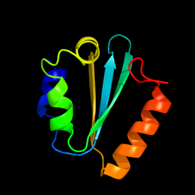
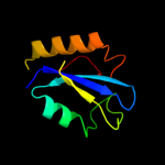
1 c1yd2A_
99.9
33
PDB header: dna binding proteinChain: A: PDB Molecule: uvrabc system protein c;PDBTitle: crystal structure of the giy-yig n-terminal endonuclease domain of2 uvrc from thermotoga maritima: point mutant y19f bound to the3 catalytic divalent cation
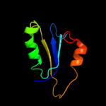
2 c1yd6A_
99.9
31
PDB header: dna binding proteinChain: A: PDB Molecule: uvrc;PDBTitle: crystal structure of the giy-yig n-terminal endonuclease2 domain of uvrc from bacillus caldotenax
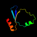
3 c2wshC_
99.9
23
PDB header: hydrolaseChain: C: PDB Molecule: endonuclease ii;PDBTitle: structure of bacteriophage t4 endoii e118a mutant
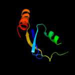
4 d1mk0a_
98.6
15
Fold: GIY-YIG endonucleaseSuperfamily: GIY-YIG endonucleaseFamily: GIY-YIG endonuclease5 c3nicA_
96.8
26
PDB header: hydrolase/dnaChain: A: PDB Molecule: eco29kir;PDBTitle: dna binding and cleavage by the giy-yig endonuclease r.eco29ki2 inactive variant y49f
6 c1zg2A_
94.0
15
PDB header: structural genomics, unknown functionChain: A: PDB Molecule: hypothetical upf0213 protein bh0048;PDBTitle: solution nmr structure of the upf0213 protein bh0048 from2 bacillus halodurans. northeast structural genomics target3 bhr2.
7 c1ywlA_
93.8
19
PDB header: structural genomics, unknown functionChain: A: PDB Molecule: hypothetical upf0213 protein ef2693;PDBTitle: solution nmr structure of the protein ef2693 from e.2 faecalis: northeast structural genomics consortium target3 efr36
8 d1pd0a4
68.5
25
Fold: Gelsolin-likeSuperfamily: C-terminal, gelsolin-like domain of Sec23/24Family: C-terminal, gelsolin-like domain of Sec23/249 d1utaa_
41.4
14
Fold: Ferredoxin-likeSuperfamily: Sporulation related repeatFamily: Sporulation related repeat10 d1xdpa4
38.0
33
Fold: Phospholipase D/nucleaseSuperfamily: Phospholipase D/nucleaseFamily: Polyphosphate kinase C-terminal domain11 c2gj3A_
37.8
19
PDB header: transferaseChain: A: PDB Molecule: nitrogen fixation regulatory protein;PDBTitle: crystal structure of the fad-containing pas domain of the2 protein nifl from azotobacter vinelandii.
12 d2o8ra4
36.6
21
Fold: Phospholipase D/nucleaseSuperfamily: Phospholipase D/nucleaseFamily: Polyphosphate kinase C-terminal domain13 c1x60A_
34.5
14
PDB header: hydrolaseChain: A: PDB Molecule: sporulation-specific n-acetylmuramoyl-l-alaninePDBTitle: solution structure of the peptidoglycan binding domain of2 b. subtilis cell wall lytic enzyme cwlc
14 c1pd0A_
32.7
26
PDB header: transport proteinChain: A: PDB Molecule: protein transport protein sec24;PDBTitle: crystal structure of the copii coat subunit, sec24,2 complexed with a peptide from the snare protein sed53 (yeast syntaxin-5)
15 c2qkpD_
31.8
28
PDB header: structural genomics, unknown functionChain: D: PDB Molecule: uncharacterized protein;PDBTitle: crystal structure of c-terminal domain of smu_1151c from streptococcus2 mutans
16 c2rivB_
30.7
33
PDB header: signaling proteinChain: B: PDB Molecule: thyroxine-binding globulin;PDBTitle: crystal structure of the reactive loop cleaved human thyroxine binding2 globulin
17 c3egxB_
28.2
25
PDB header: protein transportChain: B: PDB Molecule: protein transport protein sec24a;PDBTitle: crystal structure of the mammalian copii-coat protein2 sec23a/24a complexed with the snare protein sec22b and3 bound to the transport signal sequence of the snare protein4 bet1
18 c3luqC_
27.4
24
PDB header: transferaseChain: C: PDB Molecule: sensor protein;PDBTitle: the crystal structure of a pas domain from a sensory box2 histidine kinase regulator from geobacter sulfurreducens to3 2.5a
19 c3eh2B_
27.2
30
PDB header: protein transportChain: B: PDB Molecule: protein transport protein sec24c;PDBTitle: crystal structure of the human copii-coat protein sec24c
20 c1m2vB_
26.8
25
PDB header: protein transportChain: B: PDB Molecule: protein transport protein sec24;PDBTitle: crystal structure of the yeast sec23/24 heterodimer
21 c3eg9B_
not modelled
26.5
30
PDB header: protein transportChain: B: PDB Molecule: sec24 related gene family, member d;PDBTitle: crystal structure of the mammalian copii-coat protein2 sec23/24 bound to the transport signal sequence of membrin
22 c1hleB_
not modelled
25.4
13
PDB header: hydrolase inhibitor(serine proteinase)Chain: B: PDB Molecule: horse leukocyte elastase inhibitor;PDBTitle: crystal structure of cleaved equine leucocyte elastase2 inhibitor determined at 1.95 angstroms resolution
23 c1xdoB_
not modelled
23.0
33
PDB header: transferaseChain: B: PDB Molecule: polyphosphate kinase;PDBTitle: crystal structure of escherichia coli polyphosphate kinase
24 d1gcca_
not modelled
20.8
33
Fold: DNA-binding domainSuperfamily: DNA-binding domainFamily: GCC-box binding domain25 c2o8rA_
not modelled
20.4
21
PDB header: transferaseChain: A: PDB Molecule: polyphosphate kinase;PDBTitle: crystal structure of polyphosphate kinase from2 porphyromonas gingivalis
26 c3mxqC_
not modelled
20.1
13
PDB header: transferaseChain: C: PDB Molecule: sensor protein;PDBTitle: crystal structure of sensory box sensor histidine kinase from vibrio2 cholerae
27 c3mr0B_
not modelled
20.1
10
PDB header: transcription regulatorChain: B: PDB Molecule: sensory box histidine kinase/response regulator;PDBTitle: crystal structure of sensory box histidine kinase/response regulator2 from burkholderia thailandensis e264
28 d1g1pa_
not modelled
19.7
71
Fold: Knottins (small inhibitors, toxins, lectins)Superfamily: omega toxin-likeFamily: Conotoxin29 c1jjoE_
not modelled
18.7
25
PDB header: signaling proteinChain: E: PDB Molecule: neuroserpin;PDBTitle: crystal structure of mouse neuroserpin (cleaved form)
30 c2jheB_
not modelled
18.6
17
PDB header: transcriptionChain: B: PDB Molecule: transcription regulator tyrr;PDBTitle: n-terminal domain of tyrr transcription factor (residues 1 -2 190)
31 d1nwza_
not modelled
17.8
15
Fold: Profilin-likeSuperfamily: PYP-like sensor domain (PAS domain)Family: PYP-like32 d1lkta_
not modelled
17.6
29
Fold: Head-binding domain of phage P22 tailspike proteinSuperfamily: Head-binding domain of phage P22 tailspike proteinFamily: Head-binding domain of phage P22 tailspike protein33 c2w0nA_
not modelled
17.0
17
PDB header: transferaseChain: A: PDB Molecule: sensor protein dcus;PDBTitle: plasticity of pas domain and potential role for signal2 transduction in the histidine-kinase dcus
34 c3f02C_
not modelled
16.9
17
PDB header: hydrolase inhibitorChain: C: PDB Molecule: neuroserpin;PDBTitle: cleaved human neuroserpin
35 c3mfxA_
not modelled
16.8
14
PDB header: transcriptionChain: A: PDB Molecule: sensory box/ggdef family protein;PDBTitle: crystal structure of the sensory box domain of the sensory-2 box/ggdef protein so_1695 from shewanella oneidensis,3 northeast structural genomics consortium target sor288b
36 c2h4qB_
not modelled
15.8
13
PDB header: hydrolase inhibitorChain: B: PDB Molecule: heterochromatin-associated protein ment;PDBTitle: crystal structure of a m-loop deletion variant of ment in2 the cleaved conformation
37 c9paiB_
not modelled
15.3
19
PDB header: hydrolase inhibitorChain: B: PDB Molecule: protein (plasminogen activator inhibitor-1) residues 365-PDBTitle: cleaved substrate variant of plasminogen activator inhibitor-1
38 d1v9ya_
not modelled
15.0
19
Fold: Profilin-likeSuperfamily: PYP-like sensor domain (PAS domain)Family: Heme-binding PAS domain39 c1v9yA_
not modelled
15.0
19
PDB header: signaling proteinChain: A: PDB Molecule: heme pas sensor protein;PDBTitle: crystal structure of the heme pas sensor domain of ec dos (ferric2 form)
40 c1f0cB_
not modelled
15.0
20
PDB header: viral proteinChain: B: PDB Molecule: ice inhibitor;PDBTitle: structure of the viral serpin crma
41 d2gjva1
not modelled
14.8
27
Fold: Phage tail protein-likeSuperfamily: Phage tail protein-likeFamily: STM4215-like42 c3gr1A_
not modelled
14.4
15
PDB header: membrane proteinChain: A: PDB Molecule: protein prgh;PDBTitle: periplamic domain of the t3ss inner membrane protein prgh2 from s.typhimurium (fragment 170-392)
43 c2gjvF_
not modelled
13.8
27
PDB header: structural genomics, unknown functionChain: F: PDB Molecule: putative cytoplasmic protein;PDBTitle: crystal structure of a protein of unknown function from salmonella2 typhimurium
44 c1yylM_
not modelled
13.8
56
PDB header: viral protein/immune systemChain: M: PDB Molecule: cd4m33, scorpion-toxin mimic of cd4;PDBTitle: crystal structure of cd4m33, a scorpion-toxin mimic of cd4, in complex2 with hiv-1 yu2 gp120 envelope glycoprotein and anti-hiv-1 antibody3 17b
45 c1yylS_
not modelled
13.7
56
PDB header: viral protein/immune systemChain: S: PDB Molecule: cd4m33, scorpion-toxin mimic of cd4;PDBTitle: crystal structure of cd4m33, a scorpion-toxin mimic of cd4, in complex2 with hiv-1 yu2 gp120 envelope glycoprotein and anti-hiv-1 antibody3 17b
46 c2r78D_
not modelled
13.2
11
PDB header: transferaseChain: D: PDB Molecule: sensor protein;PDBTitle: crystal structure of a domain of the sensory box sensor2 histidine kinase/response regulator from geobacter3 sulfurreducens
47 c7apiB_
not modelled
12.9
33
PDB header: proteinase inhibitorChain: B: PDB Molecule: alpha 1-antitrypsin;PDBTitle: the s variant of human alpha1-antitrypsin, structure and implications2 for function and metabolism
48 c3eg9A_
not modelled
12.4
14
PDB header: protein transportChain: A: PDB Molecule: protein transport protein sec23a;PDBTitle: crystal structure of the mammalian copii-coat protein2 sec23/24 bound to the transport signal sequence of membrin
49 c2vkyB_
not modelled
12.2
29
PDB header: viral proteinChain: B: PDB Molecule: tail protein, piigcn4;PDBTitle: headbinding domain of phage p22 tailspike c-terminally2 fused to isoleucine zipper piigcn4 (chimera i)
50 c3mjqB_
not modelled
12.1
15
PDB header: structural genomics, unknown functionChain: B: PDB Molecule: uncharacterized protein;PDBTitle: crystal structure of the pas domain of q24qt8_deshy protein from2 desulfitobacterium hafniense. northeast structural genomics3 consortium target dhr85c.
51 d2fcja1
not modelled
11.8
16
Fold: Toprim domainSuperfamily: Toprim domainFamily: Toprim domain52 d1d0na6
not modelled
11.1
19
Fold: Gelsolin-likeSuperfamily: Actin depolymerizing proteinsFamily: Gelsolin-like53 c2i9nA_
not modelled
9.8
22
PDB header: de novo proteinChain: A: PDB Molecule: mhb4a peptide;PDBTitle: design of bivalent miniprotein consisting of two2 independent elements, a b-hairpin peptide and a-helix3 peptide, tethered by four glycines
54 d2omoa1
not modelled
9.6
9
Fold: Ferredoxin-likeSuperfamily: Dimeric alpha+beta barrelFamily: PA3566-like55 d1svya_
not modelled
9.1
10
Fold: Gelsolin-likeSuperfamily: Actin depolymerizing proteinsFamily: Gelsolin-like56 c3c7tB_
not modelled
9.0
13
PDB header: hydrolaseChain: B: PDB Molecule: ecdysteroid-phosphate phosphatase;PDBTitle: crystal structure of the ecdysone phosphate phosphatase, eppase, from2 bombix mori in complex with tungstate
57 c2dnaA_
not modelled
9.0
18
PDB header: structural genomics, unknown functionChain: A: PDB Molecule: unnamed protein product;PDBTitle: solution structure of rsgi ruh-056, a uba domain from mouse2 cdna
58 c3ll4B_
not modelled
9.0
27
PDB header: hydrolaseChain: B: PDB Molecule: uncharacterized protein ykr043c;PDBTitle: structure of the h13a mutant of ykr043c in complex with fructose-1,6-2 bisphosphate
59 d2axtb1
not modelled
8.9
20
Fold: Photosystem II antenna protein-likeSuperfamily: Photosystem II antenna protein-likeFamily: Photosystem II antenna protein-like60 c2i9oA_
not modelled
8.8
22
PDB header: de novo proteinChain: A: PDB Molecule: mhb8a peptide;PDBTitle: design of bivalent miniprotein consisting of two2 independent elements, a b-hairpin peptide and a-helix3 peptide, tethered by eight glycines
61 c1vddC_
not modelled
8.6
39
PDB header: recombinationChain: C: PDB Molecule: recombination protein recr;PDBTitle: crystal structure of recombinational repair protein recr
62 c3eehA_
not modelled
8.5
13
PDB header: transferaseChain: A: PDB Molecule: putative light and redox sensing histidine kinase;PDBTitle: the crystal structure of the domain of the putative light and redox2 sensing histidine kinase from haloarcula marismortui
63 d2fh1a3
not modelled
8.4
13
Fold: Gelsolin-likeSuperfamily: Actin depolymerizing proteinsFamily: Gelsolin-like64 c3d4iD_
not modelled
7.9
16
PDB header: hydrolaseChain: D: PDB Molecule: sts-2 protein;PDBTitle: crystal structure of the 2h-phosphatase domain of sts-2
65 d1jr3d2
not modelled
7.9
43
Fold: P-loop containing nucleoside triphosphate hydrolasesSuperfamily: P-loop containing nucleoside triphosphate hydrolasesFamily: Extended AAA-ATPase domain66 c3q5dA_
not modelled
7.9
5
PDB header: hydrolaseChain: A: PDB Molecule: atlastin-1;PDBTitle: crystal structure of human atlastin-1 (residues 1-447) bound to gdp,2 crystal form 1
67 d1oj4a2
not modelled
7.8
18
Fold: Ferredoxin-likeSuperfamily: GHMP Kinase, C-terminal domainFamily: 4-(cytidine 5'-diphospho)-2C-methyl-D-erythritol kinase IspE68 d1cuoa_
not modelled
7.8
9
Fold: Cupredoxin-likeSuperfamily: CupredoxinsFamily: Plastocyanin/azurin-like69 c1m1jA_
not modelled
7.7
21
PDB header: blood clottingChain: A: PDB Molecule: fibrinogen alpha subunit;PDBTitle: crystal structure of native chicken fibrinogen with two different2 bound ligands
70 d2axwa1
not modelled
7.5
14
Fold: Common fold of diphtheria toxin/transcription factors/cytochrome fSuperfamily: Bacterial adhesinsFamily: Pilus subunits71 c3caxA_
not modelled
7.5
24
PDB header: structural genomics, unknown functionChain: A: PDB Molecule: uncharacterized protein pf0695;PDBTitle: crystal structure of uncharacterized protein pf0695
72 c2ikqA_
not modelled
7.4
15
PDB header: signaling protein, immune systemChain: A: PDB Molecule: suppressor of t-cell receptor signaling 1;PDBTitle: crystal structure of mouse sts-1 pgm domain in complex with phosphate
73 d1b2pa_
not modelled
7.4
25
Fold: beta-Prism IISuperfamily: alpha-D-mannose-specific plant lectinsFamily: alpha-D-mannose-specific plant lectins74 d2ga9d1
not modelled
7.3
21
Fold: Poly(A) polymerase catalytic subunit-likeSuperfamily: Poly(A) polymerase catalytic subunit-likeFamily: Poxvirus poly(A) polymerase catalytic subunit-like75 d1mbya_
not modelled
7.2
29
Fold: Polo-box domainSuperfamily: Polo-box domainFamily: Swapped Polo-box domain76 c3knuD_
not modelled
7.1
21
PDB header: transferaseChain: D: PDB Molecule: trna (guanine-n(1)-)-methyltransferase;PDBTitle: crystal structure of trna (guanine-n1)-methyltransferase from2 anaplasma phagocytophilum
77 d1x6va1
not modelled
7.1
24
Fold: PUA domain-likeSuperfamily: PUA domain-likeFamily: ATP sulfurylase N-terminal domain78 c3quvB_
not modelled
7.0
29
PDB header: transferaseChain: B: PDB Molecule: trna (guanine-n(1)-)-methyltransferase;PDBTitle: crystal structure of a trna-guanine-n1-methyltransferase from2 mycobacterium abscessus
79 d1p9pa_
not modelled
7.0
29
Fold: alpha/beta knotSuperfamily: alpha/beta knotFamily: tRNA(m1G37)-methyltransferase TrmD80 d1fzta_
not modelled
7.0
18
Fold: Phosphoglycerate mutase-likeSuperfamily: Phosphoglycerate mutase-likeFamily: Cofactor-dependent phosphoglycerate mutase81 d2pp4a1
not modelled
6.9
17
Fold: TAFH domain-likeSuperfamily: TAFH domain-likeFamily: TAFH domain-like82 d2cvba1
not modelled
6.8
33
Fold: Thioredoxin foldSuperfamily: Thioredoxin-likeFamily: Glutathione peroxidase-like83 d1uala_
not modelled
6.8
36
Fold: alpha/beta knotSuperfamily: alpha/beta knotFamily: tRNA(m1G37)-methyltransferase TrmD84 d1pvda1
not modelled
6.7
14
Fold: DHS-like NAD/FAD-binding domainSuperfamily: DHS-like NAD/FAD-binding domainFamily: Pyruvate oxidase and decarboxylase, middle domain85 c2hpwA_
not modelled
6.6
26
PDB header: luminescent proteinChain: A: PDB Molecule: green fluorescent protein;PDBTitle: green fluorescent protein from clytia gregaria
86 c2fvnA_
not modelled
6.4
15
PDB header: cell adhesionChain: A: PDB Molecule: protein afad;PDBTitle: the fibrillar tip complex of the afa/dr adhesins from2 pathogen e. coli displays synergistic binding to 5 1 and v3 3 integrins
87 c2kvcA_
not modelled
6.4
22
PDB header: unknown functionChain: A: PDB Molecule: putative uncharacterized protein;PDBTitle: solution structure of the mycobacterium tuberculosis protein rv0543c,2 a member of the duf3349 superfamily. seattle structural genomics3 center for infectious disease target mytud.17112.a
88 c2kpmA_
not modelled
6.3
28
PDB header: structural genomics, unknown functionChain: A: PDB Molecule: uncharacterized protein;PDBTitle: solution nmr structure of uncharacterized protein from gene2 locus ne0665 of nitrosomonas europaea. northeast structural3 genomics target ner103a
89 c2c9wA_
not modelled
6.2
33
PDB header: transcription regulationChain: A: PDB Molecule: suppressor of cytokine signaling 2;PDBTitle: crystal structure of socs-2 in complex with elongin-b and2 elongin-c at 1.9a resolution
90 c3ky7A_
not modelled
6.2
29
PDB header: transferaseChain: A: PDB Molecule: trna (guanine-n(1)-)-methyltransferase;PDBTitle: 2.35 angstrom resolution crystal structure of a putative trna2 (guanine-7-)-methyltransferase (trmd) from staphylococcus aureus3 subsp. aureus mrsa252
91 d1sq5a_
not modelled
6.1
16
Fold: P-loop containing nucleoside triphosphate hydrolasesSuperfamily: P-loop containing nucleoside triphosphate hydrolasesFamily: Phosphoribulokinase/pantothenate kinase92 c2kpqA_
not modelled
6.1
15
PDB header: structural genomics, unknown functionChain: A: PDB Molecule: uncharacterized protein;PDBTitle: nmr structure of agrobacterium tumefaciens protein atu1219:2 northeast structural genomics consortium target att14
93 d2fe0a1
not modelled
5.8
28
Fold: Smp-1-likeSuperfamily: Smp-1-likeFamily: Smp-1-like94 d1udsa2
not modelled
5.7
30
Fold: Ribonuclease PH domain 2-likeSuperfamily: Ribonuclease PH domain 2-likeFamily: Ribonuclease PH domain 2-like95 c3p3dA_
not modelled
5.6
14
PDB header: nuclear proteinChain: A: PDB Molecule: nucleoporin 53;PDBTitle: crystal structure of the nup53 rrm domain from pichia guilliermondii
96 d2bw0a1
not modelled
5.6
14
Fold: FMT C-terminal domain-likeSuperfamily: FMT C-terminal domain-likeFamily: Post formyltransferase domain97 d1npha2
not modelled
5.5
7
Fold: Gelsolin-likeSuperfamily: Actin depolymerizing proteinsFamily: Gelsolin-like98 d1bifa2
not modelled
5.5
29
Fold: Phosphoglycerate mutase-likeSuperfamily: Phosphoglycerate mutase-likeFamily: 6-phosphofructo-2-kinase/fructose-2,6-bisphosphatase, phosphatase domain99 d1vdda_
not modelled
5.4
39
Fold: Recombination protein RecRSuperfamily: Recombination protein RecRFamily: Recombination protein RecR