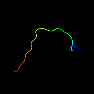
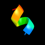
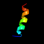
Phyre Home Retrieve Phyre Job Id
Emaill.a.kelley@imperial.ac.uk
DescriptionP64459
DateThu Jan 5 12:08:32 GMT 2012
Unique Job IDc51c91935c960550

Summary 

Top model
Image coloured by rainbow N → C terminus
Model (left) based on template c3bboF_
Top template information
PDB header:ribosome
Chain: F: PDB Molecule:ribosomal protein l3;
PDBTitle: homology model for the spinach chloroplast 50s subunit2 fitted to 9.4a cryo-em map of the 70s chlororibosome
Confidence and coverage
Confidence: 28.1% Coverage: 26%
20 residues ( 26% of your sequence) have been modelled with 28.1% confidence by the single highest scoring template.
You may wish to submit your sequence to Phyrealarm. This will automatically scan your sequence every week for new potential templates as they appear in the Phyre2 library.
Please note: You must be registered and logged in to use Phyrealarm.
3D viewing
Interactive 3D view in Jmol

Sequence analysis 

Secondary structure and disorder prediction 

   1........10.........20.........30.........40.........50.........60
Sequence  MFTKALSVVLLTCALFSGQLMAGHKGHEFVWVKNVDHQLRHEADSDELRAVAEESAEGLR
Secondary structure 


SS confidence 


Disorder  ???


?


????


Disorder confidence 


   .........70......
Sequence  EHFYWQKSRKPEAGQR
Secondary structure 


SS confidence 


Disorder 


???????????
Disorder confidence 


Confidence Key
High(9)                    Low (0)
?Disordered
Alpha helix
Beta strand

Domain analysis 

Hover over an aligned region to see model and summary info

Please note, only up to the top 20 hits are modelled to reduce computer load

RankAligned region

PDB 3bbo chain F

3D model

Region: 16 - 35
Aligned: 20
Modelled: 20
Confidence: 28.1%
Identity: 20%
PDB header:ribosome
Chain: F: PDB Molecule:ribosomal protein l3;
PDBTitle: homology model for the spinach chloroplast 50s subunit2 fitted to 9.4a cryo-em map of the 70s chlororibosome

Phyre2

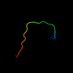
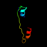
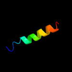
PDB 2gyc chain B domain 1

3D model

Region: 16 - 35
Aligned: 20
Modelled: 20
Confidence: 26.0%
Identity: 45%
Fold: Reductase/isomerase/elongation factor common domain
Superfamily: Translation proteins
Family: Ribosomal protein L3

Phyre2

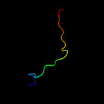
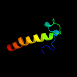
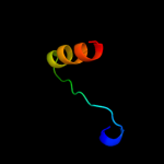
PDB 2zjr chain B domain 1

3D model

Region: 16 - 35
Aligned: 20
Modelled: 20
Confidence: 22.4%
Identity: 50%
Fold: Reductase/isomerase/elongation factor common domain
Superfamily: Translation proteins
Family: Ribosomal protein L3

Phyre2

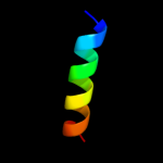
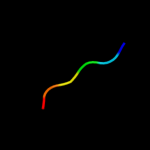
PDB 2j01 chain E domain 1

3D model

Region: 16 - 35
Aligned: 20
Modelled: 20
Confidence: 20.4%
Identity: 50%
Fold: Reductase/isomerase/elongation factor common domain
Superfamily: Translation proteins
Family: Ribosomal protein L3

Phyre2

PDB 2yqk chain A

3D model

Region: 55 - 74
Aligned: 20
Modelled: 20
Confidence: 19.7%
Identity: 35%
PDB header:transcription/apoptosis
Chain: A: PDB Molecule:arginine-glutamic acid dipeptide repeats protein;
PDBTitle: solution structure of the sant domain in arginine-glutamic2 acid dipeptide (re) repeats

Phyre2

PDB 2ftc chain C

3D model

Region: 16 - 35
Aligned: 20
Modelled: 20
Confidence: 16.7%
Identity: 20%
PDB header:ribosome
Chain: C: PDB Molecule:mitochondrial 39s ribosomal protein l3;
PDBTitle: structural model for the large subunit of the mammalian mitochondrial2 ribosome

Phyre2

PDB 1vqo chain B domain 1

3D model

Region: 16 - 35
Aligned: 20
Modelled: 20
Confidence: 16.2%
Identity: 20%
Fold: Reductase/isomerase/elongation factor common domain
Superfamily: Translation proteins
Family: Ribosomal protein L3

Phyre2

PDB 3jyw chain C

3D model

Region: 16 - 35
Aligned: 20
Modelled: 20
Confidence: 15.2%
Identity: 20%
PDB header:ribosome
Chain: C: PDB Molecule:60s ribosomal protein l3;
PDBTitle: structure of the 60s proteins for eukaryotic ribosome based on cryo-em2 map of thermomyces lanuginosus ribosome at 8.9a resolution

Phyre2

PDB 1s1i chain C

3D model

Region: 16 - 35
Aligned: 20
Modelled: 20
Confidence: 12.6%
Identity: 20%
PDB header:ribosome
Chain: C: PDB Molecule:60s ribosomal protein l3;
PDBTitle: structure of the ribosomal 80s-eef2-sordarin complex from2 yeast obtained by docking atomic models for rna and protein3 components into a 11.7 a cryo-em map. this file, 1s1i,4 contains 60s subunit. the 40s ribosomal subunit is in file5 1s1h.

Phyre2

PDB 2zkr chain B

3D model

Region: 16 - 35
Aligned: 20
Modelled: 20
Confidence: 10.5%
Identity: 25%
PDB header:ribosomal protein/rna
Chain: B: PDB Molecule:rna expansion segment es4;
PDBTitle: structure of a mammalian ribosomal 60s subunit within an2 80s complex obtained by docking homology models of the rna3 and proteins into an 8.7 a cryo-em map

Phyre2

PDB 2kwz chain A

3D model

Region: 8 - 15
Aligned: 8
Modelled: 8
Confidence: 10.4%
Identity: 75%
PDB header:viral protein
Chain: A: PDB Molecule:protease ns2-3;
PDBTitle: solution structure of ns2 [60-99]

Phyre2

PDB 1ni5 chain A

3D model

Region: 7 - 50
Aligned: 40
Modelled: 44
Confidence: 6.8%
Identity: 28%
PDB header:cell cycle
Chain: A: PDB Molecule:putative cell cycle protein mesj;
PDBTitle: structure of the mesj pp-atpase from escherichia coli

Phyre2

PDB 3du6 chain A

3D model

Region: 19 - 64
Aligned: 46
Modelled: 46
Confidence: 6.5%
Identity: 15%
PDB header:transferase
Chain: A: PDB Molecule:telomerase reverse transcriptase;
PDBTitle: structure of the catalytic subunit of telomerase, tert

Phyre2

PDB 1odp chain A

3D model

Region: 45 - 61
Aligned: 17
Modelled: 17
Confidence: 5.9%
Identity: 47%
PDB header:lipid transport
Chain: A: PDB Molecule:apoa-i peptide;
PDBTitle: peptide of human apoa-i residues 166-185. nmr, 5 structures2 at ph 6.6, 37 degrees celsius and peptide:sds mole ratio3 of 1:40

Phyre2

PDB 1odq chain A

3D model

Region: 45 - 61
Aligned: 17
Modelled: 17
Confidence: 5.9%
Identity: 47%
PDB header:lipid transport
Chain: A: PDB Molecule:apoa-i peptide;
PDBTitle: peptide of human apoa-i residues 166-185. nmr, 5 structures2 at ph 3.7, 37 degrees celsius and peptide:sds mole ratio3 of 1:40

Phyre2

PDB 1odr chain A

3D model

Region: 45 - 61
Aligned: 17
Modelled: 17
Confidence: 5.9%
Identity: 47%
PDB header:lipid transport
Chain: A: PDB Molecule:apoa-i peptide;
PDBTitle: peptide of human apoa-i residues 166-185. nmr, 5 structures2 at ph 6.0, 37 degrees celsius and peptide:dpc mole ratio3 of 1:40

Phyre2

PDB 3mtu chain D

3D model

Region: 52 - 72
Aligned: 21
Modelled: 21
Confidence: 5.8%
Identity: 38%
PDB header:contractile protein
Chain: D: PDB Molecule:tropomyosin alpha-1 chain, microtubule-associated protein
PDBTitle: structure of the tropomyosin overlap complex from chicken smooth2 muscle

Phyre2

PDB 2lci chain A

3D model

Region: 32 - 57
Aligned: 26
Modelled: 26
Confidence: 5.6%
Identity: 42%
PDB header:de novo protein
Chain: A: PDB Molecule:protein or36;
PDBTitle: solution nmr structure of de novo designed protein, p-loop ntpase2 fold, northeast structural genomics consortium target or36 (casd3 target)

Phyre2

PDB 3fd9 chain C

3D model

Region: 61 - 66
Aligned: 6
Modelled: 6
Confidence: 5.6%
Identity: 83%
PDB header:unknown function
Chain: C: PDB Molecule:uncharacterized protein;
PDBTitle: crystal structure of the transcriptional anti-activator exsd2 from pseudomonas aeruginosa

Phyre2
1

c3bboF_
2

d2gycb1
3

d2zjrb1
4

d2j01e1
5

c2yqkA_
6

c2ftcC_
7

d1vqob1
8

c3jywC_
9

c1s1iC_
10

c2zkrb_
11

c2kwzA_
12

c1ni5A_
13

c3du6A_
14

c1odpA_
15

c1odqA_
16

c1odrA_
17

c3mtuD_
18

c2lciA_
19

c3fd9C_


Detailed template information 

#
Template Alignment Coverage3D Model Confidence
% i.d. Template Information
1c3bboF_


28.1 20 PDB header:ribosome
Chain: F: PDB Molecule:ribosomal protein l3;
PDBTitle: homology model for the spinach chloroplast 50s subunit2 fitted to 9.4a cryo-em map of the 70s chlororibosome
2d2gycb1


26.0 45 Fold:Reductase/isomerase/elongation factor common domain
Superfamily:Translation proteins
Family:Ribosomal protein L3
3d2zjrb1


22.4 50 Fold:Reductase/isomerase/elongation factor common domain
Superfamily:Translation proteins
Family:Ribosomal protein L3
4d2j01e1


20.4 50 Fold:Reductase/isomerase/elongation factor common domain
Superfamily:Translation proteins
Family:Ribosomal protein L3
5c2yqkA_


19.7 35 PDB header:transcription/apoptosis
Chain: A: PDB Molecule:arginine-glutamic acid dipeptide repeats protein;
PDBTitle: solution structure of the sant domain in arginine-glutamic2 acid dipeptide (re) repeats
6c2ftcC_


16.7 20 PDB header:ribosome
Chain: C: PDB Molecule:mitochondrial 39s ribosomal protein l3;
PDBTitle: structural model for the large subunit of the mammalian mitochondrial2 ribosome
7d1vqob1


16.2 20 Fold:Reductase/isomerase/elongation factor common domain
Superfamily:Translation proteins
Family:Ribosomal protein L3
8c3jywC_


15.2 20 PDB header:ribosome
Chain: C: PDB Molecule:60s ribosomal protein l3;
PDBTitle: structure of the 60s proteins for eukaryotic ribosome based on cryo-em2 map of thermomyces lanuginosus ribosome at 8.9a resolution
9c1s1iC_


12.6 20 PDB header:ribosome
Chain: C: PDB Molecule:60s ribosomal protein l3;
PDBTitle: structure of the ribosomal 80s-eef2-sordarin complex from2 yeast obtained by docking atomic models for rna and protein3 components into a 11.7 a cryo-em map. this file, 1s1i,4 contains 60s subunit. the 40s ribosomal subunit is in file5 1s1h.
10c2zkrb_


10.5 25 PDB header:ribosomal protein/rna
Chain: B: PDB Molecule:rna expansion segment es4;
PDBTitle: structure of a mammalian ribosomal 60s subunit within an2 80s complex obtained by docking homology models of the rna3 and proteins into an 8.7 a cryo-em map
11c2kwzA_


10.4 75 PDB header:viral protein
Chain: A: PDB Molecule:protease ns2-3;
PDBTitle: solution structure of ns2 [60-99]
12c1ni5A_


6.8 28 PDB header:cell cycle
Chain: A: PDB Molecule:putative cell cycle protein mesj;
PDBTitle: structure of the mesj pp-atpase from escherichia coli
13c3du6A_


6.5 15 PDB header:transferase
Chain: A: PDB Molecule:telomerase reverse transcriptase;
PDBTitle: structure of the catalytic subunit of telomerase, tert
14c1odpA_


5.9 47 PDB header:lipid transport
Chain: A: PDB Molecule:apoa-i peptide;
PDBTitle: peptide of human apoa-i residues 166-185. nmr, 5 structures2 at ph 6.6, 37 degrees celsius and peptide:sds mole ratio3 of 1:40
15c1odqA_


5.9 47 PDB header:lipid transport
Chain: A: PDB Molecule:apoa-i peptide;
PDBTitle: peptide of human apoa-i residues 166-185. nmr, 5 structures2 at ph 3.7, 37 degrees celsius and peptide:sds mole ratio3 of 1:40
16c1odrA_


5.9 47 PDB header:lipid transport
Chain: A: PDB Molecule:apoa-i peptide;
PDBTitle: peptide of human apoa-i residues 166-185. nmr, 5 structures2 at ph 6.0, 37 degrees celsius and peptide:dpc mole ratio3 of 1:40
17c3mtuD_


5.8 38 PDB header:contractile protein
Chain: D: PDB Molecule:tropomyosin alpha-1 chain, microtubule-associated protein
PDBTitle: structure of the tropomyosin overlap complex from chicken smooth2 muscle
18c2lciA_


5.6 42 PDB header:de novo protein
Chain: A: PDB Molecule:protein or36;
PDBTitle: solution nmr structure of de novo designed protein, p-loop ntpase2 fold, northeast structural genomics consortium target or36 (casd3 target)
19c3fd9C_


5.6 83 PDB header:unknown function
Chain: C: PDB Molecule:uncharacterized protein;
PDBTitle: crystal structure of the transcriptional anti-activator exsd2 from pseudomonas aeruginosa

Binding site prediction 

Due to computational demand, binding site predictions are not run for batch jobs

If you want to predict binding sites, please manually submit your model of choice to 3DLigandSite


Phyre is for academic use only

Please cite: Protein structure prediction on the web: a case study using the Phyre server
Kelley LA and Sternberg MJE. Nature Protocols 4, 363 - 371 (2009) [pdf] [Import into BibTeX]
 
If you use the binding site predictions from 3DLigandSite, please also cite:
3DLigandSite: predicting ligand-binding sites using similar structures.
Wass MN, Kelley LA and Sternberg MJ Nucleic Acids Research 38, W469-73 (2010) [PubMed]
 
© Structural Bioinformatics Group
Imperial College London
Lawrence Kelley, Benjamin Jefferys
Disclaimer
Terms and Conditions
Component software
Template detection: HHpred 1.51
Secondary structure prediction: Psi-pred 2.5
Disorder prediction: Disopred 2.4
Transmembrane prediction: Memsat_SVM
Multi-template modelling and ab initio: Poing 1.0