| 1 | c1v9fA_
|
|
 |
100.0 |
28 |
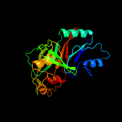
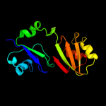
PDB header:lyase
Chain: A: PDB Molecule:ribosomal large subunit pseudouridine synthase d;
PDBTitle: crystal structure of catalytic domain of pseudouridine2 synthase rlud from escherichia coli
|
| 2 | d1v9fa_
|
|
 |
100.0 |
28 |
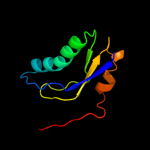
Fold:Pseudouridine synthase
Superfamily:Pseudouridine synthase
Family:Pseudouridine synthase RsuA/RluD |
| 3 | c1qyuA_
|
|
 |
100.0 |
27 |
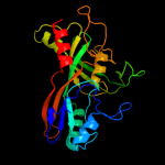
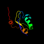
PDB header:lyase
Chain: A: PDB Molecule:ribosomal large subunit pseudouridine synthase d;
PDBTitle: structure of the catalytic domain of 23s rrna pseudouridine2 synthase rlud
|
| 4 | d1v9ka_
|
|
 |
100.0 |
34 |
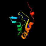
Fold:Pseudouridine synthase
Superfamily:Pseudouridine synthase
Family:Pseudouridine synthase RsuA/RluD |
| 5 | c2i82D_
|
|
 |
100.0 |
36 |
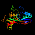
PDB header:lyase/rna
Chain: D: PDB Molecule:ribosomal large subunit pseudouridine synthase a;
PDBTitle: crystal structure of pseudouridine synthase rlua: indirect2 sequence readout through protein-induced rna structure
|
| 6 | c1vioA_
|
|
 |
100.0 |
21 |
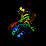
PDB header:lyase
Chain: A: PDB Molecule:ribosomal small subunit pseudouridine synthase a;
PDBTitle: crystal structure of pseudouridylate synthase
|
| 7 | c1kskA_
|
|
 |
100.0 |
19 |
PDB header:lyase
Chain: A: PDB Molecule:ribosomal small subunit pseudouridine synthase a;
PDBTitle: structure of rsua
|
| 8 | c2omlA_
|
|
 |
100.0 |
19 |
PDB header:isomerase
Chain: A: PDB Molecule:ribosomal large subunit pseudouridine synthase e;
PDBTitle: crystal structure of e. coli pseudouridine synthase rlue
|
| 9 | c2olwB_
|
|
 |
100.0 |
19 |
PDB header:isomerase
Chain: B: PDB Molecule:ribosomal large subunit pseudouridine synthase e;
PDBTitle: crystal structure of e. coli pseudouridine synthase rlue
|
| 10 | c3dh3C_
|
|
 |
100.0 |
20 |
PDB header:isomerase/rna
Chain: C: PDB Molecule:ribosomal large subunit pseudouridine synthase f;
PDBTitle: crystal structure of rluf in complex with a 22 nucleotide2 rna substrate
|
| 11 | d1kska4
|
|
 |
100.0 |
18 |
Fold:Pseudouridine synthase
Superfamily:Pseudouridine synthase
Family:Pseudouridine synthase RsuA/RluD |
| 12 | d1vioa1
|
|
 |
100.0 |
21 |
Fold:Pseudouridine synthase
Superfamily:Pseudouridine synthase
Family:Pseudouridine synthase RsuA/RluD |
| 13 | c2gmlA_
|
|
 |
100.0 |
21 |
PDB header:isomerase
Chain: A: PDB Molecule:ribosomal large subunit pseudouridine synthase f;
PDBTitle: crystal structure of catalytic domain of e.coli rluf
|
| 14 | d1k8wa5
|
|
 |
97.7 |
20 |
Fold:Pseudouridine synthase
Superfamily:Pseudouridine synthase
Family:Pseudouridine synthase II TruB |
| 15 | d1sgva2
|
|
 |
97.6 |
27 |
Fold:Pseudouridine synthase
Superfamily:Pseudouridine synthase
Family:Pseudouridine synthase II TruB |
| 16 | d2apoa2
|
|
 |
97.5 |
22 |
Fold:Pseudouridine synthase
Superfamily:Pseudouridine synthase
Family:Pseudouridine synthase II TruB |
| 17 | d2ey4a2
|
|
 |
97.5 |
23 |
Fold:Pseudouridine synthase
Superfamily:Pseudouridine synthase
Family:Pseudouridine synthase II TruB |
| 18 | c2ey4A_
|
|
 |
97.4 |
23 |
PDB header:isomerase/biosynthetic protein
Chain: A: PDB Molecule:probable trna pseudouridine synthase b;
PDBTitle: crystal structure of a cbf5-nop10-gar1 complex
|
| 19 | d1r3ea2
|
|
 |
97.4 |
20 |
Fold:Pseudouridine synthase
Superfamily:Pseudouridine synthase
Family:Pseudouridine synthase II TruB |
| 20 | c3uaiA_
|
|
 |
97.1 |
26 |
PDB header:isomerase/chaperone
Chain: A: PDB Molecule:h/aca ribonucleoprotein complex subunit 4;
PDBTitle: structure of the shq1-cbf5-nop10-gar1 complex from saccharomyces2 cerevisiae
|
| 21 | c2apoA_ |
|
not modelled |
97.0 |
21 |
PDB header:isomerase/rna binding protein
Chain: A: PDB Molecule:probable trna pseudouridine synthase b;
PDBTitle: crystal structure of the methanococcus jannaschii cbf52 nop10 complex
|
| 22 | c1k8wA_ |
|
not modelled |
97.0 |
20 |
PDB header:lyase/rna
Chain: A: PDB Molecule:trna pseudouridine synthase b;
PDBTitle: crystal structure of the e. coli pseudouridine synthase2 trub bound to a t stem-loop rna
|
| 23 | c1sgvA_ |
|
not modelled |
96.6 |
27 |
PDB header:lyase
Chain: A: PDB Molecule:trna pseudouridine synthase b;
PDBTitle: structure of trna psi55 pseudouridine synthase (trub)
|
| 24 | c1ze2B_ |
|
not modelled |
95.1 |
28 |
PDB header:lyase/rna
Chain: B: PDB Molecule:trna pseudouridine synthase b;
PDBTitle: conformational change of pseudouridine 55 synthase upon its2 association with rna substrate
|
| 25 | c3n1tE_ |
|
not modelled |
24.0 |
42 |
PDB header:hydrolase
Chain: E: PDB Molecule:hit-like protein hint;
PDBTitle: crystal structure of the h101a mutant echint gmp complex
|
| 26 | d1rzya_ |
|
not modelled |
22.8 |
25 |
Fold:HIT-like
Superfamily:HIT-like
Family:HIT (HINT, histidine triad) family of protein kinase-interacting proteins |
| 27 | c2zodB_ |
|
not modelled |
21.9 |
23 |
PDB header:transferase
Chain: B: PDB Molecule:selenide, water dikinase;
PDBTitle: crystal structure of selenophosphate synthetase from2 aquifex aeolicus
|
| 28 | c3l7xA_ |
|
not modelled |
21.4 |
33 |
PDB header:cell cycle
Chain: A: PDB Molecule:putative hit-like protein involved in cell-cycle
PDBTitle: the crystal structure of smu.412c from streptococcus mutans ua159
|
| 29 | d1emsa1 |
|
not modelled |
20.0 |
17 |
Fold:HIT-like
Superfamily:HIT-like
Family:HIT (HINT, histidine triad) family of protein kinase-interacting proteins |
| 30 | c1mzwB_ |
|
not modelled |
18.7 |
31 |
PDB header:isomerase
Chain: B: PDB Molecule:u4/u6 snrnp 60kda protein;
PDBTitle: crystal structure of a u4/u6 snrnp complex between human2 spliceosomal cyclophilin h and a u4/u6-60k peptide
|
| 31 | d1kpfa_ |
|
not modelled |
16.6 |
25 |
Fold:HIT-like
Superfamily:HIT-like
Family:HIT (HINT, histidine triad) family of protein kinase-interacting proteins |
| 32 | d1fita_ |
|
not modelled |
15.3 |
18 |
Fold:HIT-like
Superfamily:HIT-like
Family:HIT (HINT, histidine triad) family of protein kinase-interacting proteins |
| 33 | c3o0mB_ |
|
not modelled |
15.0 |
25 |
PDB header:hydrolase
Chain: B: PDB Molecule:hit family protein;
PDBTitle: crystal structure of a zn-bound histidine triad family protein from2 mycobacterium smegmatis
|
| 34 | c1emsB_ |
|
not modelled |
14.7 |
17 |
PDB header:antitumor protein
Chain: B: PDB Molecule:nit-fragile histidine triad fusion protein;
PDBTitle: crystal structure of the c. elegans nitfhit protein
|
| 35 | c2zauB_ |
|
not modelled |
13.0 |
24 |
PDB header:transferase
Chain: B: PDB Molecule:selenide, water dikinase;
PDBTitle: crystal structure of an n-terminally truncated2 selenophosphate synthetase from aquifex aeolicus
|
| 36 | d1guqa2 |
|
not modelled |
12.2 |
25 |
Fold:HIT-like
Superfamily:HIT-like
Family:Hexose-1-phosphate uridylyltransferase |
| 37 | c3p0tB_ |
|
not modelled |
12.0 |
33 |
PDB header:unknown function
Chain: B: PDB Molecule:uncharacterized protein;
PDBTitle: crystal structure of an hit-like protein from mycobacterium2 paratuberculosis
|
| 38 | d1qwga_ |
|
not modelled |
11.1 |
14 |
Fold:TIM beta/alpha-barrel
Superfamily:(2r)-phospho-3-sulfolactate synthase ComA
Family:(2r)-phospho-3-sulfolactate synthase ComA |
| 39 | d2oika1 |
|
not modelled |
10.2 |
50 |
Fold:HIT-like
Superfamily:HIT-like
Family:HIT (HINT, histidine triad) family of protein kinase-interacting proteins |
| 40 | c3lb5B_ |
|
not modelled |
10.1 |
23 |
PDB header:cell cycle
Chain: B: PDB Molecule:hit-like protein involved in cell-cycle regulation;
PDBTitle: crystal structure of hit-like protein involved in cell-cycle2 regulation from bartonella henselae with unknown ligand
|
| 41 | c2jv2A_ |
|
not modelled |
10.0 |
7 |
PDB header:unknown function
Chain: A: PDB Molecule:putative uncharacterized protein ph1500;
PDBTitle: solution structure of the n-terminal domain of ph1500
|
| 42 | d2aaaa1 |
|
not modelled |
10.0 |
29 |
Fold:Glycosyl hydrolase domain
Superfamily:Glycosyl hydrolase domain
Family:alpha-Amylases, C-terminal beta-sheet domain |
| 43 | c1xquA_ |
|
not modelled |
9.6 |
42 |
PDB header:hydrolase
Chain: A: PDB Molecule:hit family hydrolase;
PDBTitle: hit family hydrolase from clostridium thermocellum cth-393
|
| 44 | d1xqua_ |
|
not modelled |
9.6 |
42 |
Fold:HIT-like
Superfamily:HIT-like
Family:HIT (HINT, histidine triad) family of protein kinase-interacting proteins |
| 45 | c3zrhA_ |
|
not modelled |
9.4 |
31 |
PDB header:hydrolase
Chain: A: PDB Molecule:ubiquitin thioesterase zranb1;
PDBTitle: crystal structure of the lys29, lys33-linkage-specific trabid otu2 deubiquitinase domain reveals an ankyrin-repeat ubiquitin binding3 domain (ankubd)
|
| 46 | d1gr0a1 |
|
not modelled |
9.3 |
21 |
Fold:NAD(P)-binding Rossmann-fold domains
Superfamily:NAD(P)-binding Rossmann-fold domains
Family:Glyceraldehyde-3-phosphate dehydrogenase-like, N-terminal domain |
| 47 | c2lf6A_ |
|
not modelled |
9.2 |
24 |
PDB header:signaling protein
Chain: A: PDB Molecule:effector protein hopab1;
PDBTitle: solution nmr structure of hopabpph1448_220_320 from pseudomonas2 syringae pv. phaseolicola str. 1448a, midwest center for structural3 genomics target apc40132.4 and northeast structural genomics4 consortium target pst3a
|
| 48 | d2zoda2 |
|
not modelled |
8.4 |
17 |
Fold:PurM C-terminal domain-like
Superfamily:PurM C-terminal domain-like
Family:PurM C-terminal domain-like |
| 49 | d1vjpa1 |
|
not modelled |
7.8 |
12 |
Fold:NAD(P)-binding Rossmann-fold domains
Superfamily:NAD(P)-binding Rossmann-fold domains
Family:Glyceraldehyde-3-phosphate dehydrogenase-like, N-terminal domain |
| 50 | c2do5A_ |
|
not modelled |
7.8 |
35 |
PDB header:structural genomics, unknown function
Chain: A: PDB Molecule:splicing factor 3b subunit 2;
PDBTitle: solution structure of the sap domain of human splicing2 factor 3b subunit 2
|
| 51 | c3ksvA_ |
|
not modelled |
7.7 |
8 |
PDB header:unknown function
Chain: A: PDB Molecule:uncharacterized protein;
PDBTitle: hypothetical protein from leishmania major
|
| 52 | c2oi2A_ |
|
not modelled |
7.5 |
13 |
PDB header:transferase
Chain: A: PDB Molecule:mevalonate kinase;
PDBTitle: streptococcus pneumoniae mevalonate kinase in complex with2 diphosphomevalonate
|
| 53 | c2eo4A_ |
|
not modelled |
7.4 |
36 |
PDB header:hydrolase
Chain: A: PDB Molecule:150aa long hypothetical histidine triad nucleotide-binding
PDBTitle: crystal structure of hypothetical histidine triad nucleotide-binding2 protein st2152 from sulfolobus tokodaii strain7
|
| 54 | c3oj7A_ |
|
not modelled |
6.9 |
33 |
PDB header:metal binding protein
Chain: A: PDB Molecule:putative histidine triad family protein;
PDBTitle: crystal structure of a histidine triad family protein from entamoeba2 histolytica, bound to sulfate
|
| 55 | d1k8ga2 |
|
not modelled |
6.8 |
27 |
Fold:OB-fold
Superfamily:Nucleic acid-binding proteins
Family:Single strand DNA-binding domain, SSB |
| 56 | c2lkyA_ |
|
not modelled |
6.8 |
33 |
PDB header:structural genomics, unknown function
Chain: A: PDB Molecule:uncharacterized protein;
PDBTitle: solution structure of msmeg_1053, the second duf3349 annotated protein2 in the genome of mycobacterium smegmatis, seattle structural genomics3 center for infectious disease target mysma.17112.b
|
| 57 | c1u83A_ |
|
not modelled |
6.6 |
14 |
PDB header:lyase
Chain: A: PDB Molecule:phosphosulfolactate synthase;
PDBTitle: psl synthase from bacillus subtilis
|
| 58 | d1u83a_ |
|
not modelled |
6.6 |
14 |
Fold:TIM beta/alpha-barrel
Superfamily:(2r)-phospho-3-sulfolactate synthase ComA
Family:(2r)-phospho-3-sulfolactate synthase ComA |
| 59 | c3imiB_ |
|
not modelled |
6.4 |
33 |
PDB header:structural genomics, unknown function
Chain: B: PDB Molecule:hit family protein;
PDBTitle: 2.01 angstrom resolution crystal structure of a hit family protein2 from bacillus anthracis str. 'ames ancestor'
|
| 60 | c3anoA_ |
|
not modelled |
6.4 |
18 |
PDB header:transferase
Chain: A: PDB Molecule:ap-4-a phosphorylase;
PDBTitle: crystal structure of a novel diadenosine 5',5'''-p1,p4-tetraphosphate2 phosphorylase from mycobacterium tuberculosis h37rv
|
| 61 | c1e0aB_ |
|
not modelled |
6.4 |
28 |
PDB header:signalling protein
Chain: B: PDB Molecule:serine/threonine-protein kinase pak-alpha;
PDBTitle: cdc42 complexed with the gtpase binding domain of p212 activated kinase
|
| 62 | c2kvcA_ |
|
not modelled |
6.4 |
31 |
PDB header:unknown function
Chain: A: PDB Molecule:putative uncharacterized protein;
PDBTitle: solution structure of the mycobacterium tuberculosis protein rv0543c,2 a member of the duf3349 superfamily. seattle structural genomics3 center for infectious disease target mytud.17112.a
|
| 63 | d1u2ca2 |
|
not modelled |
6.2 |
27 |
Fold:Dystroglycan, domain 2
Superfamily:Dystroglycan, domain 2
Family:Dystroglycan, domain 2 |
| 64 | d1jb7a2 |
|
not modelled |
6.1 |
27 |
Fold:OB-fold
Superfamily:Nucleic acid-binding proteins
Family:Single strand DNA-binding domain, SSB |
| 65 | d1w91a1 |
|
not modelled |
6.1 |
36 |
Fold:Glycosyl hydrolase domain
Superfamily:Glycosyl hydrolase domain
Family:Composite domain of glycosyl hydrolase families 5, 30, 39 and 51 |
| 66 | d2al1a1 |
|
not modelled |
6.1 |
11 |
Fold:TIM beta/alpha-barrel
Superfamily:Enolase C-terminal domain-like
Family:Enolase |
| 67 | d2guya1 |
|
not modelled |
6.0 |
27 |
Fold:Glycosyl hydrolase domain
Superfamily:Glycosyl hydrolase domain
Family:alpha-Amylases, C-terminal beta-sheet domain |
| 68 | c2odbB_ |
|
not modelled |
6.0 |
31 |
PDB header:protein binding
Chain: B: PDB Molecule:serine/threonine-protein kinase pak 6;
PDBTitle: the crystal structure of human cdc42 in complex with the crib domain2 of human p21-activated kinase 6 (pak6)
|
| 69 | c1u1iC_ |
|
not modelled |
5.7 |
16 |
PDB header:isomerase
Chain: C: PDB Molecule:myo-inositol-1-phosphate synthase;
PDBTitle: myo-inositol phosphate synthase mips from a. fulgidus
|
| 70 | d1z84a2 |
|
not modelled |
5.4 |
17 |
Fold:HIT-like
Superfamily:HIT-like
Family:Hexose-1-phosphate uridylyltransferase |
| 71 | d2hx5a1 |
|
not modelled |
5.1 |
14 |
Fold:Thioesterase/thiol ester dehydrase-isomerase
Superfamily:Thioesterase/thiol ester dehydrase-isomerase
Family:4HBT-like |