Phyre Home Retrieve Phyre Job Id
Emaill.a.kelley@imperial.ac.uk
DescriptionP52613
DateThu Jan 5 12:05:52 GMT 2012
Unique Job IDa04a733af6bfa916

Summary 

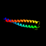
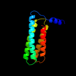
Top model
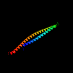
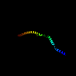
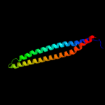
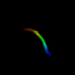
Image coloured by rainbow N → C terminus
Model (left) based on template c3ajwA_
Top template information
PDB header:protein transport
Chain: A: PDB Molecule:flagellar flij protein;
PDBTitle: structure of flij, a soluble component of flagellar type iii export2 apparatus
Confidence and coverage
Confidence: 99.6% Coverage: 91%
134 residues ( 91% of your sequence) have been modelled with 99.6% confidence by the single highest scoring template.
3D viewing
Interactive 3D view in Jmol

Sequence analysis 

Secondary structure and disorder prediction 

   1........10.........20.........30.........40.........50.........60
Sequence  MAEHGALATLKDLAEKEVEDAARLLGEMRRGCQQAEEQLKMLIDYQNEYRNNLNSDMSAG
Secondary structure 


SS confidence 


Disorder  ???????


???
Disorder confidence 


   .........70.........80.........90.........100.........110.........120
Sequence  ITSNRWINYQQFIQTLEKAITQHRQQLNQWTQKVDIALNSWREKKQRLQAWQTLQERQST
Secondary structure 

SS confidence 


Disorder  ??


Disorder confidence 


   .........130.........140.......
Sequence  AALLAENRLDQKKMDEFAQRAAMRKPE
Secondary structure 


SS confidence 


Disorder 


????
Disorder confidence 


Confidence Key
High(9)                    Low (0)
?Disordered
Alpha helix
Beta strand

Domain analysis 

Hover over an aligned region to see model and summary info

Please note, only up to the top 20 hits are modelled to reduce computer load

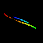
RankAligned region

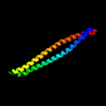
PDB 3ajw chain A

3D model

Region: 3 - 138
Aligned: 134
Modelled: 136
Confidence: 99.6%
Identity: 87%
PDB header:protein transport
Chain: A: PDB Molecule:flagellar flij protein;
PDBTitle: structure of flij, a soluble component of flagellar type iii export2 apparatus

Phyre2

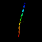
PDB 3k29 chain A

3D model

Region: 5 - 143
Aligned: 139
Modelled: 139
Confidence: 92.3%
Identity: 19%
PDB header:unknown function
Chain: A: PDB Molecule:putative uncharacterized protein;
PDBTitle: structure of a putative ysco homolog ct670 from chlamydia trachomatis

Phyre2

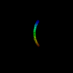
PDB 3u59 chain C

3D model

Region: 11 - 113
Aligned: 97
Modelled: 103
Confidence: 80.2%
Identity: 12%
PDB header:contractile protein
Chain: C: PDB Molecule:tropomyosin beta chain;
PDBTitle: n-terminal 98-aa fragment of smooth muscle tropomyosin beta

Phyre2

PDB 2fxm chain B

3D model

Region: 6 - 113
Aligned: 108
Modelled: 108
Confidence: 77.3%
Identity: 9%
PDB header:contractile protein
Chain: B: PDB Molecule:myosin heavy chain, cardiac muscle beta isoform;
PDBTitle: structure of the human beta-myosin s2 fragment

Phyre2

PDB 1deq chain F

3D model

Region: 5 - 115
Aligned: 109
Modelled: 111
Confidence: 74.3%
Identity: 10%
PDB header:
PDB COMPND:

Phyre2

PDB 3oja chain B

3D model

Region: 5 - 139
Aligned: 135
Modelled: 135
Confidence: 66.9%
Identity: 9%
PDB header:protein binding
Chain: B: PDB Molecule:anopheles plasmodium-responsive leucine-rich repeat protein
PDBTitle: crystal structure of lrim1/apl1c complex

Phyre2

PDB 2d3e chain D

3D model

Region: 11 - 135
Aligned: 125
Modelled: 125
Confidence: 65.8%
Identity: 13%
PDB header:contractile protein
Chain: D: PDB Molecule:general control protein gcn4 and tropomyosin 1
PDBTitle: crystal structure of the c-terminal fragment of rabbit2 skeletal alpha-tropomyosin

Phyre2

PDB 3ol1 chain A

3D model

Region: 12 - 109
Aligned: 98
Modelled: 98
Confidence: 46.3%
Identity: 16%
PDB header:structural protein
Chain: A: PDB Molecule:vimentin;
PDBTitle: crystal structure of vimentin (fragment 144-251) from homo sapiens,2 northeast structural genomics consortium target hr4796b

Phyre2

PDB 2y3a chain B

3D model

Region: 5 - 121
Aligned: 117
Modelled: 117
Confidence: 41.3%
Identity: 7%
PDB header:transferase
Chain: B: PDB Molecule:phosphatidylinositol 3-kinase regulatory subunit beta;
PDBTitle: crystal structure of p110beta in complex with icsh2 of p85beta and2 the drug gdc-0941

Phyre2

PDB 1l8d chain B

3D model

Region: 7 - 109
Aligned: 102
Modelled: 103
Confidence: 33.4%
Identity: 8%
PDB header:replication
Chain: B: PDB Molecule:dna double-strand break repair rad50 atpase;
PDBTitle: rad50 coiled-coil zn hook

Phyre2

PDB 4a55 chain B

3D model

Region: 7 - 130
Aligned: 124
Modelled: 124
Confidence: 33.1%
Identity: 5%
PDB header:transferase
Chain: B: PDB Molecule:phosphatidylinositol 3-kinase regulatory subunit alpha;
PDBTitle: crystal structure of p110alpha in complex with ish2 of p85alpha and2 the inhibitor pik-108

Phyre2

PDB 2b9c chain A

3D model

Region: 2 - 129
Aligned: 122
Modelled: 128
Confidence: 29.2%
Identity: 11%
PDB header:contractile protein
Chain: A: PDB Molecule:striated-muscle alpha tropomyosin;
PDBTitle: structure of tropomyosin's mid-region: bending and binding2 sites for actin

Phyre2

PDB 3ipk chain A

3D model

Region: 7 - 112
Aligned: 101
Modelled: 101
Confidence: 26.2%
Identity: 7%
PDB header:cell adhesion
Chain: A: PDB Molecule:agi/ii;
PDBTitle: crystal structure of a3vp1 of agi/ii of streptococcus mutans

Phyre2

PDB 1y4c chain A

3D model

Region: 14 - 138
Aligned: 125
Modelled: 125
Confidence: 26.0%
Identity: 11%
PDB header:de novo protein
Chain: A: PDB Molecule:maltose binding protein fused with designed
PDBTitle: designed helical protein fusion mbp

Phyre2

PDB 2gl2 chain B

3D model

Region: 16 - 121
Aligned: 100
Modelled: 106
Confidence: 20.3%
Identity: 7%
PDB header:cell adhesion
Chain: B: PDB Molecule:adhesion a;
PDBTitle: crystal structure of the tetra muntant (t66g,r67g,f68g,2 y69g) of bacterial adhesin fada

Phyre2

PDB 2rd0 chain B

3D model

Region: 7 - 117
Aligned: 111
Modelled: 111
Confidence: 13.1%
Identity: 6%
PDB header:transferase/oncoprotein
Chain: B: PDB Molecule:phosphatidylinositol 3-kinase regulatory subunit alpha;
PDBTitle: structure of a human p110alpha/p85alpha complex

Phyre2

PDB 3o0z chain D

3D model

Region: 6 - 138
Aligned: 127
Modelled: 133
Confidence: 11.2%
Identity: 12%
PDB header:transferase
Chain: D: PDB Molecule:rho-associated protein kinase 1;
PDBTitle: crystal structure of a coiled-coil domain from human rock i

Phyre2

PDB 1ei3 chain E

3D model

Region: 7 - 101
Aligned: 94
Modelled: 95
Confidence: 11.1%
Identity: 10%
PDB header:
PDB COMPND:

Phyre2

PDB 1deq chain O

3D model

Region: 7 - 90
Aligned: 84
Modelled: 84
Confidence: 9.7%
Identity: 11%
PDB header:
PDB COMPND:

Phyre2

PDB 1deq chain D

3D model

Region: 12 - 95
Aligned: 78
Modelled: 84
Confidence: 9.5%
Identity: 9%
PDB header:
PDB COMPND:

Phyre2
1

c3ajwA_
2

c3k29A_
3

c3u59C_
4

c2fxmB_
5

c1deqF_
6

c3ojaB_
7

c2d3eD_
8

c3ol1A_
9

c2y3aB_
10

c1l8dB_
11

c4a55B_
12

c2b9cA_
13

c3ipkA_
14

c1y4cA_
15

c2gl2B_
16

c2rd0B_
17

c3o0zD_
18

c1ei3E_
19

c1deqO_
20

c1deqD_
21


22


23


24


25


26


27


28


29


Detailed template information 

#
Template Alignment Coverage3D Model Confidence
% i.d. Template Information
1c3ajwA_


99.6 87 PDB header:protein transport
Chain: A: PDB Molecule:flagellar flij protein;
PDBTitle: structure of flij, a soluble component of flagellar type iii export2 apparatus
2c3k29A_


92.3 19 PDB header:unknown function
Chain: A: PDB Molecule:putative uncharacterized protein;
PDBTitle: structure of a putative ysco homolog ct670 from chlamydia trachomatis
3c3u59C_


80.2 12 PDB header:contractile protein
Chain: C: PDB Molecule:tropomyosin beta chain;
PDBTitle: n-terminal 98-aa fragment of smooth muscle tropomyosin beta
4c2fxmB_


77.3 9 PDB header:contractile protein
Chain: B: PDB Molecule:myosin heavy chain, cardiac muscle beta isoform;
PDBTitle: structure of the human beta-myosin s2 fragment
5c1deqF_


74.3 10 PDB header:
PDB COMPND:
6c3ojaB_


66.9 9 PDB header:protein binding
Chain: B: PDB Molecule:anopheles plasmodium-responsive leucine-rich repeat protein
PDBTitle: crystal structure of lrim1/apl1c complex
7c2d3eD_


65.8 13 PDB header:contractile protein
Chain: D: PDB Molecule:general control protein gcn4 and tropomyosin 1
PDBTitle: crystal structure of the c-terminal fragment of rabbit2 skeletal alpha-tropomyosin
8c3ol1A_


46.3 16 PDB header:structural protein
Chain: A: PDB Molecule:vimentin;
PDBTitle: crystal structure of vimentin (fragment 144-251) from homo sapiens,2 northeast structural genomics consortium target hr4796b
9c2y3aB_


41.3 7 PDB header:transferase
Chain: B: PDB Molecule:phosphatidylinositol 3-kinase regulatory subunit beta;
PDBTitle: crystal structure of p110beta in complex with icsh2 of p85beta and2 the drug gdc-0941
10c1l8dB_


33.4 8 PDB header:replication
Chain: B: PDB Molecule:dna double-strand break repair rad50 atpase;
PDBTitle: rad50 coiled-coil zn hook
11c4a55B_


33.1 5 PDB header:transferase
Chain: B: PDB Molecule:phosphatidylinositol 3-kinase regulatory subunit alpha;
PDBTitle: crystal structure of p110alpha in complex with ish2 of p85alpha and2 the inhibitor pik-108
12c2b9cA_


29.2 11 PDB header:contractile protein
Chain: A: PDB Molecule:striated-muscle alpha tropomyosin;
PDBTitle: structure of tropomyosin's mid-region: bending and binding2 sites for actin
13c3ipkA_


26.2 7 PDB header:cell adhesion
Chain: A: PDB Molecule:agi/ii;
PDBTitle: crystal structure of a3vp1 of agi/ii of streptococcus mutans
14c1y4cA_


26.0 11 PDB header:de novo protein
Chain: A: PDB Molecule:maltose binding protein fused with designed
PDBTitle: designed helical protein fusion mbp
15c2gl2B_


20.3 7 PDB header:cell adhesion
Chain: B: PDB Molecule:adhesion a;
PDBTitle: crystal structure of the tetra muntant (t66g,r67g,f68g,2 y69g) of bacterial adhesin fada
16c2rd0B_


13.1 6 PDB header:transferase/oncoprotein
Chain: B: PDB Molecule:phosphatidylinositol 3-kinase regulatory subunit alpha;
PDBTitle: structure of a human p110alpha/p85alpha complex
17c3o0zD_


11.2 12 PDB header:transferase
Chain: D: PDB Molecule:rho-associated protein kinase 1;
PDBTitle: crystal structure of a coiled-coil domain from human rock i
18c1ei3E_


11.1 10 PDB header:
PDB COMPND:
19c1deqO_


9.7 11 PDB header:
PDB COMPND:
20c1deqD_


9.5 9 PDB header:
PDB COMPND:
21c2efrB_


not modelled 9.4 18 PDB header:contractile protein
Chain: B: PDB Molecule:general control protein gcn4 and tropomyosin 1 alpha chain;
PDBTitle: crystal structure of the c-terminal tropomyosin fragment with n- and2 c-terminal extensions of the leucine zipper at 1.8 angstroms3 resolution
22c3ghgK_


not modelled 8.7 9 PDB header:blood clotting
Chain: K: PDB Molecule:fibrinogen beta chain;
PDBTitle: crystal structure of human fibrinogen
23c3hizB_


not modelled 8.3 6 PDB header:transferase/oncoprotein
Chain: B: PDB Molecule:phosphatidylinositol 3-kinase regulatory subunit
PDBTitle: crystal structure of p110alpha h1047r mutant in complex with2 nish2 of p85alpha
24c3dtpA_


not modelled 8.1 10 PDB header:contractile protein
Chain: A: PDB Molecule:myosin 2 heavy chain chimera of smooth and
PDBTitle: tarantula heavy meromyosin obtained by flexible docking to2 tarantula muscle thick filament cryo-em 3d-map
25c1jchC_


not modelled 7.9 12 PDB header:ribosome inhibitor, hydrolase
Chain: C: PDB Molecule:colicin e3;
PDBTitle: crystal structure of colicin e3 in complex with its immunity protein
26c1gk4A_


not modelled 7.0 18 PDB header:vimentin
Chain: A: PDB Molecule:vimentin;
PDBTitle: human vimentin coil 2b fragment (cys2)
27c1gk6B_


not modelled 6.2 12 PDB header:vimentin
Chain: B: PDB Molecule:vimentin;
PDBTitle: human vimentin coil 2b fragment linked to gcn4 leucine2 zipper (z2b)
28c2v71A_


not modelled 5.8 10 PDB header:nuclear protein
Chain: A: PDB Molecule:nuclear distribution protein nude-like 1;
PDBTitle: coiled-coil region of nudel
29c1g6uB_


not modelled 5.6 15 PDB header:de novo protein
Chain: B: PDB Molecule:domain swapped dimer;
PDBTitle: crystal structure of a domain swapped dimer

Binding site prediction 

Due to computational demand, binding site predictions are not run for batch jobs

If you want to predict binding sites, please manually submit your model of choice to 3DLigandSite


Phyre is for academic use only

Please cite: Protein structure prediction on the web: a case study using the Phyre server
Kelley LA and Sternberg MJE. Nature Protocols 4, 363 - 371 (2009) [pdf] [Import into BibTeX]
 
If you use the binding site predictions from 3DLigandSite, please also cite:
3DLigandSite: predicting ligand-binding sites using similar structures.
Wass MN, Kelley LA and Sternberg MJ Nucleic Acids Research 38, W469-73 (2010) [PubMed]
 
© Structural Bioinformatics Group
Imperial College London
Lawrence Kelley, Benjamin Jefferys
Disclaimer
Terms and Conditions
Component software
Template detection: HHpred 1.51
Secondary structure prediction: Psi-pred 2.5
Disorder prediction: Disopred 2.4
Transmembrane prediction: Memsat_SVM
Multi-template modelling and ab initio: Poing 1.0