Phyre Home Retrieve Phyre Job Id
Emaill.a.kelley@imperial.ac.uk
DescriptionP0A6T5
DateThu Jan 5 11:03:53 GMT 2012
Unique Job ID8f0142c6b26f270d

Summary 

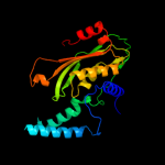
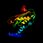
Top model
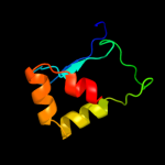
Image coloured by rainbow N → C terminus
Model (left) based on template d1a8ra_
Top template information
Fold:T-fold
Superfamily:Tetrahydrobiopterin biosynthesis enzymes-like
Family:GTP cyclohydrolase I
Confidence and coverage
Confidence:100.0% Coverage:100%
221 residues (100% of your sequence) have been modelled with 100.0% confidence by the single highest scoring template.
3D viewing
Interactive 3D view in Jmol

Sequence analysis 

Secondary structure and disorder prediction 

   1........10.........20.........30.........40.........50.........60
Sequence  MPSLSKEAALVHEALVARGLETPLRPPVHEMDNETRKSLIAGHMTEIMQLLNLDLADDSL
Secondary structure 


SS confidence 


Disorder  ???????


?????????????????


?

Disorder confidence 


   .........70.........80.........90.........100.........110.........120
Sequence  METPHRIAKMYVDEIFSGLDYANFPKITLIENKMKVDEMVTVRDITLTSTCEHHFVTIDG
Secondary structure 


SS confidence 


Disorder 


?


Disorder confidence 


   .........130.........140.........150.........160.........170.........180
Sequence  KATVAYIPKDSVIGLSKINRIVQFFAQRPQVQERLTQQILIALQTLLGTNNVAVSIDAVH
Secondary structure 


SS confidence 


Disorder 


?


Disorder confidence 


   .........190.........200.........210.........220..
Sequence  YCVKARGIRDATSATTTTSLGGLFKSSQNTRHEFLRAVRHHN
Secondary structure 


SS confidence 


Disorder 


??


?????
Disorder confidence 


Confidence Key
High(9)                    Low (0)
?Disordered
Alpha helix
Beta strand

Domain analysis 

Hover over an aligned region to see model and summary info

Please note, only up to the top 20 hits are modelled to reduce computer load

RankAligned region

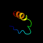
PDB 1a8r chain A

3D model

Region: 2 - 222
Aligned: 221
Modelled: 221
Confidence: 100.0%
Identity: 100%
Fold: T-fold
Superfamily: Tetrahydrobiopterin biosynthesis enzymes-like
Family: GTP cyclohydrolase I

Phyre2

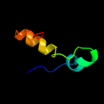
PDB 1wpl chain A

3D model

Region: 26 - 220
Aligned: 194
Modelled: 195
Confidence: 100.0%
Identity: 36%
Fold: T-fold
Superfamily: Tetrahydrobiopterin biosynthesis enzymes-like
Family: GTP cyclohydrolase I

Phyre2

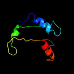
PDB 1is7 chain F

3D model

Region: 26 - 220
Aligned: 194
Modelled: 195
Confidence: 100.0%
Identity: 36%
PDB header:hydrolase/protein binding
Chain: F: PDB Molecule:gtp cyclohydrolase i;
PDBTitle: crystal structure of rat gtpchi/gfrp stimulatory complex

Phyre2

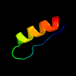
PDB 1wur chain A domain 1

3D model

Region: 34 - 219
Aligned: 185
Modelled: 186
Confidence: 100.0%
Identity: 35%
Fold: T-fold
Superfamily: Tetrahydrobiopterin biosynthesis enzymes-like
Family: GTP cyclohydrolase I

Phyre2

PDB 1wm9 chain D

3D model

Region: 34 - 219
Aligned: 185
Modelled: 186
Confidence: 100.0%
Identity: 35%
PDB header:hydrolase
Chain: D: PDB Molecule:gtp cyclohydrolase i;
PDBTitle: structure of gtp cyclohydrolase i from thermus thermophilus hb8

Phyre2

PDB 3bp1 chain A

3D model

Region: 117 - 177
Aligned: 59
Modelled: 61
Confidence: 93.1%
Identity: 14%
PDB header:oxidoreductase
Chain: A: PDB Molecule:nadph-dependent 7-cyano-7-deazaguanine reductase;
PDBTitle: crystal structure of putative 7-cyano-7-deazaguanine2 reductase quef from vibrio cholerae o1 biovar eltor

Phyre2

PDB 3ugs chain B

3D model

Region: 123 - 147
Aligned: 25
Modelled: 25
Confidence: 40.1%
Identity: 8%
PDB header:transferase
Chain: B: PDB Molecule:undecaprenyl pyrophosphate synthase;
PDBTitle: crystal structure of a probable undecaprenyl diphosphate synthase2 (upps) from campylobacter jejuni

Phyre2

PDB 3pg6 chain D

3D model

Region: 117 - 149
Aligned: 32
Modelled: 33
Confidence: 39.7%
Identity: 22%
PDB header:ligase
Chain: D: PDB Molecule:e3 ubiquitin-protein ligase dtx3l;
PDBTitle: the carboxyl terminal domain of human deltex 3-like

Phyre2

PDB 1ku2 chain A domain 1

3D model

Region: 35 - 62
Aligned: 28
Modelled: 28
Confidence: 33.6%
Identity: 11%
Fold: DNA/RNA-binding 3-helical bundle
Superfamily: Sigma3 and sigma4 domains of RNA polymerase sigma factors
Family: Sigma3 domain

Phyre2

PDB 3lk6 chain A

3D model

Region: 33 - 118
Aligned: 86
Modelled: 86
Confidence: 33.4%
Identity: 3%
PDB header:hydrolase
Chain: A: PDB Molecule:lipoprotein ybbd;
PDBTitle: beta-n-hexosaminidase n318d mutant (ybbd_n318d) from bacillus subtilis

Phyre2

PDB 2xcz chain A

3D model

Region: 150 - 211
Aligned: 60
Modelled: 62
Confidence: 29.6%
Identity: 22%
PDB header:immune system
Chain: A: PDB Molecule:possible atls1-like light-inducible protein;
PDBTitle: crystal structure of macrophage migration inhibitory factor2 homologue from prochlorococcus marinus

Phyre2

PDB 1vqo chain J domain 1

3D model

Region: 150 - 184
Aligned: 31
Modelled: 35
Confidence: 28.8%
Identity: 10%
Fold: Ribosomal protein L13
Superfamily: Ribosomal protein L13
Family: Ribosomal protein L13

Phyre2

PDB 2d2r chain A

3D model

Region: 123 - 147
Aligned: 25
Modelled: 25
Confidence: 27.0%
Identity: 20%
PDB header:transferase
Chain: A: PDB Molecule:undecaprenyl pyrophosphate synthase;
PDBTitle: crystal structure of helicobacter pylori undecaprenyl pyrophosphate2 synthase

Phyre2

PDB 1cmc chain A

3D model

Region: 21 - 57
Aligned: 35
Modelled: 37
Confidence: 25.6%
Identity: 20%
Fold: Ribbon-helix-helix
Superfamily: Ribbon-helix-helix
Family: Met repressor, MetJ (MetR)

Phyre2

PDB 1dpt chain A

3D model

Region: 150 - 218
Aligned: 66
Modelled: 69
Confidence: 25.6%
Identity: 15%
Fold: Tautomerase/MIF
Superfamily: Tautomerase/MIF
Family: MIF-related

Phyre2

PDB 1jp3 chain A

3D model

Region: 123 - 147
Aligned: 25
Modelled: 25
Confidence: 22.1%
Identity: 32%
PDB header:transferase
Chain: A: PDB Molecule:undecaprenyl pyrophosphate synthase;
PDBTitle: structure of e.coli undecaprenyl pyrophosphate synthase

Phyre2

PDB 3ira chain A

3D model

Region: 102 - 167
Aligned: 63
Modelled: 66
Confidence: 19.8%
Identity: 17%
PDB header:structural genomics, unknown function
Chain: A: PDB Molecule:conserved protein;
PDBTitle: the crystal structure of one domain of the conserved protein from2 methanosarcina mazei go1

Phyre2

PDB 1vhx chain A

3D model

Region: 126 - 170
Aligned: 45
Modelled: 45
Confidence: 19.2%
Identity: 22%
Fold: Ribonuclease H-like motif
Superfamily: Ribonuclease H-like
Family: Putative Holliday junction resolvase RuvX

Phyre2

PDB 3tev chain A

3D model

Region: 33 - 118
Aligned: 86
Modelled: 86
Confidence: 19.1%
Identity: 5%
PDB header:hydrolase
Chain: A: PDB Molecule:glycosyl hyrolase, family 3;
PDBTitle: the crystal structure of glycosyl hydrolase from deinococcus2 radiodurans r1

Phyre2

PDB 2vea chain A domain 1

3D model

Region: 133 - 176
Aligned: 43
Modelled: 44
Confidence: 19.1%
Identity: 9%
Fold: Profilin-like
Superfamily: GAF domain-like
Family: GAF domain

Phyre2
1

d1a8ra_
2

d1wpla_
3

c1is7F_
4

d1wura1
5

c1wm9D_
6

c3bp1A_
7

c3ugsB_
8

c3pg6D_
9

d1ku2a1
10

c3lk6A_
11

c2xczA_
12

d1vqoj1
13

c2d2rA_
14

d1cmca_
15

d1dpta_
16

c1jp3A_
17

c3iraA_
18

d1vhxa_
19

c3tevA_
20

d2veaa1
21


22


23


24


25


26


27


28


29


30


31


32


33


34


35


36


37


38


39


40


41


42


43


44


45


46


47


48


49


50


51


52


53


54


55


56


57


58


59


60


61


62


63


64


65


66


67


68


69


70


71


72


73


74


75


76


77


78


79


80


81


82


83


84


85


86


87


88


89


90


91


92


93


94


95


96


Detailed template information 

#
Template Alignment Coverage3D Model Confidence
% i.d. Template Information
1d1a8ra_


100.0 100 Fold:T-fold
Superfamily:Tetrahydrobiopterin biosynthesis enzymes-like
Family:GTP cyclohydrolase I
2d1wpla_


100.0 36 Fold:T-fold
Superfamily:Tetrahydrobiopterin biosynthesis enzymes-like
Family:GTP cyclohydrolase I
3c1is7F_


100.0 36 PDB header:hydrolase/protein binding
Chain: F: PDB Molecule:gtp cyclohydrolase i;
PDBTitle: crystal structure of rat gtpchi/gfrp stimulatory complex
4d1wura1


100.0 35 Fold:T-fold
Superfamily:Tetrahydrobiopterin biosynthesis enzymes-like
Family:GTP cyclohydrolase I
5c1wm9D_


100.0 35 PDB header:hydrolase
Chain: D: PDB Molecule:gtp cyclohydrolase i;
PDBTitle: structure of gtp cyclohydrolase i from thermus thermophilus hb8
6c3bp1A_


93.1 14 PDB header:oxidoreductase
Chain: A: PDB Molecule:nadph-dependent 7-cyano-7-deazaguanine reductase;
PDBTitle: crystal structure of putative 7-cyano-7-deazaguanine2 reductase quef from vibrio cholerae o1 biovar eltor
7c3ugsB_


40.1 8 PDB header:transferase
Chain: B: PDB Molecule:undecaprenyl pyrophosphate synthase;
PDBTitle: crystal structure of a probable undecaprenyl diphosphate synthase2 (upps) from campylobacter jejuni
8c3pg6D_


39.7 22 PDB header:ligase
Chain: D: PDB Molecule:e3 ubiquitin-protein ligase dtx3l;
PDBTitle: the carboxyl terminal domain of human deltex 3-like
9d1ku2a1


33.6 11 Fold:DNA/RNA-binding 3-helical bundle
Superfamily:Sigma3 and sigma4 domains of RNA polymerase sigma factors
Family:Sigma3 domain
10c3lk6A_


33.4 3 PDB header:hydrolase
Chain: A: PDB Molecule:lipoprotein ybbd;
PDBTitle: beta-n-hexosaminidase n318d mutant (ybbd_n318d) from bacillus subtilis
11c2xczA_


29.6 22 PDB header:immune system
Chain: A: PDB Molecule:possible atls1-like light-inducible protein;
PDBTitle: crystal structure of macrophage migration inhibitory factor2 homologue from prochlorococcus marinus
12d1vqoj1


28.8 10 Fold:Ribosomal protein L13
Superfamily:Ribosomal protein L13
Family:Ribosomal protein L13
13c2d2rA_


27.0 20 PDB header:transferase
Chain: A: PDB Molecule:undecaprenyl pyrophosphate synthase;
PDBTitle: crystal structure of helicobacter pylori undecaprenyl pyrophosphate2 synthase
14d1cmca_


25.6 20 Fold:Ribbon-helix-helix
Superfamily:Ribbon-helix-helix
Family:Met repressor, MetJ (MetR)
15d1dpta_


25.6 15 Fold:Tautomerase/MIF
Superfamily:Tautomerase/MIF
Family:MIF-related
16c1jp3A_


22.1 32 PDB header:transferase
Chain: A: PDB Molecule:undecaprenyl pyrophosphate synthase;
PDBTitle: structure of e.coli undecaprenyl pyrophosphate synthase
17c3iraA_


19.8 17 PDB header:structural genomics, unknown function
Chain: A: PDB Molecule:conserved protein;
PDBTitle: the crystal structure of one domain of the conserved protein from2 methanosarcina mazei go1
18d1vhxa_


19.2 22 Fold:Ribonuclease H-like motif
Superfamily:Ribonuclease H-like
Family:Putative Holliday junction resolvase RuvX
19c3tevA_


19.1 5 PDB header:hydrolase
Chain: A: PDB Molecule:glycosyl hyrolase, family 3;
PDBTitle: the crystal structure of glycosyl hydrolase from deinococcus2 radiodurans r1
20d2veaa1


19.1 9 Fold:Profilin-like
Superfamily:GAF domain-like
Family:GAF domain
21d2nn6f2


not modelled 18.5 44 Fold:Ribonuclease PH domain 2-like
Superfamily:Ribonuclease PH domain 2-like
Family:Ribonuclease PH domain 2-like
22d1ueha_


not modelled 18.2 32 Fold:Undecaprenyl diphosphate synthase
Superfamily:Undecaprenyl diphosphate synthase
Family:Undecaprenyl diphosphate synthase
23c3d5bN_


not modelled 17.8 20 PDB header:ribosome
Chain: N: PDB Molecule:50s ribosomal protein l13;
PDBTitle: structural basis for translation termination on the 70s ribosome. this2 file contains the 50s subunit of one 70s ribosome. the entire crystal3 structure contains two 70s ribosomes as described in remark 400.
24c3cf5G_


not modelled 17.8 17 PDB header:ribosome/antibiotic
Chain: G: PDB Molecule:50s ribosomal protein l13;
PDBTitle: thiopeptide antibiotic thiostrepton bound to the large ribosomal2 subunit of deinococcus radiodurans
25d2zjrg1


not modelled 17.8 17 Fold:Ribosomal protein L13
Superfamily:Ribosomal protein L13
Family:Ribosomal protein L13
26d1uiza_


not modelled 17.2 15 Fold:Tautomerase/MIF
Superfamily:Tautomerase/MIF
Family:MIF-related
27d1j3aa_


not modelled 17.0 16 Fold:Ribosomal protein L13
Superfamily:Ribosomal protein L13
Family:Ribosomal protein L13
28d1f75a_


not modelled 16.2 32 Fold:Undecaprenyl diphosphate synthase
Superfamily:Undecaprenyl diphosphate synthase
Family:Undecaprenyl diphosphate synthase
29c3bmxB_


not modelled 14.9 5 PDB header:hydrolase
Chain: B: PDB Molecule:uncharacterized lipoprotein ybbd;
PDBTitle: beta-n-hexosaminidase (ybbd) from bacillus subtilis
30d1fima_


not modelled 14.7 12 Fold:Tautomerase/MIF
Superfamily:Tautomerase/MIF
Family:MIF-related
31d1l0oc_


not modelled 14.5 8 Fold:DNA/RNA-binding 3-helical bundle
Superfamily:Sigma3 and sigma4 domains of RNA polymerase sigma factors
Family:Sigma4 domain
32c1l0oC_


not modelled 14.5 8 PDB header:protein binding
Chain: C: PDB Molecule:sigma factor;
PDBTitle: crystal structure of the bacillus stearothermophilus anti-2 sigma factor spoiiab with the sporulation sigma factor3 sigmaf
33d1sjya_


not modelled 13.2 13 Fold:Nudix
Superfamily:Nudix
Family:MutT-like
34c3gacD_


not modelled 12.7 14 PDB header:cytokine
Chain: D: PDB Molecule:macrophage migration inhibitory factor-like
PDBTitle: structure of mif with hpp
35c3b64A_


not modelled 12.3 17 PDB header:cytokine
Chain: A: PDB Molecule:macrophage migration inhibitory factor-like
PDBTitle: macrophage migration inhibitory factor (mif) from2 /leishmania major
36d1aopa2


not modelled 12.1 11 Fold:Ferredoxin-like
Superfamily:Nitrite/Sulfite reductase N-terminal domain-like
Family:Duplicated SiR/NiR-like domains 1 and 3
37c2o90A_


not modelled 11.6 15 PDB header:lyase
Chain: A: PDB Molecule:dihydroneopterin aldolase;
PDBTitle: atomic resolution crystal structure of e.coli2 dihydroneopterin aldolase in complex with neopterin
38d2j01n1


not modelled 11.4 20 Fold:Ribosomal protein L13
Superfamily:Ribosomal protein L13
Family:Ribosomal protein L13
39c2vfwB_


not modelled 11.0 16 PDB header:transferase
Chain: B: PDB Molecule:short-chain z-isoprenyl diphosphate synthetase;
PDBTitle: rv1086 native
40d1gd0a_


not modelled 10.6 9 Fold:Tautomerase/MIF
Superfamily:Tautomerase/MIF
Family:MIF-related
41d1tr9a_


not modelled 10.5 8 Fold:TIM beta/alpha-barrel
Superfamily:(Trans)glycosidases
Family:NagZ-like
42d2ba0g2


not modelled 10.5 22 Fold:Ribonuclease PH domain 2-like
Superfamily:Ribonuclease PH domain 2-like
Family:Ribonuclease PH domain 2-like
43d1vhma_


not modelled 10.3 20 Fold:Profilin-like
Superfamily:GAF domain-like
Family:GAF domain
44c3fwtA_


not modelled 10.2 11 PDB header:cytokine
Chain: A: PDB Molecule:macrophage migration inhibitory factor-like
PDBTitle: crystal structure of leishmania major mif2
45d1lfpa_


not modelled 10.0 29 Fold:YebC-like
Superfamily:YebC-like
Family:YebC-like
46d2o3bb1


not modelled 9.9 23 Fold:Nuclease A inhibitor (NuiA)
Superfamily:Nuclease A inhibitor (NuiA)
Family:Nuclease A inhibitor (NuiA)
47d1b9la_


not modelled 9.8 28 Fold:T-fold
Superfamily:Tetrahydrobiopterin biosynthesis enzymes-like
Family:DHN aldolase/epimerase
48d1u8va1


not modelled 9.6 16 Fold:Bromodomain-like
Superfamily:Acyl-CoA dehydrogenase C-terminal domain-like
Family:Medium chain acyl-CoA dehydrogenase-like, C-terminal domain
49c2kduB_


not modelled 9.3 67 PDB header:metal binding protein/exocytosis
Chain: B: PDB Molecule:protein unc-13 homolog a;
PDBTitle: structural basis of the munc13-1/ca2+-calmodulin2 interaction: a novel 1-26 calmodulin binding motif with a3 bipartite binding mode
50c3us8A_


not modelled 9.1 15 PDB header:oxidoreductase
Chain: A: PDB Molecule:isocitrate dehydrogenase [nadp];
PDBTitle: crystal structure of an isocitrate dehydrogenase from sinorhizobium2 meliloti 1021
51d1vixa1


not modelled 9.1 14 Fold:Phosphorylase/hydrolase-like
Superfamily:Zn-dependent exopeptidases
Family:Bacterial dinuclear zinc exopeptidases
52d1vmea1


not modelled 9.1 5 Fold:Flavodoxin-like
Superfamily:Flavoproteins
Family:Flavodoxin-related
53c3izcK_


not modelled 9.1 16 PDB header:ribosome
Chain: K: PDB Molecule:60s ribosomal protein rpl16 (l13p);
PDBTitle: localization of the large subunit ribosomal proteins into a 6.1 a2 cryo-em map of saccharomyces cerevisiae translating 80s ribosome
54c3nyyA_


not modelled 8.8 21 PDB header:hydrolase
Chain: A: PDB Molecule:putative glycyl-glycine endopeptidase lytm;
PDBTitle: crystal structure of a putative glycyl-glycine endopeptidase lytm2 (rumgna_02482) from ruminococcus gnavus atcc 29149 at 1.60 a3 resolution
55d1u9da_


not modelled 8.6 31 Fold:Tautomerase/MIF
Superfamily:Tautomerase/MIF
Family:VC0714-like
56d2veaa2


not modelled 8.4 16 Fold:Profilin-like
Superfamily:GAF domain-like
Family:Phytochrome-specific domain
57d2je6a2


not modelled 8.4 28 Fold:Ribonuclease PH domain 2-like
Superfamily:Ribonuclease PH domain 2-like
Family:Ribonuclease PH domain 2-like
58d1nbua_


not modelled 8.4 13 Fold:T-fold
Superfamily:Tetrahydrobiopterin biosynthesis enzymes-like
Family:DHN aldolase/epimerase
59c2vg2C_


not modelled 8.3 17 PDB header:transferase
Chain: C: PDB Molecule:undecaprenyl pyrophosphate synthetase;
PDBTitle: rv2361 with ipp
60c1vixA_


not modelled 8.3 13 PDB header:hydrolase
Chain: A: PDB Molecule:peptidase t;
PDBTitle: crystal structure of a putative peptidase t
61d1mw7a_


not modelled 8.2 7 Fold:YebC-like
Superfamily:YebC-like
Family:YebC-like
62c1s1iM_


not modelled 8.2 16 PDB header:ribosome
Chain: M: PDB Molecule:60s ribosomal protein l16-a;
PDBTitle: structure of the ribosomal 80s-eef2-sordarin complex from2 yeast obtained by docking atomic models for rna and protein3 components into a 11.7 a cryo-em map. this file, 1s1i,4 contains 60s subunit. the 40s ribosomal subunit is in file5 1s1h.
63d1n62b2


not modelled 8.2 12 Fold:Molybdenum cofactor-binding domain
Superfamily:Molybdenum cofactor-binding domain
Family:Molybdenum cofactor-binding domain
64c2uxqB_


not modelled 7.9 14 PDB header:oxidoreductase
Chain: B: PDB Molecule:isocitrate dehydrogenase native;
PDBTitle: isocitrate dehydrogenase from the psychrophilic bacterium2 desulfotalea psychrophila: biochemical properties and3 crystal structure analysis
65c3hlyA_


not modelled 7.6 24 PDB header:flavoprotein
Chain: A: PDB Molecule:flavodoxin-like domain;
PDBTitle: crystal structure of the flavodoxin-like domain from2 synechococcus sp q5mzp6_synp6 protein. northeast structural3 genomics consortium target snr135d.
66c2f7tA_


not modelled 7.5 16 PDB header:dna binding protein
Chain: A: PDB Molecule:mos1 transposase;
PDBTitle: crystal structure of the catalytic domain of mos1 mariner2 transposase
67d1rp3a1


not modelled 7.2 10 Fold:DNA/RNA-binding 3-helical bundle
Superfamily:Sigma3 and sigma4 domains of RNA polymerase sigma factors
Family:Sigma3 domain
68d5nula_


not modelled 7.0 9 Fold:Flavodoxin-like
Superfamily:Flavoproteins
Family:Flavodoxin-related
69d1d8ja_


not modelled 6.9 21 Fold:DNA/RNA-binding 3-helical bundle
Superfamily:"Winged helix" DNA-binding domain
Family:The central core domain of TFIIE beta
70d2fz5a1


not modelled 6.9 9 Fold:Flavodoxin-like
Superfamily:Flavoproteins
Family:Flavodoxin-related
71d1t0la_


not modelled 6.8 13 Fold:Isocitrate/Isopropylmalate dehydrogenase-like
Superfamily:Isocitrate/Isopropylmalate dehydrogenase-like
Family:Dimeric isocitrate & isopropylmalate dehydrogenases
72c2x3mA_


not modelled 6.7 19 PDB header:unknown function
Chain: A: PDB Molecule:hypothetical protein orf239;
PDBTitle: crystal structure of hypothetical protein orf239 from pyrobaculum2 spherical virus
73c3fniA_


not modelled 6.6 14 PDB header:oxidoreductase
Chain: A: PDB Molecule:putative diflavin flavoprotein a 3;
PDBTitle: crystal structure of a diflavin flavoprotein a3 (all3895) from nostoc2 sp., northeast structural genomics consortium target nsr431a
74c3l8jA_


not modelled 6.5 22 PDB header:protein binding
Chain: A: PDB Molecule:programmed cell death protein 10;
PDBTitle: crystal structure of ccm3, a cerebral cavernous malformation protein2 critical for vascular integrity
75d1lwda_


not modelled 6.4 18 Fold:Isocitrate/Isopropylmalate dehydrogenase-like
Superfamily:Isocitrate/Isopropylmalate dehydrogenase-like
Family:Dimeric isocitrate & isopropylmalate dehydrogenases
76d1nbwa2


not modelled 6.4 32 Fold:Ribonuclease H-like motif
Superfamily:Actin-like ATPase domain
Family:ATPase domain of dehydratase reactivase alpha subunit
77d1vj7a2


not modelled 6.4 9 Fold:Nucleotidyltransferase
Superfamily:Nucleotidyltransferase
Family:RelA/SpoT domain
78c3f6sI_


not modelled 6.3 24 PDB header:electron transport
Chain: I: PDB Molecule:flavodoxin;
PDBTitle: desulfovibrio desulfuricans (atcc 29577) oxidized flavodoxin2 alternate conformers
79c3sqlB_


not modelled 6.3 12 PDB header:hydrolase
Chain: B: PDB Molecule:glycosyl hydrolase family 3;
PDBTitle: crystal structure of glycoside hydrolase from synechococcus
80c3nr7A_


not modelled 6.1 18 PDB header:dna binding protein
Chain: A: PDB Molecule:dna-binding protein h-ns;
PDBTitle: crystal structure of s. typhimurium h-ns 1-83
81c3czxA_


not modelled 6.0 24 PDB header:hydrolase
Chain: A: PDB Molecule:putative n-acetylmuramoyl-l-alanine amidase;
PDBTitle: the crystal structure of the putative n-acetylmuramoyl-l-2 alanine amidase from neisseria meningitidis
82c3jywM_


not modelled 5.8 16 PDB header:ribosome
Chain: M: PDB Molecule:60s ribosomal protein l16(a);
PDBTitle: structure of the 60s proteins for eukaryotic ribosome based on cryo-em2 map of thermomyces lanuginosus ribosome at 8.9a resolution
83d2a0sa1


not modelled 5.7 29 Fold:T-fold
Superfamily:Tetrahydrobiopterin biosynthesis enzymes-like
Family:6-pyruvoyl tetrahydropterin synthase
84c3c5tB_


not modelled 5.6 9 PDB header:signaling protein/signaling protein
Chain: B: PDB Molecule:exendin-4;
PDBTitle: crystal structure of the ligand-bound glucagon-like peptide-1 receptor2 extracellular domain
85d1qwda_


not modelled 5.6 8 Fold:Lipocalins
Superfamily:Lipocalins
Family:Retinol binding protein-like
86d2nn6e2


not modelled 5.6 22 Fold:Ribonuclease PH domain 2-like
Superfamily:Ribonuclease PH domain 2-like
Family:Ribonuclease PH domain 2-like
87d1pdaa1


not modelled 5.6 31 Fold:Periplasmic binding protein-like II
Superfamily:Periplasmic binding protein-like II
Family:Phosphate binding protein-like
88d1dhna_


not modelled 5.4 16 Fold:T-fold
Superfamily:Tetrahydrobiopterin biosynthesis enzymes-like
Family:DHN aldolase/epimerase
89c2fnzA_


not modelled 5.4 32 PDB header:oxidoreductase
Chain: A: PDB Molecule:lactate dehydrogenase;
PDBTitle: crystal structure of the lactate dehydrogenase from cryptosporidium2 parvum complexed with cofactor (b-nicotinamide adenine dinucleotide)3 and inhibitor (oxamic acid)
90d2nn6a2


not modelled 5.2 11 Fold:Ribonuclease PH domain 2-like
Superfamily:Ribonuclease PH domain 2-like
Family:Ribonuclease PH domain 2-like
91d1otfa_


not modelled 5.2 32 Fold:Tautomerase/MIF
Superfamily:Tautomerase/MIF
Family:4-oxalocrotonate tautomerase-like
92c1bvyF_


not modelled 5.2 10 PDB header:oxidoreductase
Chain: F: PDB Molecule:protein (cytochrome p450 bm-3);
PDBTitle: complex of the heme and fmn-binding domains of the2 cytochrome p450(bm-3)
93d1bvyf_


not modelled 5.2 10 Fold:Flavodoxin-like
Superfamily:Flavoproteins
Family:Flavodoxin-related
94c2ftcH_


not modelled 5.1 23 PDB header:ribosome
Chain: H: PDB Molecule:39s ribosomal protein l13, mitochondrial;
PDBTitle: structural model for the large subunit of the mammalian mitochondrial2 ribosome
95c2h8bB_


not modelled 5.1 71 PDB header:hormone/growth factor
Chain: B: PDB Molecule:insulin-like 3;
PDBTitle: solution structure of insl3
96d2fgca2


not modelled 5.0 23 Fold:Ferredoxin-like
Superfamily:ACT-like
Family:IlvH-like

Binding site prediction 

Due to computational demand, binding site predictions are not run for batch jobs

If you want to predict binding sites, please manually submit your model of choice to 3DLigandSite


Phyre is for academic use only

Please cite: Protein structure prediction on the web: a case study using the Phyre server
Kelley LA and Sternberg MJE. Nature Protocols 4, 363 - 371 (2009) [pdf] [Import into BibTeX]
 
If you use the binding site predictions from 3DLigandSite, please also cite:
3DLigandSite: predicting ligand-binding sites using similar structures.
Wass MN, Kelley LA and Sternberg MJ Nucleic Acids Research 38, W469-73 (2010) [PubMed]
 
© Structural Bioinformatics Group
Imperial College London
Lawrence Kelley, Benjamin Jefferys
Disclaimer
Terms and Conditions
Component software
Template detection: HHpred 1.51
Secondary structure prediction: Psi-pred 2.5
Disorder prediction: Disopred 2.4
Transmembrane prediction: Memsat_SVM
Multi-template modelling and ab initio: Poing 1.0