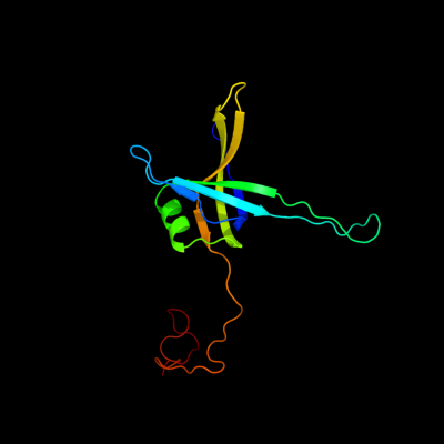
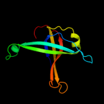
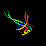
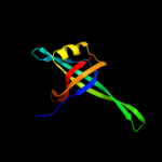
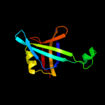
| 1 | d1qvca_
|
|
 |
100.0 |
100 |
Fold:OB-fold
Superfamily:Nucleic acid-binding proteins
Family:Single strand DNA-binding domain, SSB |
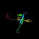
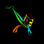
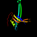
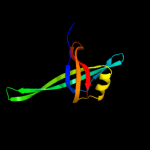
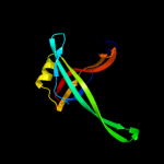
| 2 | d1eyga_
|
|
 |
100.0 |
100 |
Fold:OB-fold
Superfamily:Nucleic acid-binding proteins
Family:Single strand DNA-binding domain, SSB |
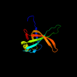
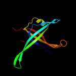
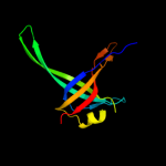
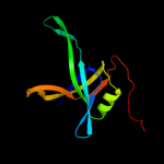
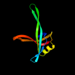
| 3 | c3tqyA_
|
|
 |
100.0 |
68 |
PDB header:transferase
Chain: A: PDB Molecule:single-stranded dna-binding protein;
PDBTitle: structure of a single-stranded dna-binding protein (ssb), from2 coxiella burnetii
|
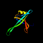
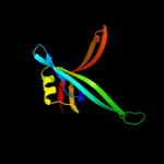
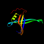
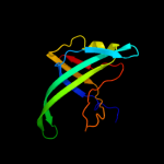
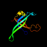
| 4 | c2iheA_
|
|
 |
100.0 |
32 |
PDB header:dna binding protein
Chain: A: PDB Molecule:single-stranded dna-binding protein;
PDBTitle: crystal structure of wild-type single-stranded dna binding protein2 from thermus aquaticus
|
| 5 | c1eqqD_
|
|
 |
100.0 |
100 |
PDB header:replication/rna
Chain: D: PDB Molecule:single stranded dna binding protein;
PDBTitle: single stranded dna binding protein and ssdna complex
|
| 6 | c2ihfA_
|
|
 |
100.0 |
34 |
PDB header:dna binding protein
Chain: A: PDB Molecule:single-stranded dna-binding protein;
PDBTitle: crystal structure of deletion mutant delta 228-252 r190a of the2 single-stranded dna binding protein from thermus aquaticus
|
| 7 | c2vw9B_
|
|
 |
100.0 |
39 |
PDB header:dna-binding protein
Chain: B: PDB Molecule:single-stranded dna binding protein;
PDBTitle: single stranded dna binding protein complex from2 helicobacter pylori
|
| 8 | c3pgzB_
|
|
 |
99.9 |
56 |
PDB header:dna binding protein
Chain: B: PDB Molecule:single-stranded dna-binding protein;
PDBTitle: crystal structure of a single strand binding protein (ssb) from2 bartonella henselae
|
| 9 | d1ue1a_
|
|
 |
99.9 |
32 |
Fold:OB-fold
Superfamily:Nucleic acid-binding proteins
Family:Single strand DNA-binding domain, SSB |
| 10 | d3ulla_
|
|
 |
99.9 |
33 |
Fold:OB-fold
Superfamily:Nucleic acid-binding proteins
Family:Single strand DNA-binding domain, SSB |
| 11 | c3eivB_
|
|
 |
99.9 |
30 |
PDB header:dna binding protein
Chain: B: PDB Molecule:single-stranded dna-binding protein 2;
PDBTitle: crystal structure of single-stranded dna-binding protein2 from streptomyces coelicolor
|
| 12 | c1se8A_
|
|
 |
99.9 |
35 |
PDB header:dna binding protein
Chain: A: PDB Molecule:single-strand binding protein;
PDBTitle: structure of single-stranded dna-binding protein (ssb) from d.2 radiodurans
|
| 13 | d1se8a_
|
|
 |
99.9 |
35 |
Fold:OB-fold
Superfamily:Nucleic acid-binding proteins
Family:Single strand DNA-binding domain, SSB |
| 14 | c1ue7A_
|
|
 |
99.9 |
33 |
PDB header:dna binding protein
Chain: A: PDB Molecule:single-strand binding protein;
PDBTitle: crystal structure of the single-stranded dna-binding2 protein from mycobacterium tuberculosis
|
| 15 | d1v1qa_
|
|
 |
99.9 |
14 |
Fold:OB-fold
Superfamily:Nucleic acid-binding proteins
Family:Single strand DNA-binding domain, SSB |
| 16 | c3lgjA_
|
|
 |
99.9 |
51 |
PDB header:dna binding protein
Chain: A: PDB Molecule:single-stranded dna-binding protein;
PDBTitle: crystal structure of single-stranded binding protein (ssb) from2 bartonella henselae
|
| 17 | c1z9fA_
|
|
 |
99.9 |
48 |
PDB header:dna binding protein
Chain: A: PDB Molecule:single-strand binding protein;
PDBTitle: crystal structure of single stranded dna-binding protein (tm0604) from2 thermotoga maritima at 2.60 a resolution
|
| 18 | c3kojA_
|
|
 |
99.8 |
27 |
PDB header:dna binding protein
Chain: A: PDB Molecule:uncharacterized protein ycf41;
PDBTitle: crystal structure of the ssb domain of q5n255_synp6 protein2 from synechococcus sp. northeast structural genomics3 consortium target snr59a.
|
| 19 | c3k8aA_
|
|
 |
99.8 |
20 |
PDB header:dna binding protein
Chain: A: PDB Molecule:putative primosomal replication protein;
PDBTitle: neisseria gonorrhoeae prib
|
| 20 | d1txya_
|
|
 |
99.8 |
15 |
Fold:OB-fold
Superfamily:Nucleic acid-binding proteins
Family:Single strand DNA-binding domain, SSB |
| 21 | c3fhwB_ |
|
not modelled |
99.8 |
16 |
PDB header:dna binding protein
Chain: B: PDB Molecule:primosomal replication protein n;
PDBTitle: crystal structure of the protein prib from bordetella parapertussis.2 northeast structural genomics consortium target bpr162.
|
| 22 | c3en2A_ |
|
not modelled |
99.7 |
21 |
PDB header:structural genomics, unknown function
Chain: A: PDB Molecule:probable primosomal replication protein n;
PDBTitle: three-dimensional structure of the protein prib from2 ralstonia solanacearum at the resolution 2.3a. northeast3 structural genomics consortium target rsr213c.
|
| 23 | c3stbC_ |
|
not modelled |
98.1 |
24 |
PDB header:rna binding protein/immune system
Chain: C: PDB Molecule:rna-editing complex protein mp42;
PDBTitle: a complex of two editosome proteins and two nanobodies
|
| 24 | c3k81D_ |
|
not modelled |
98.1 |
24 |
PDB header:immune system, rna binding protein
Chain: D: PDB Molecule:mp18 rna editing complex protein;
PDBTitle: structure of the central interaction protein from the trypanosoma2 brucei editosome in complex with single domain antibodies
|
| 25 | c3e0eA_ |
|
not modelled |
96.9 |
19 |
PDB header:replication
Chain: A: PDB Molecule:replication protein a;
PDBTitle: crystal structure of a domain of replication protein a from2 methanococcus maripaludis. northeast structural genomics3 targe mrr110b
|
| 26 | c3f2cA_ |
|
not modelled |
95.5 |
18 |
PDB header:transferase/dna
Chain: A: PDB Molecule:geobacillus kaustophilus dna polc;
PDBTitle: dna polymerase polc from geobacillus kaustophilus complex with dna,2 dgtp and mn
|
| 27 | c3dm3A_ |
|
not modelled |
95.4 |
17 |
PDB header:replication
Chain: A: PDB Molecule:replication factor a;
PDBTitle: crystal structure of a domain of a replication factor a2 protein, from methanocaldococcus jannaschii. northeast3 structural genomics target mjr118e
|
| 28 | c2k50A_ |
|
not modelled |
94.5 |
13 |
PDB header:structural genomics, unknown function
Chain: A: PDB Molecule:replication factor a related protein;
PDBTitle: solution nmr structure of the replication factor a related2 protein from methanobacterium thermoautotrophicum.3 northeast structural genomics target tr91a.
|
| 29 | c1ynxA_ |
|
not modelled |
93.1 |
21 |
PDB header:dna binding protein
Chain: A: PDB Molecule:replication factor-a protein 1;
PDBTitle: solution structure of dna binding domain a (dbd-a) of2 s.cerevisiae replication protein a (rpa)
|
| 30 | c1fguA_ |
|
not modelled |
92.2 |
11 |
PDB header:replication
Chain: A: PDB Molecule:replication protein a 70 kda dna-binding subunit;
PDBTitle: ssdna-binding domain of the large subunit of replication2 protein a
|
| 31 | d1jmca1 |
|
not modelled |
90.7 |
14 |
Fold:OB-fold
Superfamily:Nucleic acid-binding proteins
Family:Single strand DNA-binding domain, SSB |
| 32 | d1o7ia_ |
|
not modelled |
90.6 |
23 |
Fold:OB-fold
Superfamily:Nucleic acid-binding proteins
Family:Single strand DNA-binding domain, SSB |
| 33 | d1gm5a2 |
|
not modelled |
89.1 |
12 |
Fold:OB-fold
Superfamily:Nucleic acid-binding proteins
Family:RecG "wedge" domain |
| 34 | c2k75A_ |
|
not modelled |
87.9 |
21 |
PDB header:dna binding protein
Chain: A: PDB Molecule:uncharacterized protein ta0387;
PDBTitle: solution nmr structure of the ob domain of ta0387 from2 thermoplasma acidophilum. northeast structural genomics3 consortium target tar80b.
|
| 35 | d1jmca2 |
|
not modelled |
84.6 |
19 |
Fold:OB-fold
Superfamily:Nucleic acid-binding proteins
Family:Single strand DNA-binding domain, SSB |
| 36 | d1c0aa1 |
|
not modelled |
77.4 |
19 |
Fold:OB-fold
Superfamily:Nucleic acid-binding proteins
Family:Anticodon-binding domain |
| 37 | c2xgtB_ |
|
not modelled |
76.4 |
18 |
PDB header:ligase
Chain: B: PDB Molecule:asparaginyl-trna synthetase, cytoplasmic;
PDBTitle: asparaginyl-trna synthetase from brugia malayi complexed2 with the sulphamoyl analogue of asparaginyl-adenylate
|
| 38 | c2hqlB_ |
|
not modelled |
76.2 |
24 |
PDB header:dna binding protein
Chain: B: PDB Molecule:hypothetical protein mg376 homolog;
PDBTitle: crystal structure of a small single-stranded dna binding2 protein from mycoplasma pneumoniae
|
| 39 | c3kf6A_ |
|
not modelled |
75.9 |
23 |
PDB header:structural protein
Chain: A: PDB Molecule:protein stn1;
PDBTitle: crystal structure of s. pombe stn1-ten1 complex
|
| 40 | d1eova1 |
|
not modelled |
75.6 |
13 |
Fold:OB-fold
Superfamily:Nucleic acid-binding proteins
Family:Anticodon-binding domain |
| 41 | c2vl6C_ |
|
not modelled |
74.7 |
19 |
PDB header:dna binding protein
Chain: C: PDB Molecule:minichromosome maintenance protein mcm;
PDBTitle: structural analysis of the sulfolobus solfataricus mcm2 protein n-terminal domain
|
| 42 | d1b8aa1 |
|
not modelled |
74.3 |
16 |
Fold:OB-fold
Superfamily:Nucleic acid-binding proteins
Family:Anticodon-binding domain |
| 43 | d1wjja_ |
|
not modelled |
70.8 |
10 |
Fold:OB-fold
Superfamily:Nucleic acid-binding proteins
Family:Single strand DNA-binding domain, SSB |
| 44 | d1bbua1 |
|
not modelled |
70.2 |
13 |
Fold:OB-fold
Superfamily:Nucleic acid-binding proteins
Family:Anticodon-binding domain |
| 45 | d1e1oa1 |
|
not modelled |
66.6 |
13 |
Fold:OB-fold
Superfamily:Nucleic acid-binding proteins
Family:Anticodon-binding domain |
| 46 | d1l0wa1 |
|
not modelled |
62.6 |
19 |
Fold:OB-fold
Superfamily:Nucleic acid-binding proteins
Family:Anticodon-binding domain |
| 47 | c1ltlE_ |
|
not modelled |
61.7 |
22 |
PDB header:replication
Chain: E: PDB Molecule:dna replication initiator (cdc21/cdc54);
PDBTitle: the dodecamer structure of mcm from archaeal m.2 thermoautotrophicum
|
| 48 | c1eqrC_ |
|
not modelled |
53.7 |
19 |
PDB header:ligase
Chain: C: PDB Molecule:aspartyl-trna synthetase;
PDBTitle: crystal structure of free aspartyl-trna synthetase from2 escherichia coli
|
| 49 | c1gm5A_ |
|
not modelled |
52.8 |
12 |
PDB header:helicase
Chain: A: PDB Molecule:recg;
PDBTitle: structure of recg bound to three-way dna junction
|
| 50 | d1ltla_ |
|
not modelled |
49.8 |
22 |
Fold:OB-fold
Superfamily:Nucleic acid-binding proteins
Family:DNA replication initiator (cdc21/cdc54) N-terminal domain |
| 51 | c3m4qA_ |
|
not modelled |
45.4 |
17 |
PDB header:ligase
Chain: A: PDB Molecule:asparaginyl-trna synthetase, putative;
PDBTitle: entamoeba histolytica asparaginyl-trna synthetase (asnrs)
|
| 52 | c1wydB_ |
|
not modelled |
45.0 |
22 |
PDB header:ligase
Chain: B: PDB Molecule:hypothetical aspartyl-trna synthetase;
PDBTitle: crystal structure of aspartyl-trna synthetase from sulfolobus tokodaii
|
| 53 | c2pi2A_ |
|
not modelled |
44.3 |
18 |
PDB header:replication, dna binding protein
Chain: A: PDB Molecule:replication protein a 32 kda subunit;
PDBTitle: full-length replication protein a subunits rpa14 and rpa32
|
| 54 | d1vqoj1 |
|
not modelled |
41.9 |
25 |
Fold:Ribosomal protein L13
Superfamily:Ribosomal protein L13
Family:Ribosomal protein L13 |
| 55 | c3d5bN_ |
|
not modelled |
41.2 |
42 |
PDB header:ribosome
Chain: N: PDB Molecule:50s ribosomal protein l13;
PDBTitle: structural basis for translation termination on the 70s ribosome. this2 file contains the 50s subunit of one 70s ribosome. the entire crystal3 structure contains two 70s ribosomes as described in remark 400.
|
| 56 | c3kf8C_ |
|
not modelled |
39.8 |
19 |
PDB header:structural protein
Chain: C: PDB Molecule:protein stn1;
PDBTitle: crystal structure of c. tropicalis stn1-ten1 complex
|
| 57 | d2j01n1 |
|
not modelled |
39.7 |
42 |
Fold:Ribosomal protein L13
Superfamily:Ribosomal protein L13
Family:Ribosomal protein L13 |
| 58 | c3i7fA_ |
|
not modelled |
39.6 |
11 |
PDB header:ligase
Chain: A: PDB Molecule:aspartyl-trna synthetase;
PDBTitle: aspartyl trna synthetase from entamoeba histolytica
|
| 59 | c3cf5G_ |
|
not modelled |
39.4 |
35 |
PDB header:ribosome/antibiotic
Chain: G: PDB Molecule:50s ribosomal protein l13;
PDBTitle: thiopeptide antibiotic thiostrepton bound to the large ribosomal2 subunit of deinococcus radiodurans
|
| 60 | d2zjrg1 |
|
not modelled |
39.4 |
35 |
Fold:Ribosomal protein L13
Superfamily:Ribosomal protein L13
Family:Ribosomal protein L13 |
| 61 | c2zkrj_ |
|
not modelled |
38.0 |
25 |
PDB header:ribosomal protein/rna
Chain: J: PDB Molecule:rna expansion segment es15 part ii;
PDBTitle: structure of a mammalian ribosomal 60s subunit within an2 80s complex obtained by docking homology models of the rna3 and proteins into an 8.7 a cryo-em map
|
| 62 | d1j3aa_ |
|
not modelled |
36.0 |
35 |
Fold:Ribosomal protein L13
Superfamily:Ribosomal protein L13
Family:Ribosomal protein L13 |
| 63 | c4a1aI_ |
|
not modelled |
34.8 |
25 |
PDB header:ribosome
Chain: I: PDB Molecule:60s ribosomal protein l13a;
PDBTitle: t.thermophila 60s ribosomal subunit in complex with2 initiation factor 6. this file contains 5s rrna,3 5.8s rrna and proteins of molecule 3.
|
| 64 | c1b8aB_ |
|
not modelled |
33.8 |
17 |
PDB header:ligase
Chain: B: PDB Molecule:protein (aspartyl-trna synthetase);
PDBTitle: aspartyl-trna synthetase
|
| 65 | c3izcK_ |
|
not modelled |
33.6 |
30 |
PDB header:ribosome
Chain: K: PDB Molecule:60s ribosomal protein rpl16 (l13p);
PDBTitle: localization of the large subunit ribosomal proteins into a 6.1 a2 cryo-em map of saccharomyces cerevisiae translating 80s ribosome
|
| 66 | c2ftcH_ |
|
not modelled |
33.4 |
53 |
PDB header:ribosome
Chain: H: PDB Molecule:39s ribosomal protein l13, mitochondrial;
PDBTitle: structural model for the large subunit of the mammalian mitochondrial2 ribosome
|
| 67 | d2pi2a1 |
|
not modelled |
32.2 |
18 |
Fold:OB-fold
Superfamily:Nucleic acid-binding proteins
Family:Single strand DNA-binding domain, SSB |
| 68 | d1n9wa1 |
|
not modelled |
31.6 |
21 |
Fold:OB-fold
Superfamily:Nucleic acid-binding proteins
Family:Anticodon-binding domain |
| 69 | c3bjuB_ |
|
not modelled |
31.3 |
13 |
PDB header:ligase
Chain: B: PDB Molecule:lysyl-trna synthetase;
PDBTitle: crystal structure of tetrameric form of human lysyl-trna2 synthetase
|
| 70 | d1qzga_ |
|
not modelled |
30.9 |
20 |
Fold:OB-fold
Superfamily:Nucleic acid-binding proteins
Family:Single strand DNA-binding domain, SSB |
| 71 | c3jywM_ |
|
not modelled |
29.9 |
30 |
PDB header:ribosome
Chain: M: PDB Molecule:60s ribosomal protein l16(a);
PDBTitle: structure of the 60s proteins for eukaryotic ribosome based on cryo-em2 map of thermomyces lanuginosus ribosome at 8.9a resolution
|
| 72 | c3bboL_ |
|
not modelled |
29.6 |
26 |
PDB header:ribosome
Chain: L: PDB Molecule:ribosomal protein l13;
PDBTitle: homology model for the spinach chloroplast 50s subunit2 fitted to 9.4a cryo-em map of the 70s chlororibosome
|
| 73 | c1xjvA_ |
|
not modelled |
27.2 |
13 |
PDB header:transcription/dna
Chain: A: PDB Molecule:protection of telomeres 1;
PDBTitle: crystal structure of human pot1 bound to telomeric single-2 stranded dna (ttagggttag)
|
| 74 | d1krta_ |
|
not modelled |
26.5 |
11 |
Fold:OB-fold
Superfamily:Nucleic acid-binding proteins
Family:Anticodon-binding domain |
| 75 | c3iz5K_ |
|
not modelled |
26.4 |
30 |
PDB header:ribosome
Chain: K: PDB Molecule:60s ribosomal protein l13a (l13p);
PDBTitle: localization of the large subunit ribosomal proteins into a 5.5 a2 cryo-em map of triticum aestivum translating 80s ribosome
|
| 76 | d1fnda1 |
|
not modelled |
25.7 |
20 |
Fold:Reductase/isomerase/elongation factor common domain
Superfamily:Riboflavin synthase domain-like
Family:Ferredoxin reductase FAD-binding domain-like |
| 77 | c1e22A_ |
|
not modelled |
25.7 |
13 |
PDB header:ligase
Chain: A: PDB Molecule:lysyl-trna synthetase;
PDBTitle: lysyl-trna synthetase (lysu) hexagonal form complexed with2 lysine and the non-hydrolysable atp analogue amp-pcp
|
| 78 | c1jb7A_ |
|
not modelled |
25.0 |
10 |
PDB header:dna-binding protein/dna
Chain: A: PDB Molecule:telomere-binding protein alpha subunit;
PDBTitle: dna g-quartets in a 1.86 a resolution structure of an oxytricha nova2 telomeric protein-dna complex
|
| 79 | c1ph4A_ |
|
not modelled |
25.0 |
10 |
PDB header:dna binding protein/dna
Chain: A: PDB Molecule:telomere-binding protein alpha subunit;
PDBTitle: crystal structure of the oxytricha nova telomere end-binding protein2 complexed with noncognate ssdna ggggttttggcg
|
| 80 | c2l3sA_ |
|
not modelled |
24.3 |
11 |
PDB header:structural protein
Chain: A: PDB Molecule:autoinhibited crk protein;
PDBTitle: structure of the autoinhibited crk
|
| 81 | d1jb7a1 |
|
not modelled |
20.4 |
10 |
Fold:OB-fold
Superfamily:Nucleic acid-binding proteins
Family:Single strand DNA-binding domain, SSB |
| 82 | c1asyA_ |
|
not modelled |
19.8 |
18 |
PDB header:complex (aminoacyl-trna synthase/trna)
Chain: A: PDB Molecule:aspartyl-trna synthetase;
PDBTitle: class ii aminoacyl transfer rna synthetases: crystal2 structure of yeast aspartyl-trna synthetase complexed with3 trna asp
|
| 83 | d1gawa1 |
|
not modelled |
19.1 |
24 |
Fold:Reductase/isomerase/elongation factor common domain
Superfamily:Riboflavin synthase domain-like
Family:Ferredoxin reductase FAD-binding domain-like |
| 84 | c1efwA_ |
|
not modelled |
19.1 |
20 |
PDB header:ligase/rna
Chain: A: PDB Molecule:aspartyl-trna synthetase;
PDBTitle: crystal structure of aspartyl-trna synthetase from thermus2 thermophilus complexed to trnaasp from escherichia coli
|
| 85 | d1xjva1 |
|
not modelled |
18.7 |
13 |
Fold:OB-fold
Superfamily:Nucleic acid-binding proteins
Family:Single strand DNA-binding domain, SSB |
| 86 | d1htwa_ |
|
not modelled |
18.5 |
41 |
Fold:P-loop containing nucleoside triphosphate hydrolases
Superfamily:P-loop containing nucleoside triphosphate hydrolases
Family:YjeE-like |
| 87 | d1zcea1 |
|
not modelled |
17.4 |
11 |
Fold:PUA domain-like
Superfamily:PUA domain-like
Family:Atu2648/PH1033-like |
| 88 | d1xjva2 |
|
not modelled |
17.3 |
24 |
Fold:OB-fold
Superfamily:Nucleic acid-binding proteins
Family:Single strand DNA-binding domain, SSB |
| 89 | d2isba1 |
|
not modelled |
17.1 |
30 |
Fold:The "swivelling" beta/beta/alpha domain
Superfamily:FumA C-terminal domain-like
Family:FumA C-terminal domain-like |
| 90 | c3f9vA_ |
|
not modelled |
16.5 |
21 |
PDB header:hydrolase
Chain: A: PDB Molecule:minichromosome maintenance protein mcm;
PDBTitle: crystal structure of a near full-length archaeal mcm: functional2 insights for an aaa+ hexameric helicase
|
| 91 | c3e9hB_ |
|
not modelled |
15.0 |
13 |
PDB header:ligase
Chain: B: PDB Molecule:lysyl-trna synthetase;
PDBTitle: lysyl-trna synthetase from bacillus stearothermophilus2 complexed with l-lysylsulfamoyl adenosine
|
| 92 | c3kf8D_ |
|
not modelled |
14.7 |
26 |
PDB header:structural protein
Chain: D: PDB Molecule:protein ten1;
PDBTitle: crystal structure of c. tropicalis stn1-ten1 complex
|
| 93 | d2ar1a1 |
|
not modelled |
14.3 |
16 |
Fold:PUA domain-like
Superfamily:PUA domain-like
Family:Atu2648/PH1033-like |
| 94 | d2gbsa1 |
|
not modelled |
14.0 |
16 |
Fold:PUA domain-like
Superfamily:PUA domain-like
Family:Atu2648/PH1033-like |
| 95 | c2kenA_ |
|
not modelled |
13.8 |
9 |
PDB header:structural genomics, unknown function
Chain: A: PDB Molecule:conserved protein;
PDBTitle: solution nmr structure of the ob domain (residues 67-166)2 of mm0293 from methanosarcina mazei. northeast structural3 genomics consortium target mar214a.
|
| 96 | c2wkdA_ |
|
not modelled |
13.5 |
17 |
PDB header:dna binding protein
Chain: A: PDB Molecule:orf34p2;
PDBTitle: crystal structure of a double ile-to-met mutant of protein2 orf34 from lactococcus phage p2
|
| 97 | c1s1iM_ |
|
not modelled |
13.0 |
30 |
PDB header:ribosome
Chain: M: PDB Molecule:60s ribosomal protein l16-a;
PDBTitle: structure of the ribosomal 80s-eef2-sordarin complex from2 yeast obtained by docking atomic models for rna and protein3 components into a 11.7 a cryo-em map. this file, 1s1i,4 contains 60s subunit. the 40s ribosomal subunit is in file5 1s1h.
|
| 98 | c3eopB_ |
|
not modelled |
13.0 |
11 |
PDB header:unknown function
Chain: B: PDB Molecule:thymocyte nuclear protein 1;
PDBTitle: crystal structure of the duf55 domain of human thymocyte nuclear2 protein 1
|
| 99 | d1c3ha_ |
|
not modelled |
12.8 |
36 |
Fold:TNF-like
Superfamily:TNF-like
Family:TNF-like |