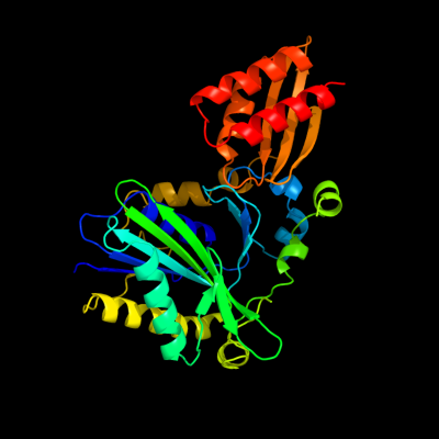
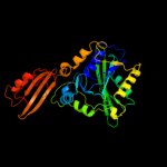
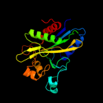
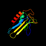
1 c1x2gB_
100.0
96
PDB header: ligaseChain: B: PDB Molecule: lipoate-protein ligase a;PDBTitle: crystal structure of lipate-protein ligase a from2 escherichia coli
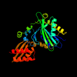
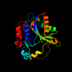
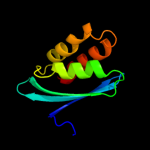
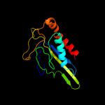
2 c1vqzA_
100.0
31
PDB header: ligaseChain: A: PDB Molecule: lipoate-protein ligase, putative;PDBTitle: crystal structure of a putative lipoate-protein ligase a (sp_1160)2 from streptococcus pneumoniae tigr4 at 1.99 a resolution
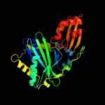
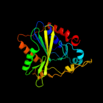
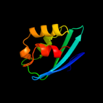
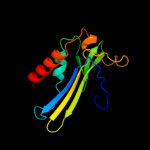
3 c2e5aA_
100.0
33
PDB header: ligaseChain: A: PDB Molecule: lipoyltransferase 1;PDBTitle: crystal structure of bovine lipoyltransferase in complex2 with lipoyl-amp
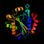
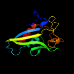
4 d1x2ga2
100.0
99
Fold: Class II aaRS and biotin synthetasesSuperfamily: Class II aaRS and biotin synthetasesFamily: LplA-like5 d1vqza2
100.0
36
Fold: Class II aaRS and biotin synthetasesSuperfamily: Class II aaRS and biotin synthetasesFamily: LplA-like6 d2c8ma1
100.0
30
Fold: Class II aaRS and biotin synthetasesSuperfamily: Class II aaRS and biotin synthetasesFamily: LplA-like7 d2p5ia1
100.0
18
Fold: Class II aaRS and biotin synthetasesSuperfamily: Class II aaRS and biotin synthetasesFamily: LplA-like8 d2p0la1
100.0
18
Fold: Class II aaRS and biotin synthetasesSuperfamily: Class II aaRS and biotin synthetasesFamily: LplA-like9 c2qhsA_
100.0
19
PDB header: transferaseChain: A: PDB Molecule: lipoyltransferase;PDBTitle: structural basis of octanoic acid recognition by lipoate-protein2 ligase b
10 c2qhvA_
100.0
19
PDB header: transferaseChain: A: PDB Molecule: lipoyltransferase;PDBTitle: structural basis of octanoic acid recognition by lipoate-protein2 ligase b
11 d1w66a1
100.0
19
Fold: Class II aaRS and biotin synthetasesSuperfamily: Class II aaRS and biotin synthetasesFamily: LplA-like12 d1x2ga1
99.9
98
Fold: SufE/NifUSuperfamily: SufE/NifUFamily: SP1160 C-terminal domain-like13 d1vqza1
99.8
18
Fold: SufE/NifUSuperfamily: SufE/NifUFamily: SP1160 C-terminal domain-like14 c2eayB_
97.8
18
PDB header: ligaseChain: B: PDB Molecule: biotin [acetyl-coa-carboxylase] ligase;PDBTitle: crystal structure of biotin protein ligase from aquifex2 aeolicus
15 d2zgwa2
97.8
19
Fold: Class II aaRS and biotin synthetasesSuperfamily: Class II aaRS and biotin synthetasesFamily: Biotin holoenzyme synthetase16 c2ej9A_
97.7
16
PDB header: ligaseChain: A: PDB Molecule: putative biotin ligase;PDBTitle: crystal structure of biotin protein ligase from2 methanococcus jannaschii
17 c2ewnA_
97.4
18
PDB header: ligase, transcriptionChain: A: PDB Molecule: bira bifunctional protein;PDBTitle: ecoli biotin repressor with co-repressor analog
18 c3bfmA_
97.2
15
PDB header: unknown functionChain: A: PDB Molecule: biotin protein ligase-like protein of unknown function;PDBTitle: crystal structure of a biotin protein ligase-like protein of unknown2 function (tm1040_0394) from silicibacter sp. tm1040 at 1.70 a3 resolution
19 c2dzcA_
96.7
20
PDB header: ligaseChain: A: PDB Molecule: biotin--[acetyl-coa-carboxylase] ligase;PDBTitle: crystal structure of biotin protein ligase from pyrococcus2 horikoshii, mutation r48a
20 c2cghB_
96.6
14
PDB header: ligaseChain: B: PDB Molecule: biotin ligase;PDBTitle: crystal structure of biotin ligase from mycobacterium2 tuberculosis
21 d1biaa3
not modelled
95.4
23
Fold: Class II aaRS and biotin synthetasesSuperfamily: Class II aaRS and biotin synthetasesFamily: Biotin holoenzyme synthetase22 c2qq4A_
not modelled
90.5
18
PDB header: metal binding proteinChain: A: PDB Molecule: iron-sulfur cluster biosynthesis protein iscu;PDBTitle: crystal structure of iron-sulfur cluster biosynthesis2 protein iscu (ttha1736) from thermus thermophilus hb8
23 d1t3qc1
not modelled
84.0
9
Fold: CO dehydrogenase flavoprotein C-domain-likeSuperfamily: CO dehydrogenase flavoprotein C-terminal domain-likeFamily: CO dehydrogenase flavoprotein C-terminal domain-like24 d1jroa3
not modelled
82.9
18
Fold: CO dehydrogenase flavoprotein C-domain-likeSuperfamily: CO dehydrogenase flavoprotein C-terminal domain-likeFamily: CO dehydrogenase flavoprotein C-terminal domain-like25 d1n62c1
not modelled
80.1
14
Fold: CO dehydrogenase flavoprotein C-domain-likeSuperfamily: CO dehydrogenase flavoprotein C-terminal domain-likeFamily: CO dehydrogenase flavoprotein C-terminal domain-like26 d1ffvc1
not modelled
80.1
16
Fold: CO dehydrogenase flavoprotein C-domain-likeSuperfamily: CO dehydrogenase flavoprotein C-terminal domain-likeFamily: CO dehydrogenase flavoprotein C-terminal domain-like27 d1v97a4
not modelled
74.7
11
Fold: CO dehydrogenase flavoprotein C-domain-likeSuperfamily: CO dehydrogenase flavoprotein C-terminal domain-likeFamily: CO dehydrogenase flavoprotein C-terminal domain-like28 d1rm6b1
not modelled
73.7
17
Fold: CO dehydrogenase flavoprotein C-domain-likeSuperfamily: CO dehydrogenase flavoprotein C-terminal domain-likeFamily: CO dehydrogenase flavoprotein C-terminal domain-like29 c2w3rG_
not modelled
67.0
18
PDB header: oxidoreductaseChain: G: PDB Molecule: xanthine dehydrogenase;PDBTitle: crystal structure of xanthine dehydrogenase (desulfo form)2 from rhodobacter capsulatus in complex with hypoxanthine
30 d1knxa1
not modelled
64.6
7
Fold: MurF and HprK N-domain-likeSuperfamily: HprK N-terminal domain-likeFamily: HPr kinase/phoshatase HprK N-terminal domain31 d1xjsa_
not modelled
54.2
7
Fold: SufE/NifUSuperfamily: SufE/NifUFamily: NifU/IscU domain32 d1ko7a1
not modelled
51.1
18
Fold: MurF and HprK N-domain-likeSuperfamily: HprK N-terminal domain-likeFamily: HPr kinase/phoshatase HprK N-terminal domain33 c1ybxA_
not modelled
49.8
13
PDB header: structural genomics, unknown functionChain: A: PDB Molecule: conserved hypothetical protein;PDBTitle: conserved hypothetical protein cth-383 from clostridium thermocellum
34 c3hrdC_
not modelled
49.7
14
PDB header: oxidoreductaseChain: C: PDB Molecule: nicotinate dehydrogenase fad-subunit;PDBTitle: crystal structure of nicotinate dehydrogenase
35 d1su0b_
not modelled
48.1
10
Fold: SufE/NifUSuperfamily: SufE/NifUFamily: NifU/IscU domain36 c1knxF_
not modelled
46.9
7
PDB header: transferase/hydrolaseChain: F: PDB Molecule: probable hpr(ser) kinase/phosphatase;PDBTitle: hpr kinase/phosphatase from mycoplasma pneumoniae
37 c1ko7B_
not modelled
46.8
17
PDB header: transferase,hydrolaseChain: B: PDB Molecule: hpr kinase/phosphatase;PDBTitle: x-ray structure of the hpr kinase/phosphatase from2 staphylococcus xylosus at 1.95 a resolution
38 d1yq9h1
not modelled
46.6
25
Fold: HydB/Nqo4-likeSuperfamily: HydB/Nqo4-likeFamily: Nickel-iron hydrogenase, large subunit39 d2brfa1
not modelled
44.5
19
Fold: SMAD/FHA domainSuperfamily: SMAD/FHA domainFamily: FHA domain40 c2z7eB_
not modelled
44.4
17
PDB header: biosynthetic proteinChain: B: PDB Molecule: nifu-like protein;PDBTitle: crystal structure of aquifex aeolicus iscu with bound [2fe-2 2s] cluster
41 d1e3db_
not modelled
43.3
18
Fold: HydB/Nqo4-likeSuperfamily: HydB/Nqo4-likeFamily: Nickel-iron hydrogenase, large subunit42 d1cc1l_
not modelled
43.3
19
Fold: HydB/Nqo4-likeSuperfamily: HydB/Nqo4-likeFamily: Nickel-iron hydrogenase, large subunit43 d1frfl_
not modelled
42.9
20
Fold: HydB/Nqo4-likeSuperfamily: HydB/Nqo4-likeFamily: Nickel-iron hydrogenase, large subunit44 c1rm6E_
not modelled
40.9
20
PDB header: oxidoreductaseChain: E: PDB Molecule: 4-hydroxybenzoyl-coa reductase beta subunit;PDBTitle: structure of 4-hydroxybenzoyl-coa reductase from thauera2 aromatica
45 d1wuil1
not modelled
40.3
21
Fold: HydB/Nqo4-likeSuperfamily: HydB/Nqo4-likeFamily: Nickel-iron hydrogenase, large subunit46 d2ddza1
not modelled
38.5
27
Fold: Class II aaRS and biotin synthetasesSuperfamily: Class II aaRS and biotin synthetasesFamily: PH0223-like47 c2grvC_
not modelled
37.5
9
PDB header: biosynthetic proteinChain: C: PDB Molecule: lpqw;PDBTitle: crystal structure of lpqw
48 c3myrB_
not modelled
35.0
15
PDB header: oxidoreductaseChain: B: PDB Molecule: nickel-dependent hydrogenase large subunit;PDBTitle: crystal structure of [nife] hydrogenase from allochromatium vinosum in2 its ni-a state
49 d2a1ja1
not modelled
34.9
20
Fold: SAM domain-likeSuperfamily: RuvA domain 2-likeFamily: Hef domain-like50 c1ffuF_
not modelled
33.0
18
PDB header: hydrolaseChain: F: PDB Molecule: cutm, flavoprotein of carbon monoxidePDBTitle: carbon monoxide dehydrogenase from hydrogenophaga2 pseudoflava which lacks the mo-pyranopterin moiety of the3 molybdenum cofactor
51 c1t3qF_
not modelled
32.6
8
PDB header: oxidoreductaseChain: F: PDB Molecule: quinoline 2-oxidoreductase medium subunit;PDBTitle: crystal structure of quinoline 2-oxidoreductase from pseudomonas2 putida 86
52 c3o6uB_
not modelled
32.4
30
PDB header: structural genomics, unknown functionChain: B: PDB Molecule: uncharacterized protein cpe2226;PDBTitle: crystal structure of cpe2226 protein from clostridium perfringens.2 northeast structural genomics consortium target cpr195
53 c3ry3B_
not modelled
31.5
9
PDB header: transport proteinChain: B: PDB Molecule: putative solute-binding protein;PDBTitle: putative solute-binding protein from yersinia pestis.
54 d1xoca1
not modelled
31.2
17
Fold: Periplasmic binding protein-like IISuperfamily: Periplasmic binding protein-like IIFamily: Phosphate binding protein-like55 c3lvuB_
not modelled
29.1
21
PDB header: transport proteinChain: B: PDB Molecule: abc transporter, periplasmic substrate-binding protein;PDBTitle: crystal structure of abc transporter, periplasmic substrate-binding2 protein spo2066 from silicibacter pomeroyi
56 c2kzxA_
not modelled
27.4
15
PDB header: structural genomics, unknown functionChain: A: PDB Molecule: uncharacterized protein;PDBTitle: solution nmr structure of a3dht5 from clostridium thermocellum,2 northeast structural genomics consortium target cmr116
57 d1u07a_
not modelled
27.3
15
Fold: TolA/TonB C-terminal domainSuperfamily: TolA/TonB C-terminal domainFamily: TonB58 c1h2aL_
not modelled
26.3
22
PDB header: oxidoreductaseChain: L: PDB Molecule: hydrogenase;PDBTitle: single crystals of hydrogenase from desulfovibrio vulgaris
59 d1p30a1
not modelled
25.0
15
Fold: Nucleoplasmin-like/VP (viral coat and capsid proteins)Superfamily: Group II dsDNA viruses VPFamily: Adenovirus hexon60 c3uotB_
not modelled
24.5
13
PDB header: cell cycleChain: B: PDB Molecule: mediator of dna damage checkpoint protein 1;PDBTitle: crystal structure of mdc1 fha domain in complex with a phosphorylated2 peptide from the mdc1 n-terminus
61 d1zlqa1
not modelled
23.5
9
Fold: Periplasmic binding protein-like IISuperfamily: Periplasmic binding protein-like IIFamily: Phosphate binding protein-like62 c2o7jA_
not modelled
22.8
12
PDB header: sugar binding proteinChain: A: PDB Molecule: oligopeptide abc transporter, periplasmicPDBTitle: the x-ray crystal structure of a thermophilic cellobiose2 binding protein bound with cellopentaose
63 c2kklA_
not modelled
22.4
9
PDB header: structural genomics, unknown functionChain: A: PDB Molecule: uncharacterized protein mb1858;PDBTitle: solution nmr structure of fha domain of mb1858 from2 mycobacterium bovis. northeast structural genomics3 consortium target mbr243c (24-155).
64 d1wh9a_
not modelled
22.3
14
Fold: Alpha-lytic protease prodomain-likeSuperfamily: Prokaryotic type KH domain (KH-domain type II)Family: Prokaryotic type KH domain (KH-domain type II)65 c2wpnB_
not modelled
22.0
22
PDB header: oxidoreductaseChain: B: PDB Molecule: periplasmic [nifese] hydrogenase, large subunit,PDBTitle: structure of the oxidised, as-isolated nifese hydrogenase2 from d. vulgaris hildenborough
66 c1n62C_
not modelled
21.7
14
PDB header: oxidoreductaseChain: C: PDB Molecule: carbon monoxide dehydrogenase medium chain;PDBTitle: crystal structure of the mo,cu-co dehydrogenase (codh), n-2 butylisocyanide-bound state
67 d1puga_
not modelled
21.3
14
Fold: YbaB-likeSuperfamily: YbaB-likeFamily: YbaB-like68 d1r9pa_
not modelled
20.2
17
Fold: SufE/NifUSuperfamily: SufE/NifUFamily: NifU/IscU domain69 c2k9kA_
not modelled
17.1
16
PDB header: metal transportChain: A: PDB Molecule: tonb2;PDBTitle: molecular characterization of the tonb2 protein from vibrio2 anguillarum
70 c3iz5s_
not modelled
17.0
12
PDB header: ribosomeChain: S: PDB Molecule: 60s ribosomal protein l18a (l18ae);PDBTitle: localization of the large subunit ribosomal proteins into a 5.5 a2 cryo-em map of triticum aestivum translating 80s ribosome
71 d2gskb1
not modelled
16.9
15
Fold: TolA/TonB C-terminal domainSuperfamily: TolA/TonB C-terminal domainFamily: TonB72 c3izcs_
not modelled
16.3
16
PDB header: ribosomeChain: S: PDB Molecule: 60s ribosomal protein rpl20 (l18ae);PDBTitle: localization of the large subunit ribosomal proteins into a 6.1 a2 cryo-em map of saccharomyces cerevisiae translating 80s ribosome
73 c1wygA_
not modelled
16.0
9
PDB header: oxidoreductaseChain: A: PDB Molecule: xanthine dehydrogenase/oxidase;PDBTitle: crystal structure of a rat xanthine dehydrogenase triple mutant2 (c535a, c992r and c1324s)
74 d1j8ba_
not modelled
16.0
16
Fold: YbaB-likeSuperfamily: YbaB-likeFamily: YbaB-like75 c1ztyA_
not modelled
15.9
13
PDB header: sugar binding protein, signaling proteinChain: A: PDB Molecule: chitin oligosaccharide binding protein;PDBTitle: crystal structure of the chitin oligasaccharide binding2 protein
76 c2hw4A_
not modelled
14.9
15
PDB header: structural genomics, hydrolaseChain: A: PDB Molecule: 14 kda phosphohistidine phosphatase;PDBTitle: crystal structure of human phosphohistidine phosphatase
77 c2vqzB_
not modelled
14.5
18
PDB header: transcriptionChain: B: PDB Molecule: polymerase basic protein 2;PDBTitle: structure of the cap-binding domain of influenza virus2 polymerase subunit pb2 with bound m7gtp
78 d1pugb_
not modelled
14.3
14
Fold: YbaB-likeSuperfamily: YbaB-likeFamily: YbaB-like79 c2a5wC_
not modelled
14.2
10
PDB header: oxidoreductaseChain: C: PDB Molecule: sulfite reductase, desulfoviridin-type subunit gammaPDBTitle: crystal structure of the oxidized gamma-subunit of the dissimilatory2 sulfite reductase (dsrc) from archaeoglobus fulgidus
80 d1f0xa2
not modelled
14.2
7
Fold: FAD-binding/transporter-associated domain-likeSuperfamily: FAD-binding/transporter-associated domain-likeFamily: FAD-linked oxidases, N-terminal domain81 d2hw4a1
not modelled
13.9
15
Fold: PHP14-likeSuperfamily: PHP14-likeFamily: Janus/Ocnus82 c1vjqB_
not modelled
13.9
19
PDB header: structural genomics, de novo proteinChain: B: PDB Molecule: designed protein;PDBTitle: designed protein based on backbone conformation of2 procarboxypeptidase-a (1aye) with sidechains chosen for maximal3 predicted stability.
83 d2gmha2
not modelled
13.7
17
Fold: FAD-linked reductases, C-terminal domainSuperfamily: FAD-linked reductases, C-terminal domainFamily: Electron transfer flavoprotein-ubiquinone oxidoreductase-like84 c2jpeA_
not modelled
13.7
16
PDB header: transcriptionChain: A: PDB Molecule: nuclear inhibitor of protein phosphatase 1;PDBTitle: fha domain of nipp1
85 d1wfza_
not modelled
13.3
22
Fold: SufE/NifUSuperfamily: SufE/NifUFamily: NifU/IscU domain86 c3t66A_
not modelled
12.6
11
PDB header: transport proteinChain: A: PDB Molecule: nickel abc transporter (nickel-binding protein);PDBTitle: crystal structure of nickel abc transporter from bacillus halodurans
87 d1yjma1
not modelled
12.3
18
Fold: SMAD/FHA domainSuperfamily: SMAD/FHA domainFamily: FHA domain88 c2kzwA_
not modelled
12.1
31
PDB header: structural genomics, unknown functionChain: A: PDB Molecule: uncharacterized protein;PDBTitle: solution nmr structure of q8psa4 from methanosarcina mazei, northeast2 structural genomics consortium target mar143a
89 c3ftoA_
not modelled
11.8
10
PDB header: peptide binding proteinChain: A: PDB Molecule: oligopeptide-binding protein oppa;PDBTitle: crystal structure of oppa in a open conformation
90 d2ji7a1
not modelled
11.6
12
Fold: DHS-like NAD/FAD-binding domainSuperfamily: DHS-like NAD/FAD-binding domainFamily: Pyruvate oxidase and decarboxylase, middle domain91 c2x8xX_
not modelled
11.1
21
PDB header: chaperoneChain: X: PDB Molecule: tlr1789 protein;PDBTitle: structure of the n-terminal domain of omp85 from the2 thermophilic cyanobacterium thermosynechococcus elongatus
92 d1b4ra_
not modelled
11.0
21
Fold: Immunoglobulin-like beta-sandwichSuperfamily: PKD domainFamily: PKD domain93 d2q4ma1
not modelled
10.9
15
Fold: Tubby C-terminal domain-likeSuperfamily: Tubby C-terminal domain-likeFamily: At5g01750-like94 c1zxuA_
not modelled
10.9
15
PDB header: structural genomics, unknown functionChain: A: PDB Molecule: at5g01750 protein;PDBTitle: x-ray structure of protein from arabidopsis thaliana2 at5g01750
95 c2pmyB_
not modelled
10.6
13
PDB header: structural genomics, unknown functionChain: B: PDB Molecule: ras and ef-hand domain-containing protein;PDBTitle: ef-hand domain of human rasef
96 c2grxC_
not modelled
10.5
16
PDB header: metal transportChain: C: PDB Molecule: protein tonb;PDBTitle: crystal structure of tonb in complex with fhua, e. coli2 outer membrane receptor for ferrichrome
97 c3dclC_
not modelled
10.3
27
PDB header: structural genomics, unknown functionChain: C: PDB Molecule: tm1086;PDBTitle: crystal structure of tm1086
98 c1yj5C_
not modelled
10.2
17
PDB header: transferaseChain: C: PDB Molecule: 5' polynucleotide kinase-3' phosphatase fha domain;PDBTitle: molecular architecture of mammalian polynucleotide kinase, a dna2 repair enzyme
99 d2ihta1
not modelled
9.5
12
Fold: DHS-like NAD/FAD-binding domainSuperfamily: DHS-like NAD/FAD-binding domainFamily: Pyruvate oxidase and decarboxylase, middle domain