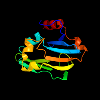
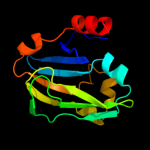
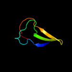
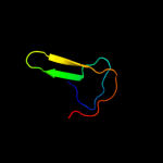
1 c3a8jF_
100.0
100
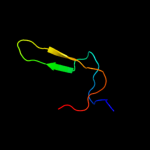
PDB header: transferase/transport proteinChain: F: PDB Molecule: glycine cleavage system h protein;PDBTitle: crystal structure of et-ehred complex
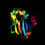
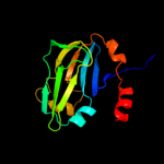
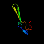
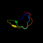
2 c3iftA_
100.0
48
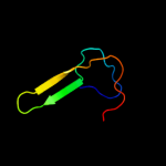
PDB header: oxidoreductaseChain: A: PDB Molecule: glycine cleavage system h protein;PDBTitle: crystal structure of glycine cleavage system protein h from2 mycobacterium tuberculosis, using x-rays from the compact light3 source.
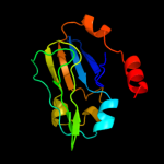
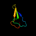
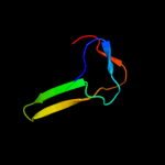
3 d1onla_
100.0
56
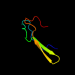
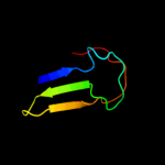
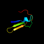
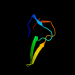
Fold: Barrel-sandwich hybridSuperfamily: Single hybrid motifFamily: Biotinyl/lipoyl-carrier proteins and domains4 c2edgA_
100.0
44
PDB header: biosynthetic proteinChain: A: PDB Molecule: glycine cleavage system h protein;PDBTitle: solution structure of the gcv_h domain from mouse glycine
5 d1hpca_
100.0
50
Fold: Barrel-sandwich hybridSuperfamily: Single hybrid motifFamily: Biotinyl/lipoyl-carrier proteins and domains6 c2ka7A_
100.0
57
PDB header: structural genomics, unknown functionChain: A: PDB Molecule: glycine cleavage system h protein;PDBTitle: nmr solution structure of tm0212 at 40 c
7 c3mxuA_
100.0
53
PDB header: oxidoreductaseChain: A: PDB Molecule: glycine cleavage system h protein;PDBTitle: crystal structure of glycine cleavage system protein h from bartonella2 henselae
8 c2b8gA_
97.8
30
PDB header: biosynthetic proteinChain: A: PDB Molecule: biotin/lipoyl attachment protein;PDBTitle: solution structure of bacillus subtilis blap biotinylated-2 form (energy minimized mean structure)
9 d1dcza_
97.7
32
Fold: Barrel-sandwich hybridSuperfamily: Single hybrid motifFamily: Biotinyl/lipoyl-carrier proteins and domains10 d1k8ma_
97.7
30
Fold: Barrel-sandwich hybridSuperfamily: Single hybrid motifFamily: Biotinyl/lipoyl-carrier proteins and domains11 d1bdoa_
97.6
34
Fold: Barrel-sandwich hybridSuperfamily: Single hybrid motifFamily: Biotinyl/lipoyl-carrier proteins and domains12 c3n6rK_
97.6
32
PDB header: ligaseChain: K: PDB Molecule: propionyl-coa carboxylase, alpha subunit;PDBTitle: crystal structure of the holoenzyme of propionyl-coa carboxylase (pcc)
13 c2ejmA_
97.6
30
PDB header: ligaseChain: A: PDB Molecule: methylcrotonoyl-coa carboxylase subunit alpha;PDBTitle: solution structure of ruh-072, an apo-biotnyl domain form2 human acetyl coenzyme a carboxylase
14 d1ghja_
97.6
18
Fold: Barrel-sandwich hybridSuperfamily: Single hybrid motifFamily: Biotinyl/lipoyl-carrier proteins and domains15 c2l5tA_
97.6
26
PDB header: transferaseChain: A: PDB Molecule: lipoamide acyltransferase;PDBTitle: solution nmr structure of e2 lipoyl domain from thermoplasma2 acidophilum
16 c2dneA_
97.5
19
PDB header: transferaseChain: A: PDB Molecule: dihydrolipoyllysine-residue acetyltransferasePDBTitle: solution structure of rsgi ruh-058, a lipoyl domain of2 human 2-oxoacid dehydrogenase
17 d1iyua_
97.5
23
Fold: Barrel-sandwich hybridSuperfamily: Single hybrid motifFamily: Biotinyl/lipoyl-carrier proteins and domains18 d1qjoa_
97.5
26
Fold: Barrel-sandwich hybridSuperfamily: Single hybrid motifFamily: Biotinyl/lipoyl-carrier proteins and domains19 c2q8iB_
97.5
26
PDB header: transferaseChain: B: PDB Molecule: dihydrolipoyllysine-residue acetyltransferase component ofPDBTitle: pyruvate dehydrogenase kinase isoform 3 in complex with antitumor drug2 radicicol
20 c2ejgD_
97.5
29
PDB header: ligaseChain: D: PDB Molecule: 149aa long hypothetical methylmalonyl-coa decarboxylasePDBTitle: crystal structure of the biotin protein ligase (mutation r48a) and2 biotin carboxyl carrier protein complex from pyrococcus horikoshii3 ot3
21 c2dncA_
not modelled
97.4
32
PDB header: transferaseChain: A: PDB Molecule: pyruvate dehydrogenase protein x component;PDBTitle: solution structure of rsgi ruh-054, a lipoyl domain from2 human 2-oxoacid dehydrogenase
22 d1y8ob1
not modelled
97.4
27
Fold: Barrel-sandwich hybridSuperfamily: Single hybrid motifFamily: Biotinyl/lipoyl-carrier proteins and domains23 d2pnrc1
not modelled
97.4
26
Fold: Barrel-sandwich hybridSuperfamily: Single hybrid motifFamily: Biotinyl/lipoyl-carrier proteins and domains24 d1o78a_
not modelled
97.4
32
Fold: Barrel-sandwich hybridSuperfamily: Single hybrid motifFamily: Biotinyl/lipoyl-carrier proteins and domains25 d1laba_
not modelled
97.3
24
Fold: Barrel-sandwich hybridSuperfamily: Single hybrid motifFamily: Biotinyl/lipoyl-carrier proteins and domains26 d1gjxa_
not modelled
97.3
32
Fold: Barrel-sandwich hybridSuperfamily: Single hybrid motifFamily: Biotinyl/lipoyl-carrier proteins and domains27 c2kccA_
not modelled
97.2
23
PDB header: ligaseChain: A: PDB Molecule: acetyl-coa carboxylase 2;PDBTitle: solution structure of biotinoyl domain from human acetyl-2 coa carboxylase 2
28 c2dn8A_
not modelled
97.1
23
PDB header: ligaseChain: A: PDB Molecule: acetyl-coa carboxylase 2;PDBTitle: solution structure of rsgi ruh-053, an apo-biotin carboxy2 carrier protein from human transcarboxylase
29 d1pmra_
not modelled
97.0
21
Fold: Barrel-sandwich hybridSuperfamily: Single hybrid motifFamily: Biotinyl/lipoyl-carrier proteins and domains30 c3d4rE_
not modelled
96.6
31
PDB header: unknown functionChain: E: PDB Molecule: domain of unknown function from the pfam-b_34464 family;PDBTitle: crystal structure of a duf2118 family protein (mmp0046) from2 methanococcus maripaludis at 2.20 a resolution
31 c2k33A_
not modelled
93.8
32
PDB header: membrane protein, transport proteinChain: A: PDB Molecule: acra;PDBTitle: solution structure of an n-glycosylated protein using in2 vitro glycosylation
32 c2qf7A_
not modelled
93.3
33
PDB header: ligaseChain: A: PDB Molecule: pyruvate carboxylase protein;PDBTitle: crystal structure of a complete multifunctional pyruvate carboxylase2 from rhizobium etli
33 c2qj8B_
not modelled
91.7
29
PDB header: hydrolaseChain: B: PDB Molecule: mlr6093 protein;PDBTitle: crystal structure of an aspartoacylase family protein (mlr6093) from2 mesorhizobium loti maff303099 at 2.00 a resolution
34 c3na6A_
not modelled
88.6
22
PDB header: hydrolaseChain: A: PDB Molecule: succinylglutamate desuccinylase/aspartoacylase;PDBTitle: crystal structure of a succinylglutamate desuccinylase (tm1040_2694)2 from silicibacter sp. tm1040 at 2.00 a resolution
35 c3fmcC_
not modelled
86.7
17
PDB header: hydrolaseChain: C: PDB Molecule: putative succinylglutamate desuccinylase / aspartoacylase;PDBTitle: crystal structure of a putative succinylglutamate desuccinylase /2 aspartoacylase family protein (sama_0604) from shewanella amazonensis3 sb2b at 1.80 a resolution
36 c3cdxB_
not modelled
81.2
17
PDB header: hydrolaseChain: B: PDB Molecule: succinylglutamatedesuccinylase/aspartoacylase;PDBTitle: crystal structure of2 succinylglutamatedesuccinylase/aspartoacylase from3 rhodobacter sphaeroides
37 c2jkuA_
not modelled
68.2
30
PDB header: ligaseChain: A: PDB Molecule: propionyl-coa carboxylase alpha chain,PDBTitle: crystal structure of the n-terminal region of the biotin2 acceptor domain of human propionyl-coa carboxylase
38 d1brwa3
not modelled
61.2
24
Fold: alpha/beta-HammerheadSuperfamily: Pyrimidine nucleoside phosphorylase C-terminal domainFamily: Pyrimidine nucleoside phosphorylase C-terminal domain39 c3h9iB_
not modelled
58.3
38
PDB header: transport proteinChain: B: PDB Molecule: cation efflux system protein cusb;PDBTitle: crystal structure of the membrane fusion protein cusb from escherichia2 coli
40 d1uoua3
not modelled
57.6
19
Fold: alpha/beta-HammerheadSuperfamily: Pyrimidine nucleoside phosphorylase C-terminal domainFamily: Pyrimidine nucleoside phosphorylase C-terminal domain41 d2tpta3
not modelled
51.9
19
Fold: alpha/beta-HammerheadSuperfamily: Pyrimidine nucleoside phosphorylase C-terminal domainFamily: Pyrimidine nucleoside phosphorylase C-terminal domain42 d2gpra_
not modelled
44.7
21
Fold: Barrel-sandwich hybridSuperfamily: Duplicated hybrid motifFamily: Glucose permease-like43 c2aukA_
not modelled
40.8
25
PDB header: transferaseChain: A: PDB Molecule: dna-directed rna polymerase beta' chain;PDBTitle: structure of e. coli rna polymerase beta' g/g' insert
44 d2f3ga_
not modelled
36.5
17
Fold: Barrel-sandwich hybridSuperfamily: Duplicated hybrid motifFamily: Glucose permease-like45 c3lnnB_
not modelled
36.2
32
PDB header: metal transportChain: B: PDB Molecule: membrane fusion protein (mfp) heavy metal cation effluxPDBTitle: crystal structure of zneb from cupriavidus metallidurans
46 d1glaf_
not modelled
35.9
33
Fold: Barrel-sandwich hybridSuperfamily: Duplicated hybrid motifFamily: Glucose permease-like47 c2f1mA_
not modelled
34.0
16
PDB header: transport proteinChain: A: PDB Molecule: acriflavine resistance protein a;PDBTitle: conformational flexibility in the multidrug efflux system protein acra
48 c3h5qA_
not modelled
32.7
19
PDB header: transferaseChain: A: PDB Molecule: pyrimidine-nucleoside phosphorylase;PDBTitle: crystal structure of a putative pyrimidine-nucleoside phosphorylase2 from staphylococcus aureus
49 c1t5eB_
not modelled
32.2
20
PDB header: transport proteinChain: B: PDB Molecule: multidrug resistance protein mexa;PDBTitle: the structure of mexa
50 c2dsjA_
not modelled
31.7
24
PDB header: transferaseChain: A: PDB Molecule: pyrimidine-nucleoside (thymidine) phosphorylase;PDBTitle: crystal structure of project id tt0128 from thermus thermophilus hb8
51 c1brwB_
not modelled
31.1
24
PDB header: transferaseChain: B: PDB Molecule: protein (pyrimidine nucleoside phosphorylase);PDBTitle: the crystal structure of pyrimidine nucleoside2 phosphorylase in a closed conformation
52 c2j0fC_
not modelled
30.3
19
PDB header: transferaseChain: C: PDB Molecule: thymidine phosphorylase;PDBTitle: structural basis for non-competitive product inhibition in2 human thymidine phosphorylase: implication for drug design
53 d1gpra_
not modelled
30.2
20
Fold: Barrel-sandwich hybridSuperfamily: Duplicated hybrid motifFamily: Glucose permease-like54 c1otpA_
not modelled
29.9
19
PDB header: phosphorylaseChain: A: PDB Molecule: thymidine phosphorylase;PDBTitle: structural and theoretical studies suggest domain movement produces an2 active conformation of thymidine phosphorylase
55 d1vf7a_
not modelled
29.6
20
Fold: HlyD-like secretion proteinsSuperfamily: HlyD-like secretion proteinsFamily: HlyD-like secretion proteins56 d1ci3m2
not modelled
26.7
32
Fold: Barrel-sandwich hybridSuperfamily: Rudiment single hybrid motifFamily: Cytochrome f, small domain57 d2p84a1
not modelled
26.6
20
Fold: YopX-likeSuperfamily: YopX-likeFamily: YopX-like58 d1m1fa_
not modelled
19.8
21
Fold: SH3-like barrelSuperfamily: Cell growth inhibitor/plasmid maintenance toxic componentFamily: Kid/PemK59 c3fppB_
not modelled
18.5
22
PDB header: membrane proteinChain: B: PDB Molecule: macrolide-specific efflux protein maca;PDBTitle: crystal structure of e.coli maca
60 c2xhaB_
not modelled
15.8
28
PDB header: transcriptionChain: B: PDB Molecule: transcription antitermination protein nusg;PDBTitle: crystal structure of domain 2 of thermotoga maritima n-utilization2 substance g (nusg)
61 c1vdzA_
not modelled
15.0
30
PDB header: hydrolaseChain: A: PDB Molecule: a-type atpase subunit a;PDBTitle: crystal structure of a-type atpase catalytic subunit a from2 pyrococcus horikoshii ot3
62 d1d7qa_
not modelled
10.6
19
Fold: OB-foldSuperfamily: Nucleic acid-binding proteinsFamily: Cold shock DNA-binding domain-like63 c2b44A_
not modelled
10.5
40
PDB header: hydrolaseChain: A: PDB Molecule: glycyl-glycine endopeptidase lytm;PDBTitle: truncated s. aureus lytm, p 32 2 1 crystal form
64 d1v95a_
not modelled
9.4
35
Fold: Anticodon-binding domain-likeSuperfamily: Class II aaRS ABD-relatedFamily: Anticodon-binding domain of Class II aaRS65 d1e2wa2
not modelled
8.6
34
Fold: Barrel-sandwich hybridSuperfamily: Rudiment single hybrid motifFamily: Cytochrome f, small domain66 c2xhcA_
not modelled
7.5
28
PDB header: transcriptionChain: A: PDB Molecule: transcription antitermination protein nusg;PDBTitle: crystal structure of thermotoga maritima n-utilization substance g2 (nusg)
67 d1ne8a_
not modelled
7.5
38
Fold: SH3-like barrelSuperfamily: Cell growth inhibitor/plasmid maintenance toxic componentFamily: Kid/PemK68 d2vv5a1
not modelled
7.1
19
Fold: Sm-like foldSuperfamily: Sm-like ribonucleoproteinsFamily: Mechanosensitive channel protein MscS (YggB), middle domain69 d1qapa2
not modelled
6.8
18
Fold: alpha/beta-HammerheadSuperfamily: Nicotinate/Quinolinate PRTase N-terminal domain-likeFamily: NadC N-terminal domain-like70 c2hsiB_
not modelled
6.3
20
PDB header: structural genomics, unknown functionChain: B: PDB Molecule: putative peptidase m23;PDBTitle: crystal structure of putative peptidase m23 from2 pseudomonas aeruginosa, new york structural genomics3 consortium
71 c2gu1A_
not modelled
6.2
24
PDB header: hydrolaseChain: A: PDB Molecule: zinc peptidase;PDBTitle: crystal structure of a zinc containing peptidase from2 vibrio cholerae
72 d1jt8a_
not modelled
6.1
26
Fold: OB-foldSuperfamily: Nucleic acid-binding proteinsFamily: Cold shock DNA-binding domain-like73 c3pajA_
not modelled
5.8
15
PDB header: transferaseChain: A: PDB Molecule: nicotinate-nucleotide pyrophosphorylase, carboxylating;PDBTitle: 2.00 angstrom resolution crystal structure of a quinolinate2 phosphoribosyltransferase from vibrio cholerae o1 biovar eltor str.3 n16961
74 d1qwya_
not modelled
5.8
40
Fold: Barrel-sandwich hybridSuperfamily: Duplicated hybrid motifFamily: Peptidoglycan hydrolase LytM75 d1o4ua2
not modelled
5.3
16
Fold: alpha/beta-HammerheadSuperfamily: Nicotinate/Quinolinate PRTase N-terminal domain-likeFamily: NadC N-terminal domain-like76 c3fggA_
not modelled
5.3
14
PDB header: structural genomics, unknown functionChain: A: PDB Molecule: uncharacterized protein bce2196;PDBTitle: crystal structure of putative ecf-type sigma factor negative effector2 from bacillus cereus
77 c3tqvA_
not modelled
5.0
12
PDB header: transferaseChain: A: PDB Molecule: nicotinate-nucleotide pyrophosphorylase;PDBTitle: structure of the nicotinate-nucleotide pyrophosphorylase from2 francisella tularensis.