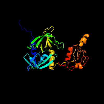
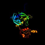
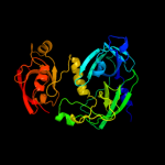
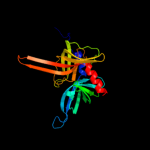
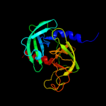
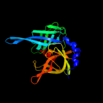
| 1 | c3stjC_
|
|
 |
100.0 |
43 |
PDB header:hydrolase
Chain: C: PDB Molecule:protease degq;
PDBTitle: crystal structure of the protease + pdz1 domain of degq from2 escherichia coli
|
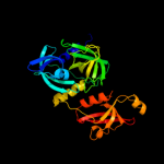
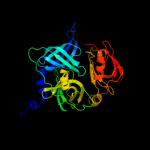
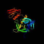
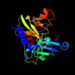
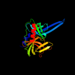
| 2 | c1ky9A_
|
|
 |
100.0 |
40 |
PDB header:hydrolase
Chain: A: PDB Molecule:protease do;
PDBTitle: crystal structure of degp (htra)
|
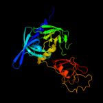
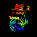
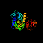
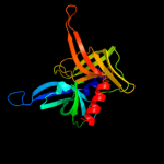
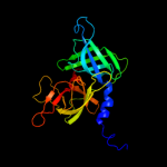
| 3 | c3gdsA_
|
|
 |
100.0 |
95 |
PDB header:hydrolase/hydrolase activator
Chain: A: PDB Molecule:protease degs;
PDBTitle: crystal structure of degs h198p/d320a mutant modified by dfp in2 complex with dnrdgnvyyf peptide
|
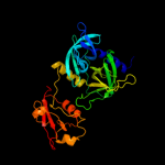
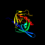
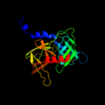
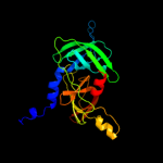
| 4 | c4a8aI_
|
|
 |
100.0 |
42 |
PDB header:hydrolase/hydrolase
Chain: I: PDB Molecule:periplasmic ph-dependent serine endoprotease degq;
PDBTitle: asymmetric cryo-em reconstruction of e. coli degq 12-mer in complex2 with lysozyme
|
| 5 | c3pv5B_
|
|
 |
100.0 |
44 |
PDB header:hydrolase
Chain: B: PDB Molecule:degq;
PDBTitle: structure of legionella fallonii degq (n189g/p190g variant)
|
| 6 | c3qo6B_
|
|
 |
100.0 |
35 |
PDB header:photosynthesis
Chain: B: PDB Molecule:protease do-like 1, chloroplastic;
PDBTitle: crystal structure analysis of the plant protease deg1
|
| 7 | c1lcyA_
|
|
 |
100.0 |
30 |
PDB header:hydrolase
Chain: A: PDB Molecule:htra2 serine protease;
PDBTitle: crystal structure of the mitochondrial serine protease htra2
|
| 8 | c3pv4A_
|
|
 |
100.0 |
44 |
PDB header:hydrolase
Chain: A: PDB Molecule:degq;
PDBTitle: structure of legionella fallonii degq (delta-pdz2 variant)
|
| 9 | c2r3yC_
|
|
 |
100.0 |
100 |
PDB header:hydrolase/hydrolase activator
Chain: C: PDB Molecule:protease degs;
PDBTitle: crystal structure of the degs protease in complex with the2 ywf activating peptide
|
| 10 | c2z9iB_
|
|
 |
100.0 |
36 |
PDB header:hydrolase
Chain: B: PDB Molecule:probable serine protease pepd;
PDBTitle: crystal structure of rv0983 from mycobacterium tuberculosis-2 proteolytically active form
|
| 11 | d1ky9a2
|
|
 |
100.0 |
43 |
Fold:Trypsin-like serine proteases
Superfamily:Trypsin-like serine proteases
Family:Prokaryotic proteases |
| 12 | c2rceI_
|
|
 |
100.0 |
97 |
PDB header:hydrolase
Chain: I: PDB Molecule:protease degs;
PDBTitle: dfp modified degs delta pdz
|
| 13 | d1l1ja_
|
|
 |
100.0 |
39 |
Fold:Trypsin-like serine proteases
Superfamily:Trypsin-like serine proteases
Family:Prokaryotic proteases |
| 14 | d2qf3a1
|
|
 |
100.0 |
97 |
Fold:Trypsin-like serine proteases
Superfamily:Trypsin-like serine proteases
Family:Prokaryotic proteases |
| 15 | c3nziA_
|
|
 |
100.0 |
35 |
PDB header:hydrolase/hydrolase substrate
Chain: A: PDB Molecule:serine protease htra1;
PDBTitle: substrate induced remodeling of the active site regulates htra12 activity
|
| 16 | c3stiC_
|
|
 |
100.0 |
52 |
PDB header:hydrolase
Chain: C: PDB Molecule:protease degq;
PDBTitle: crystal structure of the protease domain of degq from escherichia coli
|
| 17 | d2z9ia2
|
|
 |
100.0 |
39 |
Fold:Trypsin-like serine proteases
Superfamily:Trypsin-like serine proteases
Family:Prokaryotic proteases |
| 18 | c3nwuB_
|
|
 |
100.0 |
39 |
PDB header:hydrolase
Chain: B: PDB Molecule:serine protease htra1;
PDBTitle: substrate induced remodeling of the active site regulates htra12 activity
|
| 19 | d1lcya2
|
|
 |
100.0 |
34 |
Fold:Trypsin-like serine proteases
Superfamily:Trypsin-like serine proteases
Family:Prokaryotic proteases |
| 20 | c3k6zA_
|
|
 |
100.0 |
24 |
PDB header:hydrolase
Chain: A: PDB Molecule:possible membrane-associated serine protease;
PDBTitle: crystal structure of rv3671c protease, inactive form
|
| 21 | c1zyoA_ |
|
not modelled |
100.0 |
16 |
PDB header:hydrolase
Chain: A: PDB Molecule:serine protease;
PDBTitle: crystal structure of the serine protease domain of sesbania mosaic2 virus polyprotein
|
| 22 | c3mmgB_ |
|
not modelled |
99.9 |
19 |
PDB header:viral protein, hydrolase
Chain: B: PDB Molecule:nuclear inclusion protein a;
PDBTitle: crystal structure of tobacco vein mottling virus protease
|
| 23 | c2w5eB_ |
|
not modelled |
99.9 |
18 |
PDB header:hydrolase
Chain: B: PDB Molecule:putative serine protease;
PDBTitle: structural and biochemical analysis of human pathogenic2 astrovirus serine protease at 2.0 angstrom resolution
|
| 24 | d1q31a_ |
|
not modelled |
99.9 |
18 |
Fold:Trypsin-like serine proteases
Superfamily:Trypsin-like serine proteases
Family:Viral proteases |
| 25 | d1lvmb_ |
|
not modelled |
99.9 |
17 |
Fold:Trypsin-like serine proteases
Superfamily:Trypsin-like serine proteases
Family:Viral proteases |
| 26 | d1qtfa_ |
|
not modelled |
99.9 |
28 |
Fold:Trypsin-like serine proteases
Superfamily:Trypsin-like serine proteases
Family:Prokaryotic proteases |
| 27 | d1agja_ |
|
not modelled |
99.9 |
23 |
Fold:Trypsin-like serine proteases
Superfamily:Trypsin-like serine proteases
Family:Prokaryotic proteases |
| 28 | c2vidA_ |
|
not modelled |
99.9 |
19 |
PDB header:hydrolase
Chain: A: PDB Molecule:serine protease splb;
PDBTitle: serine protease splb from staphylococcus aureus at 1.8a2 resolution
|
| 29 | c2w7uC_ |
|
not modelled |
99.8 |
26 |
PDB header:hydrolase
Chain: C: PDB Molecule:serine protease spla;
PDBTitle: spla serine protease of staphylococcus aureus (2.4a)
|
| 30 | d1cqqa_ |
|
not modelled |
99.8 |
15 |
Fold:Trypsin-like serine proteases
Superfamily:Trypsin-like serine proteases
Family:Viral cysteine protease of trypsin fold |
| 31 | c2p3wB_ |
|
not modelled |
99.8 |
28 |
PDB header:protein binding
Chain: B: PDB Molecule:probable serine protease htra3;
PDBTitle: crystal structure of the htra3 pdz domain bound to a phage-derived2 ligand (fgrwv)
|
| 32 | d1lcya1 |
|
not modelled |
99.8 |
27 |
Fold:PDZ domain-like
Superfamily:PDZ domain-like
Family:HtrA-like serine proteases |
| 33 | c2as9B_ |
|
not modelled |
99.8 |
24 |
PDB header:hydrolase
Chain: B: PDB Molecule:serine protease;
PDBTitle: functional and structural characterization of spl proteases2 from staphylococcus aureus
|
| 34 | d1ky9a1 |
|
not modelled |
99.8 |
34 |
Fold:PDZ domain-like
Superfamily:PDZ domain-like
Family:HtrA-like serine proteases |
| 35 | c2b0fA_ |
|
not modelled |
99.8 |
19 |
PDB header:hydrolase/hydrolase inhibitor
Chain: A: PDB Molecule:picornain 3c (protease 3c) (p3c);
PDBTitle: nmr structure of the human rhinovirus 3c protease (serotype 14) with2 covalently bound ace-lealfq-ethylpropionate inhibitor
|
| 36 | c2joaA_ |
|
not modelled |
99.8 |
30 |
PDB header:protein binding
Chain: A: PDB Molecule:serine protease htra1;
PDBTitle: htra1 bound to an optimized peptide: nmr assignment of pdz2 domain and ligand resonances
|
| 37 | d2qaaa1 |
|
not modelled |
99.8 |
17 |
Fold:Trypsin-like serine proteases
Superfamily:Trypsin-like serine proteases
Family:Prokaryotic proteases |
| 38 | d1l1na_ |
|
not modelled |
99.8 |
15 |
Fold:Trypsin-like serine proteases
Superfamily:Trypsin-like serine proteases
Family:Viral cysteine protease of trypsin fold |
| 39 | d2o8la1 |
|
not modelled |
99.7 |
18 |
Fold:Trypsin-like serine proteases
Superfamily:Trypsin-like serine proteases
Family:Prokaryotic proteases |
| 40 | c2o8lA_ |
|
not modelled |
99.7 |
18 |
PDB header:hydrolase
Chain: A: PDB Molecule:v8 protease;
PDBTitle: structure of v8 protease from staphylococcus aureus
|
| 41 | c1qy6A_ |
|
not modelled |
99.7 |
19 |
PDB header:protease
Chain: A: PDB Molecule:serine protease;
PDBTitle: structue of v8 protease from staphylococcus aureus
|
| 42 | d2z9ia1 |
|
not modelled |
99.7 |
27 |
Fold:PDZ domain-like
Superfamily:PDZ domain-like
Family:HtrA-like serine proteases |
| 43 | c1wczA_ |
|
not modelled |
99.7 |
20 |
PDB header:hydrolase
Chain: A: PDB Molecule:glutamyl endopeptidase;
PDBTitle: crystal structure of an alkaline form of v8 protease from2 staphylococcus aureus
|
| 44 | c2ouaA_ |
|
not modelled |
99.7 |
16 |
PDB header:hydrolase
Chain: A: PDB Molecule:serine protease;
PDBTitle: crystal structure of nocardiopsis protease (napase)
|
| 45 | c2kl1A_ |
|
not modelled |
99.7 |
25 |
PDB header:protein binding
Chain: A: PDB Molecule:ylbl protein;
PDBTitle: solution structure of gtr34c from geobacillus thermodenitrificans.2 northeast structural genomics consortium target gtr34c
|
| 46 | d2h5ca1 |
|
not modelled |
99.7 |
18 |
Fold:Trypsin-like serine proteases
Superfamily:Trypsin-like serine proteases
Family:Prokaryotic proteases |
| 47 | d2sfaa_ |
|
not modelled |
99.6 |
17 |
Fold:Trypsin-like serine proteases
Superfamily:Trypsin-like serine proteases
Family:Prokaryotic proteases |
| 48 | d2sgaa_ |
|
not modelled |
99.6 |
17 |
Fold:Trypsin-like serine proteases
Superfamily:Trypsin-like serine proteases
Family:Prokaryotic proteases |
| 49 | c3i18A_ |
|
not modelled |
99.6 |
23 |
PDB header:structural genomics, unknown function
Chain: A: PDB Molecule:lmo2051 protein;
PDBTitle: crystal structure of the pdz domain of the sdrc-like protein2 (lmo2051) from listeria monocytogenes, northeast structural3 genomics consortium target lmr166b
|
| 50 | c2kjpA_ |
|
not modelled |
99.6 |
29 |
PDB header:structural genomics, unknown function
Chain: A: PDB Molecule:uncharacterized protein ylbl;
PDBTitle: solution structure of protein ylbl (bsu15050) from bacillus2 subtilis, northeast structural genomics consortium target3 sr713a
|
| 51 | d2i4sa1 |
|
not modelled |
99.6 |
14 |
Fold:PDZ domain-like
Superfamily:PDZ domain-like
Family:EpsC C-terminal domain-like |
| 52 | d1p3ca_ |
|
not modelled |
99.6 |
15 |
Fold:Trypsin-like serine proteases
Superfamily:Trypsin-like serine proteases
Family:Prokaryotic proteases |
| 53 | c2pfeA_ |
|
not modelled |
99.6 |
15 |
PDB header:hydrolase
Chain: A: PDB Molecule:alkaline serine protease;
PDBTitle: crystal structure of thermobifida fusca protease a (tfpa)
|
| 54 | d1hpga_ |
|
not modelled |
99.6 |
19 |
Fold:Trypsin-like serine proteases
Superfamily:Trypsin-like serine proteases
Family:Prokaryotic proteases |
| 55 | d1sota1 |
|
not modelled |
99.6 |
97 |
Fold:PDZ domain-like
Superfamily:PDZ domain-like
Family:HtrA-like serine proteases |
| 56 | c2ea3A_ |
|
not modelled |
99.6 |
17 |
PDB header:hydrolase
Chain: A: PDB Molecule:chymotrypsin;
PDBTitle: crystal structure of cellulomonas bogoriensis chymotrypsin
|
| 57 | d1arba_ |
|
not modelled |
99.5 |
14 |
Fold:Trypsin-like serine proteases
Superfamily:Trypsin-like serine proteases
Family:Prokaryotic proteases |
| 58 | d2i6va1 |
|
not modelled |
99.5 |
13 |
Fold:PDZ domain-like
Superfamily:PDZ domain-like
Family:EpsC C-terminal domain-like |
| 59 | d2hgaa1 |
|
not modelled |
99.5 |
21 |
Fold:PDZ domain-like
Superfamily:PDZ domain-like
Family:MTH1368 C-terminal domain-like |
| 60 | d1ky9b2 |
|
not modelled |
99.4 |
28 |
Fold:PDZ domain-like
Superfamily:PDZ domain-like
Family:HtrA-like serine proteases |
| 61 | d1fc6a3 |
|
not modelled |
99.4 |
24 |
Fold:PDZ domain-like
Superfamily:PDZ domain-like
Family:Tail specific protease PDZ domain |
| 62 | c2zplA_ |
|
not modelled |
99.4 |
17 |
PDB header:hydrolase
Chain: A: PDB Molecule:regulator of sigma e protease;
PDBTitle: crystal structure analysis of pdz domain a
|
| 63 | c3rleA_ |
|
not modelled |
99.3 |
13 |
PDB header:membrane protein
Chain: A: PDB Molecule:golgi reassembly-stacking protein 2;
PDBTitle: crystal structure of grasp55 grasp domain (residues 7-208)
|
| 64 | c2zpmA_ |
|
not modelled |
99.3 |
25 |
PDB header:hydrolase
Chain: A: PDB Molecule:regulator of sigma e protease;
PDBTitle: crystal structure analysis of pdz domain b
|
| 65 | c3cp7B_ |
|
not modelled |
99.3 |
17 |
PDB header:hydrolase
Chain: B: PDB Molecule:alkaline serine protease al20;
PDBTitle: crystal structure of a thermostable serine protease al202 from extremophilic microoganism
|
| 66 | c2eaqA_ |
|
not modelled |
99.1 |
26 |
PDB header:metal binding protein
Chain: A: PDB Molecule:lim domain only protein 7;
PDBTitle: crystal structure of pdz domain of kiaa0858 (lim), ms07932 from homo sapiens
|
| 67 | c3diwB_ |
|
not modelled |
99.1 |
29 |
PDB header:signaling protein/cell adhesion
Chain: B: PDB Molecule:tax1-binding protein 3;
PDBTitle: c-terminal beta-catenin bound tip-1 structure
|
| 68 | d1uf1a_ |
|
not modelled |
99.1 |
24 |
Fold:PDZ domain-like
Superfamily:PDZ domain-like
Family:PDZ domain |
| 69 | c2eehA_ |
|
not modelled |
99.1 |
28 |
PDB header:metal binding protein
Chain: A: PDB Molecule:pdz domain-containing protein 7;
PDBTitle: solution structure of first pdz domain of pdz domain2 containing protein 7
|
| 70 | d1x5qa1 |
|
not modelled |
99.1 |
28 |
Fold:PDZ domain-like
Superfamily:PDZ domain-like
Family:PDZ domain |
| 71 | c3shuB_ |
|
not modelled |
99.0 |
25 |
PDB header:cell adhesion
Chain: B: PDB Molecule:tight junction protein zo-1;
PDBTitle: crystal structure of zo-1 pdz3
|
| 72 | d1rgwa_ |
|
not modelled |
99.0 |
14 |
Fold:PDZ domain-like
Superfamily:PDZ domain-like
Family:PDZ domain |
| 73 | c2q3gA_ |
|
not modelled |
99.0 |
26 |
PDB header:structural genomics
Chain: A: PDB Molecule:pdz and lim domain protein 7;
PDBTitle: structure of the pdz domain of human pdlim7 bound to a c-2 terminal extension from human beta-tropomyosin
|
| 74 | c3l4fD_ |
|
not modelled |
99.0 |
17 |
PDB header:signaling protein/protein binding
Chain: D: PDB Molecule:sh3 and multiple ankyrin repeat domains protein
PDBTitle: crystal structure of betapix coiled-coil domain and shank2 pdz complex
|
| 75 | c2v90E_ |
|
not modelled |
99.0 |
24 |
PDB header:protein-binding
Chain: E: PDB Molecule:pdz domain-containing protein 3;
PDBTitle: crystal structure of the 3rd pdz domain of intestine- and2 kidney-enriched pdz domain ikepp (pdzd3)
|
| 76 | d2f5ya1 |
|
not modelled |
99.0 |
23 |
Fold:PDZ domain-like
Superfamily:PDZ domain-like
Family:PDZ domain |
| 77 | d1wfga_ |
|
not modelled |
99.0 |
17 |
Fold:PDZ domain-like
Superfamily:PDZ domain-like
Family:PDZ domain |
| 78 | c2v1wB_ |
|
not modelled |
99.0 |
20 |
PDB header:structural protein
Chain: B: PDB Molecule:pdz and lim domain protein 4;
PDBTitle: crystal structure of human lim protein ril (pdlim4) pdz2 domain bound to the c-terminal peptide of human alpha-3 actinin-1
|
| 79 | c2komA_ |
|
not modelled |
99.0 |
20 |
PDB header:signaling protein
Chain: A: PDB Molecule:partitioning defective 3 homolog;
PDBTitle: solution structure of humar par-3b pdz2 (residues 451-549)
|
| 80 | d1m5za_ |
|
not modelled |
99.0 |
18 |
Fold:PDZ domain-like
Superfamily:PDZ domain-like
Family:PDZ domain |
| 81 | c2krgA_ |
|
not modelled |
99.0 |
21 |
PDB header:signaling protein
Chain: A: PDB Molecule:na(+)/h(+) exchange regulatory cofactor nhe-rf1;
PDBTitle: solution structure of human sodium/ hydrogen exchange2 regulatory factor 1(150-358)
|
| 82 | c2yuyA_ |
|
not modelled |
99.0 |
22 |
PDB header:signaling protein
Chain: A: PDB Molecule:rho gtpase activating protein 21;
PDBTitle: solution structure of pdz domain of rho gtpase activating2 protein 21
|
| 83 | d1ueqa_ |
|
not modelled |
99.0 |
23 |
Fold:PDZ domain-like
Superfamily:PDZ domain-like
Family:PDZ domain |
| 84 | d1q3oa_ |
|
not modelled |
99.0 |
18 |
Fold:PDZ domain-like
Superfamily:PDZ domain-like
Family:PDZ domain |
| 85 | d1p1da2 |
|
not modelled |
99.0 |
13 |
Fold:PDZ domain-like
Superfamily:PDZ domain-like
Family:PDZ domain |
| 86 | d1wifa_ |
|
not modelled |
99.0 |
23 |
Fold:PDZ domain-like
Superfamily:PDZ domain-like
Family:PDZ domain |
| 87 | c2iwnA_ |
|
not modelled |
99.0 |
26 |
PDB header:signaling protein
Chain: A: PDB Molecule:multiple pdz domain protein;
PDBTitle: 3rd pdz domain of multiple pdz domain protein mpdz (casp2 target)
|
| 88 | c3f1sB_ |
|
not modelled |
99.0 |
15 |
PDB header:hydrolase inhibitor/hydrolase
Chain: B: PDB Molecule:vitamin k-dependent protein z;
PDBTitle: crystal structure of protein z complexed with protein z-dependent2 inhibitor
|
| 89 | c2f83A_ |
|
not modelled |
99.0 |
23 |
PDB header:hydrolase
Chain: A: PDB Molecule:coagulation factor xi;
PDBTitle: crystal structure at 2.9 angstroms resolution of human plasma2 coagulation factor xi zymogen
|
| 90 | c2kjdA_ |
|
not modelled |
99.0 |
21 |
PDB header:signaling protein
Chain: A: PDB Molecule:sodium/hydrogen exchange regulatory cofactor nhe-
PDBTitle: solution structure of extended pdz2 domain from nherf1 (150-2 270)
|
| 91 | d1wf7a_ |
|
not modelled |
99.0 |
19 |
Fold:PDZ domain-like
Superfamily:PDZ domain-like
Family:PDZ domain |
| 92 | c3shwA_ |
|
not modelled |
98.9 |
26 |
PDB header:cell adhesion
Chain: A: PDB Molecule:tight junction protein zo-1;
PDBTitle: crystal structure of zo-1 pdz3-sh3-guk supramodule complex with2 connexin-45 peptide
|
| 93 | d1ozia_ |
|
not modelled |
98.9 |
20 |
Fold:PDZ domain-like
Superfamily:PDZ domain-like
Family:PDZ domain |
| 94 | d1w9ea1 |
|
not modelled |
98.9 |
17 |
Fold:PDZ domain-like
Superfamily:PDZ domain-like
Family:PDZ domain |
| 95 | d2fe5a1 |
|
not modelled |
98.9 |
16 |
Fold:PDZ domain-like
Superfamily:PDZ domain-like
Family:PDZ domain |
| 96 | c2he4A_ |
|
not modelled |
98.9 |
18 |
PDB header:structural genomics, unknown function
Chain: A: PDB Molecule:na(+)/h(+) exchange regulatory cofactor nhe-rf2;
PDBTitle: the crystal structure of the second pdz domain of human2 nherf-2 (slc9a3r2) interacting with a mode 1 pdz binding3 motif
|
| 97 | c2egkC_ |
|
not modelled |
98.9 |
23 |
PDB header:protein binding
Chain: C: PDB Molecule:general receptor for phosphoinositides 1-
PDBTitle: crystal structure of tamalin pdz-intrinsic ligand fusion2 protein
|
| 98 | c3cyyA_ |
|
not modelled |
98.9 |
27 |
PDB header:peptide binding protein
Chain: A: PDB Molecule:tight junction protein zo-1;
PDBTitle: the crystal structure of zo-1 pdz2 in complex with the cx43 peptide
|
| 99 | c3osyA_ |
|
not modelled |
98.9 |
15 |
PDB header:hydrolase
Chain: A: PDB Molecule:3c protease;
PDBTitle: human enterovirus 71 3c protease
|
| 100 | c2xrcD_ |
|
not modelled |
98.9 |
16 |
PDB header:immune system
Chain: D: PDB Molecule:human complement factor i;
PDBTitle: human complement factor i
|
| 101 | c3k1rA_ |
|
not modelled |
98.9 |
21 |
PDB header:structural protein
Chain: A: PDB Molecule:harmonin;
PDBTitle: structure of harmonin npdz1 in complex with the sam-pbm of2 sans
|
| 102 | c2vsvB_ |
|
not modelled |
98.9 |
21 |
PDB header:protein-binding
Chain: B: PDB Molecule:rhophilin-2;
PDBTitle: crystal structure of the pdz domain of human rhophilin-2
|
| 103 | d1rzxa_ |
|
not modelled |
98.9 |
20 |
Fold:PDZ domain-like
Superfamily:PDZ domain-like
Family:PDZ domain |
| 104 | d1g9oa_ |
|
not modelled |
98.9 |
16 |
Fold:PDZ domain-like
Superfamily:PDZ domain-like
Family:PDZ domain |
| 105 | c2vwrA_ |
|
not modelled |
98.9 |
16 |
PDB header:protein-binding
Chain: A: PDB Molecule:ligand of numb protein x 2;
PDBTitle: crystal structure of the second pdz domain of numb-binding2 protein 2
|
| 106 | d1t2ma1 |
|
not modelled |
98.9 |
24 |
Fold:PDZ domain-like
Superfamily:PDZ domain-like
Family:PDZ domain |
| 107 | c2o2tB_ |
|
not modelled |
98.9 |
19 |
PDB header:structural protein
Chain: B: PDB Molecule:multiple pdz domain protein;
PDBTitle: the crystal structure of the 1st pdz domain of mpdz
|
| 108 | d1ry4a_ |
|
not modelled |
98.9 |
20 |
Fold:PDZ domain-like
Superfamily:PDZ domain-like
Family:PDZ domain |
| 109 | c2i04B_ |
|
not modelled |
98.9 |
24 |
PDB header:peptide binding protein
Chain: B: PDB Molecule:membrane-associated guanylate kinase, ww and pdz
PDBTitle: x-ray crystal structure of magi-1 pdz1 bound to the c-2 terminal peptide of hpv18 e6
|
| 110 | c1p1dA_ |
|
not modelled |
98.9 |
11 |
PDB header:protein binding
Chain: A: PDB Molecule:glutamate receptor interacting protein;
PDBTitle: structural insights into the inter-domain chaperoning of2 tandem pdz domains in glutamate receptor interacting3 proteins
|
| 111 | d2byga1 |
|
not modelled |
98.9 |
21 |
Fold:PDZ domain-like
Superfamily:PDZ domain-like
Family:PDZ domain |
| 112 | c1rs0A_ |
|
not modelled |
98.9 |
16 |
PDB header:hydrolase
Chain: A: PDB Molecule:complement factor b;
PDBTitle: crystal structure analysis of the bb segment of factor b2 complexed with di-isopropyl-phosphate (dip)
|
| 113 | c2ok5A_ |
|
not modelled |
98.9 |
17 |
PDB header:hydrolase
Chain: A: PDB Molecule:complement factor b;
PDBTitle: human complement factor b
|
| 114 | c2omjA_ |
|
not modelled |
98.9 |
29 |
PDB header:cell adhesion
Chain: A: PDB Molecule:rho guanine nucleotide exchange factor 12;
PDBTitle: solution structure of larg pdz domain
|
| 115 | c2qt5A_ |
|
not modelled |
98.9 |
23 |
PDB header:peptide binding protein
Chain: A: PDB Molecule:glutamate receptor-interacting protein 1;
PDBTitle: crystal structure of grip1 pdz12 in complex with the fras12 peptide
|
| 116 | c3khfA_ |
|
not modelled |
98.9 |
21 |
PDB header:transferase
Chain: A: PDB Molecule:microtubule-associated serine/threonine-protein
PDBTitle: the crystal structure of the pdz domain of human microtubule2 associated serine/threonine kinase 3 (mast3)
|
| 117 | c2jxoA_ |
|
not modelled |
98.9 |
21 |
PDB header:protein binding
Chain: A: PDB Molecule:ezrin-radixin-moesin-binding phosphoprotein 50;
PDBTitle: structure of the second pdz domain of nherf-1
|
| 118 | d1ueza_ |
|
not modelled |
98.9 |
26 |
Fold:PDZ domain-like
Superfamily:PDZ domain-like
Family:PDZ domain |
| 119 | c2ogpA_ |
|
not modelled |
98.9 |
23 |
PDB header:signaling protein
Chain: A: PDB Molecule:partitioning-defective 3 homolog;
PDBTitle: solution structure of the second pdz domain of par-3
|
| 120 | c3qglD_ |
|
not modelled |
98.9 |
20 |
PDB header:protein binding
Chain: D: PDB Molecule:sorting nexin-27;
PDBTitle: crystal structure of pdz domain of sorting nexin 27 (snx27) in complex2 with the eseskv peptide corresponding to the c-terminal tail of girk3
|