1 c1gvhA_
100.0
100
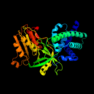
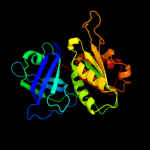
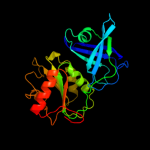
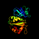
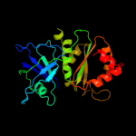
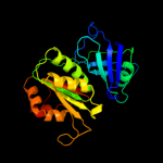
PDB header: oxidoreductaseChain: A: PDB Molecule: flavohemoprotein;PDBTitle: the x-ray structure of ferric escherichia coli2 flavohemoglobin reveals an unespected geometry of the3 distal heme pocket
2 c1cqxB_
100.0
46
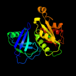
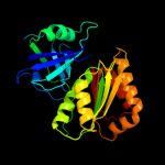
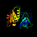
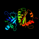
PDB header: lipid binding proteinChain: B: PDB Molecule: flavohemoprotein;PDBTitle: crystal structure of the flavohemoglobin from alcaligenes eutrophus at2 1.75 a resolution
3 c1krhA_
100.0
24
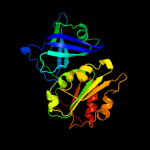
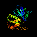
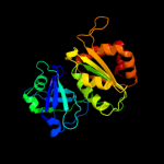
PDB header: oxidoreductaseChain: A: PDB Molecule: benzoate 1,2-dioxygenase reductase;PDBTitle: x-ray stucture of benzoate dioxygenase reductase
4 c2r6hC_
100.0
20
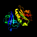
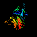
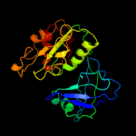
PDB header: oxidoreductaseChain: C: PDB Molecule: nadh:ubiquinone oxidoreductase, na translocating, fPDBTitle: crystal structure of the domain comprising the nad binding and the fad2 binding regions of the nadh:ubiquinone oxidoreductase, na3 translocating, f subunit from porphyromonas gingivalis
5 c2eixA_
100.0
22
PDB header: oxidoreductaseChain: A: PDB Molecule: nadh-cytochrome b5 reductase;PDBTitle: the structure of physarum polycephalum cytochrome b5 reductase
6 c1tvcA_
100.0
23
PDB header: oxidoreductaseChain: A: PDB Molecule: methane monooxygenase component c;PDBTitle: fad and nadh binding domain of methane monooxygenase2 reductase from methylococcus capsulatus (bath)
7 c3fpkB_
100.0
18
PDB header: flavoprotein, oxidoreductaseChain: B: PDB Molecule: ferredoxin-nadp reductase;PDBTitle: crystal structure of ferredoxin-nadp reductase from salmonella2 typhimurium
8 c2ok8D_
100.0
17
PDB header: oxidoreductaseChain: D: PDB Molecule: putative ferredoxin--nadp reductase;PDBTitle: ferredoxin-nadp+ reductase from plasmodium falciparum
9 c1a8pA_
100.0
13
PDB header: oxidoreductaseChain: A: PDB Molecule: nadph\:ferredoxin oxidoreductase;PDBTitle: ferredoxin reductase from azotobacter vinelandii
10 c2bgjB_
100.0
14
PDB header: oxidoreductaseChain: B: PDB Molecule: ferredoxin-nadp(h) reductase;PDBTitle: x-ray structure of the ferredoxin-nadp(h) reductase from2 rhodobacter capsulatus at 2.1 angstroms
11 c1fncA_
100.0
20
PDB header: oxidoreductase (nadp+(a),ferredoxin(a))Chain: A: PDB Molecule: ferredoxin-nadp+ reductase;PDBTitle: refined crystal structure of spinach ferredoxin reductase2 at 1.7 angstroms resolution: oxidized, reduced, and 2'-3 phospho-5'-amp bound states
12 c1umkA_
100.0
20
PDB header: oxidoreductaseChain: A: PDB Molecule: nadh-cytochrome b5 reductase;PDBTitle: the structure of human erythrocyte nadh-cytochrome b52 reductase
13 c2piaA_
100.0
22
PDB header: reductaseChain: A: PDB Molecule: phthalate dioxygenase reductase;PDBTitle: phthalate dioxygenase reductase: a modular structure for2 electron transfer from pyridine nucleotides to [2fe-2s]
14 c1qfjD_
100.0
16
PDB header: oxidoreductaseChain: D: PDB Molecule: protein (flavin reductase);PDBTitle: crystal structure of nad(p)h:flavin oxidoreductase from escherichia2 coli
15 c1qgyA_
100.0
20
PDB header: oxidoreductaseChain: A: PDB Molecule: ferredoxin--nadp+ reductase;PDBTitle: ferredoxin:nadp+ reductase mutant with lys 75 replaced by glu (k75e)
16 c2b5oA_
100.0
20
PDB header: oxidoreductaseChain: A: PDB Molecule: ferredoxin--nadp reductase;PDBTitle: ferredoxin-nadp reductase
17 c1jb9A_
100.0
21
PDB header: oxidoreductaseChain: A: PDB Molecule: ferredoxin-nadp reductase;PDBTitle: crystal structure of the ferredoxin:nadp+ reductase from maize root at2 1.7 angstroms
18 c1cneA_
100.0
19
PDB header: oxidoreductaseChain: A: PDB Molecule: nitrate reductase;PDBTitle: structural studies on corn nitrate reductase: refined2 structure of the cytochrome b reductase fragment at 2.53 angstroms, its adp complex and an active site mutant and4 modeling of the cytochrome b domain
19 c2rc5D_
100.0
21
PDB header: oxidoreductaseChain: D: PDB Molecule: ferredoxin-nadp reductase;PDBTitle: refined structure of fnr from leptospira interrogans
20 d1gvha1
100.0
100
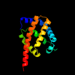
Fold: Globin-likeSuperfamily: Globin-likeFamily: Globins21 c1ep3B_
not modelled
100.0
19
PDB header: oxidoreductaseChain: B: PDB Molecule: dihydroorotate dehydrogenase b (pyrk subunit);PDBTitle: crystal structure of lactococcus lactis dihydroorotate dehydrogenase2 b. data collected under cryogenic conditions.
22 d1cqxa1
not modelled
100.0
47
Fold: Globin-likeSuperfamily: Globin-likeFamily: Globins23 c2gpjA_
not modelled
100.0
10
PDB header: fad-binding proteinChain: A: PDB Molecule: siderophore-interacting protein;PDBTitle: crystal structure of a siderophore-interacting protein (sputcn32_0076)2 from shewanella putrefaciens cn-32 at 2.20 a resolution
24 c2wy4A_
not modelled
99.9
38
PDB header: oxygen transportChain: A: PDB Molecule: single domain haemoglobin;PDBTitle: structure of bacterial globin from campylobacter jejuni at2 1.35 a resolution
25 d1gvha3
not modelled
99.9
100
Fold: Ferredoxin reductase-like, C-terminal NADP-linked domainSuperfamily: Ferredoxin reductase-like, C-terminal NADP-linked domainFamily: Flavohemoglobin, C-terminal domain26 d1vhba_
not modelled
99.9
48
Fold: Globin-likeSuperfamily: Globin-likeFamily: Globins27 c1o1nA_
not modelled
99.9
18
PDB header: oxygen storage/transportChain: A: PDB Molecule: hemoglobin alpha chain;PDBTitle: deoxy hemoglobin (a-glyglygly-c:v1m,l29w; b,d:v1m)
28 c1tllA_
not modelled
99.9
20
PDB header: oxidoreductaseChain: A: PDB Molecule: nitric-oxide synthase, brain;PDBTitle: crystal structure of rat neuronal nitric-oxide synthase2 reductase module at 2.3 a resolution.
29 c1f20A_
not modelled
99.9
18
PDB header: oxidoreductaseChain: A: PDB Molecule: nitric-oxide synthase;PDBTitle: crystal structure of rat neuronal nitric-oxide synthase fad/nadp+2 domain at 1.9a resolution.
30 c1ddiA_
not modelled
99.9
15
PDB header: oxidoreductaseChain: A: PDB Molecule: sulfite reductase [nadph] flavoprotein alpha-PDBTitle: crystal structure of sir-fp60
31 c3pt8B_
not modelled
99.9
18
PDB header: oxygen transportChain: B: PDB Molecule: hemoglobin iii;PDBTitle: structure of hbii-iii-cn from lucina pectinata at ph 5.0
32 c2olpB_
not modelled
99.9
17
PDB header: oxygen storage/transportChain: B: PDB Molecule: hemoglobin ii;PDBTitle: structure and ligand selection of hemoglobin ii from lucina pectinata
33 d1cqxa3
not modelled
99.9
37
Fold: Ferredoxin reductase-like, C-terminal NADP-linked domainSuperfamily: Ferredoxin reductase-like, C-terminal NADP-linked domainFamily: Flavohemoglobin, C-terminal domain34 d1mbaa_
not modelled
99.9
16
Fold: Globin-likeSuperfamily: Globin-likeFamily: Globins35 c3s1jC_
not modelled
99.9
22
PDB header: oxygen transport, oxygen storageChain: C: PDB Molecule: hemoglobin-like flavoprotein;PDBTitle: crystal structure of acetate-bound hell's gate globin i
36 c1j9zB_
not modelled
99.9
16
PDB header: oxidoreductaseChain: B: PDB Molecule: nadph-cytochrome p450 reductase;PDBTitle: cypor-w677g
37 d1scta_
not modelled
99.9
19
Fold: Globin-likeSuperfamily: Globin-likeFamily: Globins38 c1yhuJ_
not modelled
99.9
13
PDB header: oxygen storage/transportChain: J: PDB Molecule: giant hemoglobins b chain;PDBTitle: crystal structure of riftia pachyptila c1 hemoglobin reveals novel2 assembly of 24 subunits.
39 d1hbga_
not modelled
99.9
16
Fold: Globin-likeSuperfamily: Globin-likeFamily: Globins40 c2qtzA_
not modelled
99.9
14
PDB header: oxidoreductaseChain: A: PDB Molecule: methionine synthase reductase;PDBTitle: crystal structure of the nadp+-bound fad-containing fnr-like module of2 human methionine synthase reductase
41 d1qpwa_
not modelled
99.9
20
Fold: Globin-likeSuperfamily: Globin-likeFamily: Globins42 d1sctb_
not modelled
99.9
18
Fold: Globin-likeSuperfamily: Globin-likeFamily: Globins43 d1tvca2
not modelled
99.9
20
Fold: Ferredoxin reductase-like, C-terminal NADP-linked domainSuperfamily: Ferredoxin reductase-like, C-terminal NADP-linked domainFamily: Aromatic dioxygenase reductase-like44 d1jl6a_
not modelled
99.9
17
Fold: Globin-likeSuperfamily: Globin-likeFamily: Globins45 c3qftA_
not modelled
99.9
16
PDB header: oxidoreductaseChain: A: PDB Molecule: nadph--cytochrome p450 reductase;PDBTitle: crystal structure of nadph-cytochrome p450 reductase (fad/nadph domain2 and r457h mutant)
46 d1gcvb_
not modelled
99.9
14
Fold: Globin-likeSuperfamily: Globin-likeFamily: Globins47 c2zs0A_
not modelled
99.9
13
PDB header: oxygen storage, oxygen transportChain: A: PDB Molecule: extracellular giant hemoglobin major globin subunit a1;PDBTitle: structural basis for the heterotropic and homotropic interactions of2 invertebrate giant hemoglobin
48 d3d1ka1
not modelled
99.9
18
Fold: Globin-likeSuperfamily: Globin-likeFamily: Globins49 d1cg5a_
not modelled
99.9
19
Fold: Globin-likeSuperfamily: Globin-likeFamily: Globins50 d1itha_
not modelled
99.9
18
Fold: Globin-likeSuperfamily: Globin-likeFamily: Globins51 c2c0kB_
not modelled
99.9
13
PDB header: oxygen transportChain: B: PDB Molecule: hemoglobin;PDBTitle: the structure of hemoglobin from the botfly gasterophilus2 intestinalis
52 d1a4fa_
not modelled
99.9
19
Fold: Globin-likeSuperfamily: Globin-likeFamily: Globins53 d1outa_
not modelled
99.9
20
Fold: Globin-likeSuperfamily: Globin-likeFamily: Globins54 d1gcva_
not modelled
99.9
16
Fold: Globin-likeSuperfamily: Globin-likeFamily: Globins55 d1b0ba_
not modelled
99.9
17
Fold: Globin-likeSuperfamily: Globin-likeFamily: Globins56 c1ut0A_
not modelled
99.9
17
PDB header: oxygen transportChain: A: PDB Molecule: cytoglobin;PDBTitle: crystal structure of cytoglobin: the fourth globin type2 discovered in man displays heme hexa-coordination
57 d1urva_
not modelled
99.9
17
Fold: Globin-likeSuperfamily: Globin-likeFamily: Globins58 c3bomC_
not modelled
99.9
19
PDB header: oxygen storage/transportChain: C: PDB Molecule: hemoglobin subunit alpha-4;PDBTitle: crystal structure of trout hemoglobin at 1.35 angstrom2 resolution
59 d1lhta_
not modelled
99.9
17
Fold: Globin-likeSuperfamily: Globin-likeFamily: Globins60 d3sdha_
not modelled
99.9
19
Fold: Globin-likeSuperfamily: Globin-likeFamily: Globins61 d1hlba_
not modelled
99.9
20
Fold: Globin-likeSuperfamily: Globin-likeFamily: Globins62 c2d2mB_
not modelled
99.9
12
PDB header: oxygen storage/transportChain: B: PDB Molecule: giant hemoglobin, a2(a5) globin chain;PDBTitle: structure of an extracellular giant hemoglobin of the2 gutless beard worm oligobrachia mashikoi
63 d2d5xa1
not modelled
99.9
18
Fold: Globin-likeSuperfamily: Globin-likeFamily: Globins64 d1x9fb_
not modelled
99.9
11
Fold: Globin-likeSuperfamily: Globin-likeFamily: Globins65 d1jl7a_
not modelled
99.9
19
Fold: Globin-likeSuperfamily: Globin-likeFamily: Globins66 d1mwca_
not modelled
99.9
17
Fold: Globin-likeSuperfamily: Globin-likeFamily: Globins67 d1wmua_
not modelled
99.9
16
Fold: Globin-likeSuperfamily: Globin-likeFamily: Globins68 c3mkbD_
not modelled
99.9
14
PDB header: oxygen storage, oxygen transportChain: D: PDB Molecule: hemoglobin subunit beta;PDBTitle: crystal structure determination of shortfin mako (isurus oxyrinchus)2 hemoglobin at 1.9 angstrom resolution
69 c1yhuA_
not modelled
99.9
18
PDB header: oxygen storage/transportChain: A: PDB Molecule: hemoglobin a1 chain;PDBTitle: crystal structure of riftia pachyptila c1 hemoglobin reveals novel2 assembly of 24 subunits.
70 d1v4wa_
not modelled
99.9
19
Fold: Globin-likeSuperfamily: Globin-likeFamily: Globins71 d1a6ma_
not modelled
99.9
16
Fold: Globin-likeSuperfamily: Globin-likeFamily: Globins72 c1yhuW_
not modelled
99.9
13
PDB header: oxygen storage/transportChain: W: PDB Molecule: hemoglobin b1a chain;PDBTitle: crystal structure of riftia pachyptila c1 hemoglobin reveals novel2 assembly of 24 subunits.
73 d1x9fd_
not modelled
99.9
16
Fold: Globin-likeSuperfamily: Globin-likeFamily: Globins74 d1spga_
not modelled
99.9
20
Fold: Globin-likeSuperfamily: Globin-likeFamily: Globins75 c2bk9A_
not modelled
99.9
19
PDB header: oxygen transportChain: A: PDB Molecule: cg9734-pa;PDBTitle: drosophila melanogaster globin
76 d1xq5a_
not modelled
99.9
20
Fold: Globin-likeSuperfamily: Globin-likeFamily: Globins77 d1fhja_
not modelled
99.9
16
Fold: Globin-likeSuperfamily: Globin-likeFamily: Globins78 d1fsla_
not modelled
99.9
22
Fold: Globin-likeSuperfamily: Globin-likeFamily: Globins79 d2gdma_
not modelled
99.9
22
Fold: Globin-likeSuperfamily: Globin-likeFamily: Globins80 d1qfja2
not modelled
99.9
14
Fold: Ferredoxin reductase-like, C-terminal NADP-linked domainSuperfamily: Ferredoxin reductase-like, C-terminal NADP-linked domainFamily: Reductases81 c3qqrA_
not modelled
99.9
19
PDB header: oxygen transportChain: A: PDB Molecule: non-legume hemoglobin;PDBTitle: crystal structure of parasponia hemoglobin; differential heme2 coordination is linked to quaternary structure
82 d1mbsa_
not modelled
99.9
18
Fold: Globin-likeSuperfamily: Globin-likeFamily: Globins83 d1hbra_
not modelled
99.9
17
Fold: Globin-likeSuperfamily: Globin-likeFamily: Globins84 d1x9fc_
not modelled
99.9
15
Fold: Globin-likeSuperfamily: Globin-likeFamily: Globins85 d2qssa1
not modelled
99.9
19
Fold: Globin-likeSuperfamily: Globin-likeFamily: Globins86 c1yhuX_
not modelled
99.9
9
PDB header: oxygen storage/transportChain: X: PDB Molecule: hemoglobin b2 chain;PDBTitle: crystal structure of riftia pachyptila c1 hemoglobin reveals novel2 assembly of 24 subunits.
87 d2mm1a_
not modelled
99.9
19
Fold: Globin-likeSuperfamily: Globin-likeFamily: Globins88 c2bpoA_
not modelled
99.9
21
PDB header: reductaseChain: A: PDB Molecule: nadph-cytochrom p450 reductase;PDBTitle: crystal structure of the yeast cpr triple mutant: d74g,2 y75f, k78a.
89 d1jebb_
not modelled
99.9
15
Fold: Globin-likeSuperfamily: Globin-likeFamily: Globins90 d1h97a_
not modelled
99.9
15
Fold: Globin-likeSuperfamily: Globin-likeFamily: Globins91 d1krha2
not modelled
99.9
24
Fold: Ferredoxin reductase-like, C-terminal NADP-linked domainSuperfamily: Ferredoxin reductase-like, C-terminal NADP-linked domainFamily: Aromatic dioxygenase reductase-like92 c2zs1D_
not modelled
99.9
12
PDB header: oxygen storage, oxygen transportChain: D: PDB Molecule: extracellular giant hemoglobin major globin subunit b1;PDBTitle: structural basis for the heterotropic and homotropic interactions of2 invertebrate giant hemoglobin
93 c1x3kA_
not modelled
99.9
12
PDB header: oxygen storage/transportChain: A: PDB Molecule: hemoglobin component v;PDBTitle: crystal structure of a hemoglobin component (ta-v) from2 tokunagayusurika akamusi
94 d1jeba_
not modelled
99.9
16
Fold: Globin-likeSuperfamily: Globin-likeFamily: Globins95 d1x9fa_
not modelled
99.8
15
Fold: Globin-likeSuperfamily: Globin-likeFamily: Globins96 d1it2a_
not modelled
99.8
13
Fold: Globin-likeSuperfamily: Globin-likeFamily: Globins97 c2d2mC_
not modelled
99.8
11
PDB header: oxygen storage/transportChain: C: PDB Molecule: giant hemoglobin, b2(c) globin chain;PDBTitle: structure of an extracellular giant hemoglobin of the2 gutless beard worm oligobrachia mashikoi
98 d2lhba_
not modelled
99.8
16
Fold: Globin-likeSuperfamily: Globin-likeFamily: Globins99 d1d8ua_
not modelled
99.8
17
Fold: Globin-likeSuperfamily: Globin-likeFamily: Globins100 d1hlma_
not modelled
99.8
16
Fold: Globin-likeSuperfamily: Globin-likeFamily: Globins101 d1cg5b_
not modelled
99.8
13
Fold: Globin-likeSuperfamily: Globin-likeFamily: Globins102 d1myta_
not modelled
99.8
14
Fold: Globin-likeSuperfamily: Globin-likeFamily: Globins103 d2v1fa1
not modelled
99.8
16
Fold: Globin-likeSuperfamily: Globin-likeFamily: Globins104 d1q1fa_
not modelled
99.8
20
Fold: Globin-likeSuperfamily: Globin-likeFamily: Globins105 d1oj6a_
not modelled
99.8
21
Fold: Globin-likeSuperfamily: Globin-likeFamily: Globins106 d2d5xb1
not modelled
99.8
18
Fold: Globin-likeSuperfamily: Globin-likeFamily: Globins107 c2vywA_
not modelled
99.8
12
PDB header: oxygen bindingChain: A: PDB Molecule: hemoglobin;PDBTitle: hemoglobin (hb2) from trematode fasciola hepatica
108 d2qssb1
not modelled
99.8
18
Fold: Globin-likeSuperfamily: Globin-likeFamily: Globins109 d2dn3a1
not modelled
99.8
18
Fold: Globin-likeSuperfamily: Globin-likeFamily: Globins110 d1hdsa_
not modelled
99.8
19
Fold: Globin-likeSuperfamily: Globin-likeFamily: Globins111 d1gvha2
not modelled
99.8
100
Fold: Reductase/isomerase/elongation factor common domainSuperfamily: Riboflavin synthase domain-likeFamily: Ferredoxin reductase FAD-binding domain-like112 c2wtgA_
not modelled
99.8
17
PDB header: oxygen transportChain: A: PDB Molecule: globin-like protein;PDBTitle: high resolution 3d structure of c.elegans globin-like2 protein glb-1
113 d1i3da_
not modelled
99.8
17
Fold: Globin-likeSuperfamily: Globin-likeFamily: Globins114 c1x46A_
not modelled
99.8
18
PDB header: oxygen storage/transportChain: A: PDB Molecule: hemoglobin component vii;PDBTitle: crystal structure of a hemoglobin component (ta-vii) from2 tokunagayusurika akamusi
115 d1ch4a_
not modelled
99.8
16
Fold: Globin-likeSuperfamily: Globin-likeFamily: Globins116 d1v4wb_
not modelled
99.8
13
Fold: Globin-likeSuperfamily: Globin-likeFamily: Globins117 d1outb_
not modelled
99.8
17
Fold: Globin-likeSuperfamily: Globin-likeFamily: Globins118 d1hbrb_
not modelled
99.8
18
Fold: Globin-likeSuperfamily: Globin-likeFamily: Globins119 d1emya_
not modelled
99.8
19
Fold: Globin-likeSuperfamily: Globin-likeFamily: Globins120 d1fhjb_
not modelled
99.8
21
Fold: Globin-likeSuperfamily: Globin-likeFamily: Globins