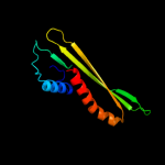
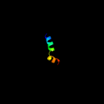
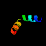
| 1 | d2iqia1
|
|
 |
97.6 |
12 |
Fold:Anticodon-binding domain-like
Superfamily:XCC0632-like
Family:XCC0632-like |
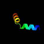
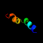
| 2 | c2k7rA_
|
|
 |
85.6 |
20 |
PDB header:replication
Chain: A: PDB Molecule:primosomal protein dnai;
PDBTitle: n-terminal domain of the bacillus subtilis helicase-loading2 protein dnai
|
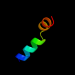
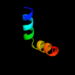
| 3 | c2r76A_
|
|
 |
81.9 |
6 |
PDB header:lipoprotein
Chain: A: PDB Molecule:rare lipoprotein b;
PDBTitle: crystal structure of the rare lipoprotein b (so_1173) from shewanella2 oneidensis, northeast structural genomics consortium target sor91a
|
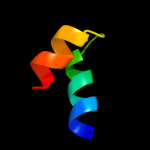
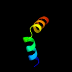
| 4 | c3qhpB_
|
|
 |
46.6 |
12 |
PDB header:transferase
Chain: B: PDB Molecule:type 1 capsular polysaccharide biosynthesis protein j
PDBTitle: crystal structure of the catalytic domain of cholesterol-alpha-2 glucosyltransferase from helicobacter pylori
|
| 5 | d2iw1a1
|
|
 |
45.2 |
16 |
Fold:UDP-Glycosyltransferase/glycogen phosphorylase
Superfamily:UDP-Glycosyltransferase/glycogen phosphorylase
Family:Glycosyl transferases group 1 |
| 6 | c2jxpA_
|
|
 |
41.9 |
9 |
PDB header:lipoprotein
Chain: A: PDB Molecule:putative lipoprotein b;
PDBTitle: solution nmr structure of uncharacterized lipoprotein b2 from nitrosomonas europaea. northeast structural genomics3 target ner45a
|
| 7 | c3iaaB_
|
|
 |
41.8 |
8 |
PDB header:transferase
Chain: B: PDB Molecule:calg2;
PDBTitle: crystal structure of calg2, calicheamicin glycosyltransferase, tdp2 bound form
|
| 8 | c2iyfA_
|
|
 |
36.9 |
12 |
PDB header:transferase
Chain: A: PDB Molecule:oleandomycin glycosyltransferase;
PDBTitle: the crystal structure of macrolide glycosyltransferases: a2 blueprint for antibiotic engineering
|
| 9 | c3ia7A_
|
|
 |
36.5 |
12 |
PDB header:transferase
Chain: A: PDB Molecule:calg4;
PDBTitle: crystal structure of calg4, the calicheamicin glycosyltransferase
|
| 10 | d2bfwa1
|
|
 |
36.0 |
15 |
Fold:UDP-Glycosyltransferase/glycogen phosphorylase
Superfamily:UDP-Glycosyltransferase/glycogen phosphorylase
Family:Glycosyl transferases group 1 |
| 11 | c2kw3C_
|
|
 |
35.7 |
12 |
PDB header:dna binding protein
Chain: C: PDB Molecule:regulatory factor x-associated protein;
PDBTitle: heterotrimeric interaction between rfx5 and rfxap
|
| 12 | c2r60A_
|
|
 |
35.3 |
0 |
PDB header:transferase
Chain: A: PDB Molecule:glycosyl transferase, group 1;
PDBTitle: structure of apo sucrose phosphate synthase (sps) of2 halothermothrix orenii
|
| 13 | c3okaA_
|
|
 |
34.4 |
12 |
PDB header:transferase
Chain: A: PDB Molecule:gdp-mannose-dependent alpha-(1-6)-phosphatidylinositol
PDBTitle: crystal structure of corynebacterium glutamicum pimb' in complex with2 gdp-man (triclinic crystal form)
|
| 14 | c3c4vB_
|
|
 |
30.8 |
16 |
PDB header:transferase
Chain: B: PDB Molecule:predicted glycosyltransferases;
PDBTitle: structure of the retaining glycosyltransferase msha:the2 first step in mycothiol biosynthesis. organism:3 corynebacterium glutamicum : complex with udp and 1l-ins-1-4 p.
|
| 15 | c2iyaB_
|
|
 |
29.8 |
12 |
PDB header:transferase
Chain: B: PDB Molecule:oleandomycin glycosyltransferase;
PDBTitle: the crystal structure of macrolide glycosyltransferases: a2 blueprint for antibiotic engineering
|
| 16 | c2jjmH_
|
|
 |
29.3 |
20 |
PDB header:transferase
Chain: H: PDB Molecule:glycosyl transferase, group 1 family protein;
PDBTitle: crystal structure of a family gt4 glycosyltransferase from2 bacillus anthracis orf ba1558.
|
| 17 | c3dzcA_
|
|
 |
28.9 |
12 |
PDB header:isomerase
Chain: A: PDB Molecule:udp-n-acetylglucosamine 2-epimerase;
PDBTitle: 2.35 angstrom resolution structure of wecb (vc0917), a udp-n-2 acetylglucosamine 2-epimerase from vibrio cholerae.
|
| 18 | c2xmpB_
|
|
 |
25.6 |
4 |
PDB header:sugar binding protein
Chain: B: PDB Molecule:trehalose-synthase tret;
PDBTitle: crystal structure of trehalose synthase tret mutant e326a2 from p.horishiki in complex with udp
|
| 19 | c2gejA_
|
|
 |
25.2 |
8 |
PDB header:transferase
Chain: A: PDB Molecule:phosphatidylinositol mannosyltransferase (pima);
PDBTitle: crystal structure of phosphatidylinositol mannosyltransferase (pima)2 from mycobacterium smegmatis in complex with gdp-man
|
| 20 | c2fu2A_
|
|
 |
24.5 |
27 |
PDB header:structural genomics, unknown function
Chain: A: PDB Molecule:hypothetical protein spy2152;
PDBTitle: crystal structure of protein spy2152 from streptococcus pyogenes
|
| 21 | c2x6rA_ |
|
not modelled |
22.5 |
4 |
PDB header:isomerase
Chain: A: PDB Molecule:trehalose-synthase tret;
PDBTitle: crystal structure of trehalose synthase tret from p.2 horikoshi produced by soaking in trehalose
|
| 22 | c3bf2A_ |
|
not modelled |
20.5 |
9 |
PDB header:lipoprotein
Chain: A: PDB Molecule:putative lipoprotein;
PDBTitle: crystal structure of the a1ksw9_neimf protein from2 neisseria meningitidis. northeast structural genomics3 consortium target mr36a
|
| 23 | c3othB_ |
|
not modelled |
20.0 |
8 |
PDB header:transferase/antibiotic
Chain: B: PDB Molecule:calg1;
PDBTitle: crystal structure of calg1, calicheamicin glycostyltransferase, tdp2 and calicheamicin alpha3i bound form
|
| 24 | d1o6ca_ |
|
not modelled |
18.3 |
8 |
Fold:UDP-Glycosyltransferase/glycogen phosphorylase
Superfamily:UDP-Glycosyltransferase/glycogen phosphorylase
Family:UDP-N-acetylglucosamine 2-epimerase |
| 25 | c3s29C_ |
|
not modelled |
18.1 |
20 |
PDB header:transferase
Chain: C: PDB Molecule:sucrose synthase 1;
PDBTitle: the crystal structure of sucrose synthase-1 from arabidopsis thaliana2 and its functional implications.
|
| 26 | c2p6pB_ |
|
not modelled |
17.0 |
8 |
PDB header:transferase
Chain: B: PDB Molecule:glycosyl transferase;
PDBTitle: x-ray crystal structure of c-c bond-forming dtdp-d-olivose-transferase2 urdgt2
|
| 27 | d2ijra1 |
|
not modelled |
15.1 |
71 |
Fold:Api92-like
Superfamily:Api92-like
Family:Api92-like |
| 28 | c2qpqC_ |
|
not modelled |
15.0 |
26 |
PDB header:transport protein
Chain: C: PDB Molecule:protein bug27;
PDBTitle: structure of bug27 from bordetella pertussis
|
| 29 | c2kw0A_ |
|
not modelled |
14.5 |
14 |
PDB header:oxidoreductase
Chain: A: PDB Molecule:ccmh protein;
PDBTitle: solution structure of n-terminal domain of ccmh from escherichia.coli
|
| 30 | d1f6da_ |
|
not modelled |
14.3 |
4 |
Fold:UDP-Glycosyltransferase/glycogen phosphorylase
Superfamily:UDP-Glycosyltransferase/glycogen phosphorylase
Family:UDP-N-acetylglucosamine 2-epimerase |
| 31 | d1v4va_ |
|
not modelled |
13.6 |
0 |
Fold:UDP-Glycosyltransferase/glycogen phosphorylase
Superfamily:UDP-Glycosyltransferase/glycogen phosphorylase
Family:UDP-N-acetylglucosamine 2-epimerase |
| 32 | c2hl7A_ |
|
not modelled |
13.3 |
17 |
PDB header:oxidoreductase
Chain: A: PDB Molecule:cytochrome c-type biogenesis protein ccmh;
PDBTitle: crystal structure of the periplasmic domain of ccmh from pseudomonas2 aeruginosa
|
| 33 | d2f9fa1 |
|
not modelled |
12.3 |
12 |
Fold:UDP-Glycosyltransferase/glycogen phosphorylase
Superfamily:UDP-Glycosyltransferase/glycogen phosphorylase
Family:Glycosyl transferases group 1 |
| 34 | c3ot5D_ |
|
not modelled |
11.7 |
20 |
PDB header:isomerase
Chain: D: PDB Molecule:udp-n-acetylglucosamine 2-epimerase;
PDBTitle: 2.2 angstrom resolution crystal structure of putative udp-n-2 acetylglucosamine 2-epimerase from listeria monocytogenes
|
| 35 | c2dvzA_ |
|
not modelled |
11.5 |
15 |
PDB header:transport protein
Chain: A: PDB Molecule:putative exported protein;
PDBTitle: structure of a periplasmic transporter
|
| 36 | c3iq2B_ |
|
not modelled |
10.9 |
17 |
PDB header:protein transport
Chain: B: PDB Molecule:sorting nexin-7;
PDBTitle: human sorting nexin 7, phox homology (px) domain
|
| 37 | c2cskA_ |
|
not modelled |
10.1 |
10 |
PDB header:protein transport
Chain: A: PDB Molecule:sorting nexin 12;
PDBTitle: solution structure of px domain from human snx12
|
| 38 | d1m6ex_ |
|
not modelled |
9.5 |
33 |
Fold:S-adenosyl-L-methionine-dependent methyltransferases
Superfamily:S-adenosyl-L-methionine-dependent methyltransferases
Family:Salicylic acid carboxyl methyltransferase (SAMT) |
| 39 | c2f5xC_ |
|
not modelled |
9.1 |
23 |
PDB header:transport protein
Chain: C: PDB Molecule:bugd;
PDBTitle: structure of periplasmic binding protein bugd
|
| 40 | c3b5iB_ |
|
not modelled |
8.7 |
33 |
PDB header:transferase
Chain: B: PDB Molecule:s-adenosyl-l-methionine:salicylic acid carboxyl
PDBTitle: crystal structure of indole-3-acetic acid methyltransferase
|
| 41 | c3d0qB_ |
|
not modelled |
8.2 |
13 |
PDB header:transferase
Chain: B: PDB Molecule:protein calg3;
PDBTitle: crystal structure of calg3 from micromonospora echinospora determined2 in space group i222
|
| 42 | c3kjlG_ |
|
not modelled |
7.5 |
36 |
PDB header:transcription
Chain: G: PDB Molecule:saga-associated factor 11;
PDBTitle: sgf11:sus1 complex
|
| 43 | d1kmda_ |
|
not modelled |
7.4 |
7 |
Fold:PX domain
Superfamily:PX domain
Family:PX domain |
| 44 | d1pvea_ |
|
not modelled |
7.2 |
17 |
Fold:XPC-binding domain
Superfamily:XPC-binding domain
Family:XPC-binding domain |
| 45 | c3kikE_ |
|
not modelled |
7.0 |
36 |
PDB header:transcription
Chain: E: PDB Molecule:saga-associated factor 11;
PDBTitle: sgf11:sus1 complex
|
| 46 | c3kikF_ |
|
not modelled |
7.0 |
36 |
PDB header:transcription
Chain: F: PDB Molecule:saga-associated factor 11;
PDBTitle: sgf11:sus1 complex
|
| 47 | c3kikH_ |
|
not modelled |
6.9 |
36 |
PDB header:transcription
Chain: H: PDB Molecule:saga-associated factor 11;
PDBTitle: sgf11:sus1 complex
|
| 48 | c2eg5C_ |
|
not modelled |
6.7 |
56 |
PDB header:transferase
Chain: C: PDB Molecule:xanthosine methyltransferase;
PDBTitle: the structure of xanthosine methyltransferase
|
| 49 | c1xteA_ |
|
not modelled |
6.7 |
4 |
PDB header:transferase
Chain: A: PDB Molecule:serine/threonine-protein kinase sgk3;
PDBTitle: crystal structure of cisk-px domain
|
| 50 | d1xtea_ |
|
not modelled |
6.7 |
4 |
Fold:PX domain
Superfamily:PX domain
Family:PX domain |
| 51 | c3kikG_ |
|
not modelled |
6.7 |
36 |
PDB header:transcription
Chain: G: PDB Molecule:saga-associated factor 11;
PDBTitle: sgf11:sus1 complex
|
| 52 | c1x3wB_ |
|
not modelled |
6.6 |
21 |
PDB header:hydrolase
Chain: B: PDB Molecule:uv excision repair protein rad23;
PDBTitle: structure of a peptide:n-glycanase-rad23 complex
|
| 53 | c3fogA_ |
|
not modelled |
6.5 |
7 |
PDB header:protein transport
Chain: A: PDB Molecule:sorting nexin-17;
PDBTitle: crystal structure of the px domain of sorting nexin-172 (snx17)
|
| 54 | d2f4mb1 |
|
not modelled |
6.3 |
17 |
Fold:XPC-binding domain
Superfamily:XPC-binding domain
Family:XPC-binding domain |
| 55 | c2i4kA_ |
|
not modelled |
6.3 |
17 |
PDB header:protein transport
Chain: A: PDB Molecule:sorting nexin-1;
PDBTitle: solution structure of the px domain of sorting nexin 1
|
| 56 | c3onoA_ |
|
not modelled |
6.3 |
7 |
PDB header:isomerase
Chain: A: PDB Molecule:ribose/galactose isomerase;
PDBTitle: crystal structure of ribose-5-phosphate isomerase lacab_rpib from2 vibrio parahaemolyticus
|
| 57 | c3kjlF_ |
|
not modelled |
6.3 |
36 |
PDB header:transcription
Chain: F: PDB Molecule:saga-associated factor 11;
PDBTitle: sgf11:sus1 complex
|
| 58 | c3kjlH_ |
|
not modelled |
5.6 |
36 |
PDB header:transcription
Chain: H: PDB Molecule:saga-associated factor 11;
PDBTitle: sgf11:sus1 complex
|
| 59 | c3kjlE_ |
|
not modelled |
5.6 |
36 |
PDB header:transcription
Chain: E: PDB Molecule:saga-associated factor 11;
PDBTitle: sgf11:sus1 complex
|
| 60 | d2fhea1 |
|
not modelled |
5.5 |
17 |
Fold:GST C-terminal domain-like
Superfamily:GST C-terminal domain-like
Family:Glutathione S-transferase (GST), C-terminal domain |
| 61 | c3c5yD_ |
|
not modelled |
5.3 |
14 |
PDB header:isomerase
Chain: D: PDB Molecule:ribose/galactose isomerase;
PDBTitle: crystal structure of a putative ribose 5-phosphate isomerase2 (saro_3514) from novosphingobium aromaticivorans dsm at 1.81 a3 resolution
|