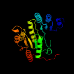
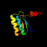
1 c3dmyA_
100.0
39
PDB header: structural genomics, unknown functionChain: A: PDB Molecule: protein fdra;PDBTitle: crystal structure of a predicated acyl-coa synthetase from e.coli
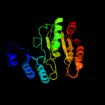
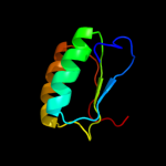
2 c2csuB_
100.0
16
PDB header: structural genomics, unknown functionChain: B: PDB Molecule: 457aa long hypothetical protein;PDBTitle: crystal structure of ph0766 from pyrococcus horikoshii ot3
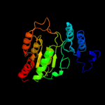
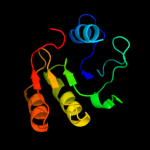
3 c3mwdB_
100.0
15
PDB header: transferaseChain: B: PDB Molecule: atp-citrate synthase;PDBTitle: truncated human atp-citrate lyase with citrate bound
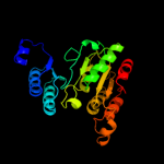
4 c2nu8D_
100.0
25
PDB header: ligaseChain: D: PDB Molecule: succinyl-coa ligase [adp-forming] subunit alpha;PDBTitle: c123at mutant of e. coli succinyl-coa synthetase
5 c2fpgA_
100.0
24
PDB header: ligaseChain: A: PDB Molecule: succinyl-coa ligase [gdp-forming] alpha-chain,PDBTitle: crystal structure of pig gtp-specific succinyl-coa2 synthetase in complex with gdp
6 c2yv2A_
100.0
28
PDB header: ligaseChain: A: PDB Molecule: succinyl-coa synthetase alpha chain;PDBTitle: crystal structure of succinyl-coa synthetase alpha chain from2 aeropyrum pernix k1
7 c1oi7A_
100.0
24
PDB header: synthetaseChain: A: PDB Molecule: succinyl-coa synthetase alpha chain;PDBTitle: the crystal structure of succinyl-coa synthetase alpha2 subunit from thermus thermophilus
8 c2yv1A_
100.0
25
PDB header: ligaseChain: A: PDB Molecule: succinyl-coa ligase [adp-forming] subunit alpha;PDBTitle: crystal structure of succinyl-coa synthetase alpha chain from2 methanocaldococcus jannaschii dsm 2661
9 d2nu7a2
100.0
27
Fold: Flavodoxin-likeSuperfamily: Succinyl-CoA synthetase domainsFamily: Succinyl-CoA synthetase domains10 d1euca2
100.0
25
Fold: Flavodoxin-likeSuperfamily: Succinyl-CoA synthetase domainsFamily: Succinyl-CoA synthetase domains11 d1oi7a2
100.0
25
Fold: Flavodoxin-likeSuperfamily: Succinyl-CoA synthetase domainsFamily: Succinyl-CoA synthetase domains12 d2csua2
100.0
16
Fold: Flavodoxin-likeSuperfamily: Succinyl-CoA synthetase domainsFamily: Succinyl-CoA synthetase domains13 d2csua3
99.9
11
Fold: Flavodoxin-likeSuperfamily: Succinyl-CoA synthetase domainsFamily: Succinyl-CoA synthetase domains14 d2nu7b1
99.8
13
Fold: Flavodoxin-likeSuperfamily: Succinyl-CoA synthetase domainsFamily: Succinyl-CoA synthetase domains15 d1eucb1
99.7
13
Fold: Flavodoxin-likeSuperfamily: Succinyl-CoA synthetase domainsFamily: Succinyl-CoA synthetase domains16 c2nu9E_
99.5
12
PDB header: ligaseChain: E: PDB Molecule: succinyl-coa synthetase beta chain;PDBTitle: c123at mutant of e. coli succinyl-coa synthetase2 orthorhombic crystal form
17 c1eucB_
99.4
14
PDB header: ligaseChain: B: PDB Molecule: succinyl-coa synthetase, beta chain;PDBTitle: crystal structure of dephosphorylated pig heart, gtp-2 specific succinyl-coa synthetase
18 d2csua1
99.4
30
Fold: NAD(P)-binding Rossmann-fold domainsSuperfamily: NAD(P)-binding Rossmann-fold domainsFamily: CoA-binding domain19 c2duwA_
99.4
19
PDB header: ligand binding proteinChain: A: PDB Molecule: putative coa-binding protein;PDBTitle: solution structure of putative coa-binding protein of2 klebsiella pneumoniae
20 d1y81a1
99.2
16
Fold: NAD(P)-binding Rossmann-fold domainsSuperfamily: NAD(P)-binding Rossmann-fold domainsFamily: CoA-binding domain21 d1iuka_
not modelled
99.2
7
Fold: NAD(P)-binding Rossmann-fold domainsSuperfamily: NAD(P)-binding Rossmann-fold domainsFamily: CoA-binding domain22 d2d59a1
99.1
19
Fold: NAD(P)-binding Rossmann-fold domainsSuperfamily: NAD(P)-binding Rossmann-fold domainsFamily: CoA-binding domain23 d1oi7a1
not modelled
99.0
19
Fold: NAD(P)-binding Rossmann-fold domainsSuperfamily: NAD(P)-binding Rossmann-fold domainsFamily: CoA-binding domain24 d2nu7a1
not modelled
99.0
18
Fold: NAD(P)-binding Rossmann-fold domainsSuperfamily: NAD(P)-binding Rossmann-fold domainsFamily: CoA-binding domain25 d1euca1
not modelled
99.0
19
Fold: NAD(P)-binding Rossmann-fold domainsSuperfamily: NAD(P)-binding Rossmann-fold domainsFamily: CoA-binding domain26 c3ff4A_
not modelled
98.6
17
PDB header: structural genomics, unknown functionChain: A: PDB Molecule: uncharacterized protein;PDBTitle: crystal structure of uncharacterized protein chu_1412
27 c3mwdA_
not modelled
97.8
7
PDB header: transferaseChain: A: PDB Molecule: atp-citrate synthase;PDBTitle: truncated human atp-citrate lyase with citrate bound
28 c3m2tA_
not modelled
97.4
11
PDB header: oxidoreductaseChain: A: PDB Molecule: probable dehydrogenase;PDBTitle: the crystal structure of dehydrogenase from chromobacterium2 violaceum
29 c3dtyA_
not modelled
97.4
20
PDB header: oxidoreductaseChain: A: PDB Molecule: oxidoreductase, gfo/idh/moca family;PDBTitle: crystal structure of an oxidoreductase from pseudomonas2 syringae
30 c2q4eB_
not modelled
97.4
13
PDB header: oxidoreductaseChain: B: PDB Molecule: probable oxidoreductase at4g09670;PDBTitle: ensemble refinement of the protein crystal structure of gene product2 from arabidopsis thaliana at4g09670
31 c3ec7C_
not modelled
97.3
13
PDB header: oxidoreductaseChain: C: PDB Molecule: putative dehydrogenase;PDBTitle: crystal structure of putative dehydrogenase from salmonella2 typhimurium lt2
32 c3fd8A_
not modelled
97.3
20
PDB header: oxidoreductaseChain: A: PDB Molecule: oxidoreductase, gfo/idh/moca family;PDBTitle: crystal structure of an oxidoreductase from enterococcus2 faecalis
33 c3rbvA_
not modelled
97.3
15
PDB header: sugar binding proteinChain: A: PDB Molecule: sugar 3-ketoreductase;PDBTitle: crystal structure of kijd10, a 3-ketoreductase from actinomadura2 kijaniata incomplex with nadp
34 c2ixaA_
not modelled
97.3
9
PDB header: hydrolaseChain: A: PDB Molecule: alpha-n-acetylgalactosaminidase;PDBTitle: a-zyme, n-acetylgalactosaminidase
35 c3e9mC_
not modelled
97.3
19
PDB header: oxidoreductaseChain: C: PDB Molecule: oxidoreductase, gfo/idh/moca family;PDBTitle: crystal structure of an oxidoreductase from enterococcus2 faecalis
36 c3moiA_
not modelled
97.3
13
PDB header: oxidoreductaseChain: A: PDB Molecule: probable dehydrogenase;PDBTitle: the crystal structure of the putative dehydrogenase from bordetella2 bronchiseptica rb50
37 c1zh8B_
not modelled
97.3
9
PDB header: oxidoreductaseChain: B: PDB Molecule: oxidoreductase;PDBTitle: crystal structure of oxidoreductase (tm0312) from thermotoga maritima2 at 2.50 a resolution
38 c3ip3D_
not modelled
97.3
10
PDB header: oxidoreductaseChain: D: PDB Molecule: oxidoreductase, putative;PDBTitle: structure of putative oxidoreductase (tm_0425) from2 thermotoga maritima
39 c3nt5B_
not modelled
97.2
10
PDB header: oxidoreductaseChain: B: PDB Molecule: inositol 2-dehydrogenase/d-chiro-inositol 3-dehydrogenase;PDBTitle: crystal structure of myo-inositol dehydrogenase from bacillus subtilis2 with bound cofactor and product inosose
40 d1zh8a1
not modelled
97.2
10
Fold: NAD(P)-binding Rossmann-fold domainsSuperfamily: NAD(P)-binding Rossmann-fold domainsFamily: Glyceraldehyde-3-phosphate dehydrogenase-like, N-terminal domain41 c2o48X_
not modelled
97.2
12
PDB header: oxidoreductaseChain: X: PDB Molecule: dimeric dihydrodiol dehydrogenase;PDBTitle: crystal structure of mammalian dimeric dihydrodiol dehydrogenase
42 c2p2sA_
not modelled
97.2
12
PDB header: oxidoreductaseChain: A: PDB Molecule: putative oxidoreductase;PDBTitle: crystal structure of putative oxidoreductase (yp_050235.1) from2 erwinia carotovora atroseptica scri1043 at 1.25 a resolution
43 c3ezyB_
not modelled
97.2
8
PDB header: structural genomics, unknown functionChain: B: PDB Molecule: dehydrogenase;PDBTitle: crystal structure of probable dehydrogenase tm_0414 from2 thermotoga maritima
44 d1h6da1
not modelled
97.2
17
Fold: NAD(P)-binding Rossmann-fold domainsSuperfamily: NAD(P)-binding Rossmann-fold domainsFamily: Glyceraldehyde-3-phosphate dehydrogenase-like, N-terminal domain45 c3oqbF_
not modelled
97.2
16
PDB header: oxidoreductaseChain: F: PDB Molecule: oxidoreductase;PDBTitle: crystal structure of putative oxidoreductase from bradyrhizobium2 japonicum usda 110
46 c1lc3A_
not modelled
97.2
22
PDB header: oxidoreductaseChain: A: PDB Molecule: biliverdin reductase a;PDBTitle: crystal structure of a biliverdin reductase enzyme-cofactor2 complex
47 d1lc0a1
not modelled
97.1
24
Fold: NAD(P)-binding Rossmann-fold domainsSuperfamily: NAD(P)-binding Rossmann-fold domainsFamily: Glyceraldehyde-3-phosphate dehydrogenase-like, N-terminal domain48 d1ydwa1
not modelled
97.1
13
Fold: NAD(P)-binding Rossmann-fold domainsSuperfamily: NAD(P)-binding Rossmann-fold domainsFamily: Glyceraldehyde-3-phosphate dehydrogenase-like, N-terminal domain49 c3btuD_
not modelled
97.1
13
PDB header: transcriptionChain: D: PDB Molecule: galactose/lactose metabolism regulatory proteinPDBTitle: crystal structure of the super-repressor mutant of gal80p2 from saccharomyces cerevisiae; gal80(s2) [e351k]
50 c3evnA_
not modelled
97.1
13
PDB header: oxidoreductaseChain: A: PDB Molecule: oxidoreductase, gfo/idh/moca family;PDBTitle: crystal structure of putative oxidoreductase from streptococcus2 agalactiae 2603v/r
51 c1evjC_
not modelled
97.1
16
PDB header: oxidoreductaseChain: C: PDB Molecule: glucose-fructose oxidoreductase;PDBTitle: crystal structure of glucose-fructose oxidoreductase (gfor)2 delta1-22 s64d
52 c3e18A_
not modelled
97.1
18
PDB header: oxidoreductaseChain: A: PDB Molecule: oxidoreductase;PDBTitle: crystal structure of nad-binding protein from listeria innocua
53 c3db2C_
not modelled
97.1
15
PDB header: oxidoreductaseChain: C: PDB Molecule: putative nadph-dependent oxidoreductase;PDBTitle: crystal structure of a putative nadph-dependent oxidoreductase2 (dhaf_2064) from desulfitobacterium hafniense dcb-2 at 1.70 a3 resolution
54 c1ofgF_
not modelled
97.0
15
PDB header: oxidoreductaseChain: F: PDB Molecule: glucose-fructose oxidoreductase;PDBTitle: glucose-fructose oxidoreductase
55 c1h6dL_
not modelled
97.0
17
PDB header: protein translocationChain: L: PDB Molecule: precursor form of glucose-fructosePDBTitle: oxidized precursor form of glucose-fructose oxidoreductase2 from zymomonas mobilis complexed with glycerol
56 c3e82A_
not modelled
97.0
24
PDB header: oxidoreductaseChain: A: PDB Molecule: putative oxidoreductase;PDBTitle: crystal structure of a putative oxidoreductase from2 klebsiella pneumoniae
57 c3q2kB_
not modelled
97.0
11
PDB header: oxidoreductaseChain: B: PDB Molecule: oxidoreductase;PDBTitle: crystal structure of the wlba dehydrogenase from bordetella pertussis2 in complex with nadh and udp-glcnaca
58 c2glxD_
not modelled
97.0
13
PDB header: oxidoreductaseChain: D: PDB Molecule: 1,5-anhydro-d-fructose reductase;PDBTitle: crystal structure analysis of bacterial 1,5-af reductase
59 c2axqA_
not modelled
97.0
12
PDB header: oxidoreductaseChain: A: PDB Molecule: saccharopine dehydrogenase;PDBTitle: apo histidine-tagged saccharopine dehydrogenase (l-glu2 forming) from saccharomyces cerevisiae
60 c3do5A_
not modelled
97.0
14
PDB header: oxidoreductaseChain: A: PDB Molecule: homoserine dehydrogenase;PDBTitle: crystal structure of putative homoserine dehydrogenase (np_069768.1)2 from archaeoglobus fulgidus at 2.20 a resolution
61 c3gfgB_
not modelled
97.0
19
PDB header: oxidoreductaseChain: B: PDB Molecule: uncharacterized oxidoreductase yvaa;PDBTitle: structure of putative oxidoreductase yvaa from bacillus subtilis in2 triclinic form
62 c2ho3D_
not modelled
96.9
12
PDB header: oxidoreductaseChain: D: PDB Molecule: oxidoreductase, gfo/idh/moca family;PDBTitle: crystal structure of oxidoreductase, gfo/idh/moca family from2 streptococcus pneumoniae
63 c3f4lF_
not modelled
96.9
22
PDB header: oxidoreductaseChain: F: PDB Molecule: putative oxidoreductase yhhx;PDBTitle: crystal structure of a probable oxidoreductase yhhx in2 triclinic form. northeast structural genomics target er647
64 d1ryda1
not modelled
96.9
16
Fold: NAD(P)-binding Rossmann-fold domainsSuperfamily: NAD(P)-binding Rossmann-fold domainsFamily: Glyceraldehyde-3-phosphate dehydrogenase-like, N-terminal domain65 c3kuxA_
not modelled
96.9
21
PDB header: oxidoreductaseChain: A: PDB Molecule: putative oxidoreductase;PDBTitle: structure of the ypo2259 putative oxidoreductase from yersinia pestis
66 d1xeaa1
not modelled
96.8
13
Fold: NAD(P)-binding Rossmann-fold domainsSuperfamily: NAD(P)-binding Rossmann-fold domainsFamily: Glyceraldehyde-3-phosphate dehydrogenase-like, N-terminal domain67 c1xeaD_
not modelled
96.8
12
PDB header: oxidoreductaseChain: D: PDB Molecule: oxidoreductase, gfo/idh/moca family;PDBTitle: crystal structure of a gfo/idh/moca family oxidoreductase2 from vibrio cholerae
68 c3c1aB_
not modelled
96.8
14
PDB header: oxidoreductaseChain: B: PDB Molecule: putative oxidoreductase;PDBTitle: crystal structure of a putative oxidoreductase (zp_00056571.1) from2 magnetospirillum magnetotacticum ms-1 at 1.85 a resolution
69 c3fhlC_
not modelled
96.8
19
PDB header: oxidoreductaseChain: C: PDB Molecule: putative oxidoreductase;PDBTitle: crystal structure of a putative oxidoreductase from bacteroides2 fragilis nctc 9343
70 c1drwA_
not modelled
96.8
7
PDB header: oxidoreductaseChain: A: PDB Molecule: dihydrodipicolinate reductase;PDBTitle: escherichia coli dhpr/nhdh complex
71 c1e5lA_
not modelled
96.7
16
PDB header: oxidoreductaseChain: A: PDB Molecule: saccharopine reductase;PDBTitle: apo saccharopine reductase from magnaporthe grisea
72 c3bioB_
not modelled
96.6
13
PDB header: oxidoreductaseChain: B: PDB Molecule: oxidoreductase, gfo/idh/moca family;PDBTitle: crystal structure of oxidoreductase (gfo/idh/moca family member) from2 porphyromonas gingivalis w83
73 c3v5nA_
not modelled
96.4
17
PDB header: oxidoreductaseChain: A: PDB Molecule: oxidoreductase;PDBTitle: the crystal structure of oxidoreductase from sinorhizobium meliloti
74 c3euwB_
not modelled
96.4
11
PDB header: oxidoreductaseChain: B: PDB Molecule: myo-inositol dehydrogenase;PDBTitle: crystal structure of a myo-inositol dehydrogenase from corynebacterium2 glutamicum atcc 13032
75 c3ceaA_
not modelled
96.4
10
PDB header: oxidoreductaseChain: A: PDB Molecule: myo-inositol 2-dehydrogenase;PDBTitle: crystal structure of myo-inositol 2-dehydrogenase (np_786804.1) from2 lactobacillus plantarum at 2.40 a resolution
76 c3oa0B_
not modelled
96.4
17
PDB header: oxidoreductaseChain: B: PDB Molecule: lipopolysaccaride biosynthesis protein wbpb;PDBTitle: crystal structure of the wlba (wbpb) dehydrogenase from thermus2 thermophilus in complex with nad and udp-glcnaca
77 d1j5pa4
not modelled
96.4
13
Fold: NAD(P)-binding Rossmann-fold domainsSuperfamily: NAD(P)-binding Rossmann-fold domainsFamily: Glyceraldehyde-3-phosphate dehydrogenase-like, N-terminal domain78 c3oa2B_
not modelled
96.4
11
PDB header: oxidoreductaseChain: B: PDB Molecule: wbpb;PDBTitle: crystal structure of the wlba (wbpb) dehydrogenase from pseudomonas2 aeruginosa in complex with nad at 1.5 angstrom resolution
79 d2nvwa1
not modelled
96.2
15
Fold: NAD(P)-binding Rossmann-fold domainsSuperfamily: NAD(P)-binding Rossmann-fold domainsFamily: Glyceraldehyde-3-phosphate dehydrogenase-like, N-terminal domain80 c2dc1A_
not modelled
96.1
11
PDB header: oxidoreductaseChain: A: PDB Molecule: l-aspartate dehydrogenase;PDBTitle: crystal structure of l-aspartate dehydrogenase from2 hyperthermophilic archaeon archaeoglobus fulgidus
81 d1diha1
not modelled
95.6
6
Fold: NAD(P)-binding Rossmann-fold domainsSuperfamily: NAD(P)-binding Rossmann-fold domainsFamily: Glyceraldehyde-3-phosphate dehydrogenase-like, N-terminal domain82 c1j5pA_
not modelled
95.6
9
PDB header: oxidoreductaseChain: A: PDB Molecule: aspartate dehydrogenase;PDBTitle: crystal structure of aspartate dehydrogenase (tm1643) from thermotoga2 maritima at 1.9 a resolution
83 d1tlta1
not modelled
95.3
16
Fold: NAD(P)-binding Rossmann-fold domainsSuperfamily: NAD(P)-binding Rossmann-fold domainsFamily: Glyceraldehyde-3-phosphate dehydrogenase-like, N-terminal domain84 c2nvwB_
not modelled
95.3
13
PDB header: transcriptionChain: B: PDB Molecule: galactose/lactose metabolism regulatory proteinPDBTitle: crystal sctucture of transcriptional regulator gal80p from2 kluyveromymes lactis
85 c3ketA_
not modelled
95.3
17
PDB header: transcription/dnaChain: A: PDB Molecule: redox-sensing transcriptional repressor rex;PDBTitle: crystal structure of a rex-family transcriptional regulatory protein2 from streptococcus agalactiae bound to a palindromic operator
86 c1tltB_
not modelled
95.2
18
PDB header: oxidoreductaseChain: B: PDB Molecule: putative oxidoreductase (virulence factor mvim homolog);PDBTitle: crystal structure of a putative oxidoreductase (virulence factor mvim2 homolog)
87 c2z2vA_
not modelled
95.0
15
PDB header: oxidoreductaseChain: A: PDB Molecule: hypothetical protein ph1688;PDBTitle: crystal structure of l-lysine dehydrogenase from2 hyperthermophilic archaeon pyrococcus horikoshii
88 c3ijpA_
not modelled
95.0
11
PDB header: oxidoreductaseChain: A: PDB Molecule: dihydrodipicolinate reductase;PDBTitle: crystal structure of dihydrodipicolinate reductase from2 bartonella henselae at 2.0a resolution
89 d1f06a1
not modelled
94.4
7
Fold: NAD(P)-binding Rossmann-fold domainsSuperfamily: NAD(P)-binding Rossmann-fold domainsFamily: Glyceraldehyde-3-phosphate dehydrogenase-like, N-terminal domain90 d1ebfa1
not modelled
94.2
13
Fold: NAD(P)-binding Rossmann-fold domainsSuperfamily: NAD(P)-binding Rossmann-fold domainsFamily: Glyceraldehyde-3-phosphate dehydrogenase-like, N-terminal domain91 d1e5qa1
not modelled
93.1
15
Fold: NAD(P)-binding Rossmann-fold domainsSuperfamily: NAD(P)-binding Rossmann-fold domainsFamily: Glyceraldehyde-3-phosphate dehydrogenase-like, N-terminal domain92 c2p4qA_
not modelled
93.1
12
PDB header: oxidoreductaseChain: A: PDB Molecule: 6-phosphogluconate dehydrogenase, decarboxylating 1;PDBTitle: crystal structure analysis of gnd1 in saccharomyces cerevisiae
93 c2dt5A_
not modelled
93.0
12
PDB header: dna binding proteinChain: A: PDB Molecule: at-rich dna-binding protein;PDBTitle: crystal structure of ttha1657 (at-rich dna-binding protein) from2 thermus thermophilus hb8
94 c3ic5A_
not modelled
93.0
10
PDB header: structural genomics, unknown functionChain: A: PDB Molecule: putative saccharopine dehydrogenase;PDBTitle: n-terminal domain of putative saccharopine dehydrogenase from ruegeria2 pomeroyi.
95 c3dapB_
not modelled
92.8
6
PDB header: oxidoreductaseChain: B: PDB Molecule: diaminopimelic acid dehydrogenase;PDBTitle: c. glutamicum dap dehydrogenase in complex with nadp+ and2 the inhibitor 5s-isoxazoline
96 c3uuwB_
not modelled
92.6
12
PDB header: oxidoreductaseChain: B: PDB Molecule: putative oxidoreductase with nad(p)-binding rossmann-foldPDBTitle: 1.63 angstrom resolution crystal structure of dehydrogenase (mvim)2 from clostridium difficile.
97 c3bezC_
not modelled
92.6
27
PDB header: hydrolaseChain: C: PDB Molecule: protease 4;PDBTitle: crystal structure of escherichia coli signal peptide peptidase (sppa),2 semet crystals
98 d2dt5a2
not modelled
92.4
9
Fold: NAD(P)-binding Rossmann-fold domainsSuperfamily: NAD(P)-binding Rossmann-fold domainsFamily: Transcriptional repressor Rex, C-terminal domain99 c1pgjA_
not modelled
92.3
17
PDB header: oxidoreductaseChain: A: PDB Molecule: 6-phosphogluconate dehydrogenase;PDBTitle: x-ray structure of 6-phosphogluconate dehydrogenase from the protozoan2 parasite t. brucei
100 c3fwnB_
not modelled
92.0
14
PDB header: oxidoreductaseChain: B: PDB Molecule: 6-phosphogluconate dehydrogenase, decarboxylating;PDBTitle: dimeric 6-phosphogluconate dehydrogenase complexed with 6-2 phosphogluconate and 2'-monophosphoadenosine-5'-diphosphate
101 c1pgqA_
not modelled
91.6
14
PDB header: oxidoreductase (choh(d)-nadp+(a))Chain: A: PDB Molecule: 6-phosphogluconate dehydrogenase;PDBTitle: crystallographic study of coenzyme, coenzyme analogue and substrate2 binding in 6-phosphogluconate dehydrogenase: implications for nadp3 specificity and the enzyme mechanism
102 c2iz1C_
not modelled
91.1
13
PDB header: oxidoreductaseChain: C: PDB Molecule: 6-phosphogluconate dehydrogenase, decarboxylating;PDBTitle: 6pdh complexed with pex inhibitor synchrotron data
103 c2ejwB_
not modelled
90.6
17
PDB header: oxidoreductaseChain: B: PDB Molecule: homoserine dehydrogenase;PDBTitle: homoserine dehydrogenase from thermus thermophilus hb8
104 c2vt2A_
not modelled
90.4
12
PDB header: transcriptionChain: A: PDB Molecule: redox-sensing transcriptional repressor rex;PDBTitle: structure and functional properties of the bacillus2 subtilis transcriptional repressor rex
105 c1yl7F_
not modelled
90.1
7
PDB header: oxidoreductaseChain: F: PDB Molecule: dihydrodipicolinate reductase;PDBTitle: the crystal structure of mycobacterium tuberculosis2 dihydrodipicolinate reductase (rv2773c) in complex with nadh (crystal3 form c)
106 c1vm6B_
not modelled
89.9
8
PDB header: oxidoreductaseChain: B: PDB Molecule: dihydrodipicolinate reductase;PDBTitle: crystal structure of dihydrodipicolinate reductase (tm1520) from2 thermotoga maritima at 2.27 a resolution
107 c3c8mA_
not modelled
89.9
11
PDB header: oxidoreductaseChain: A: PDB Molecule: homoserine dehydrogenase;PDBTitle: crystal structure of homoserine dehydrogenase from thermoplasma2 volcanium
108 c3mtjA_
not modelled
89.8
15
PDB header: oxidoreductaseChain: A: PDB Molecule: homoserine dehydrogenase;PDBTitle: the crystal structure of a homoserine dehydrogenase from thiobacillus2 denitrificans to 2.15a
109 d1vm6a3
not modelled
89.4
8
Fold: NAD(P)-binding Rossmann-fold domainsSuperfamily: NAD(P)-binding Rossmann-fold domainsFamily: Glyceraldehyde-3-phosphate dehydrogenase-like, N-terminal domain110 d1yl7a1
not modelled
89.3
8
Fold: NAD(P)-binding Rossmann-fold domainsSuperfamily: NAD(P)-binding Rossmann-fold domainsFamily: Glyceraldehyde-3-phosphate dehydrogenase-like, N-terminal domain111 c2vbiF_
not modelled
88.6
18
PDB header: lyaseChain: F: PDB Molecule: pyruvate decarboxylase;PDBTitle: holostructure of pyruvate decarboxylase from acetobacter2 pasteurianus
112 c2iexA_
not modelled
88.5
22
PDB header: lyaseChain: A: PDB Molecule: dihydroxynapthoic acid synthetase;PDBTitle: crystal structure of dihydroxynapthoic acid synthetase (gk2873) from2 geobacillus kaustophilus hta426
113 c1zpdA_
not modelled
87.7
20
PDB header: alcohol fermentationChain: A: PDB Molecule: pyruvate decarboxylase;PDBTitle: pyruvate decarboxylase from zymomonas mobilis
114 d1zpda2
not modelled
87.7
21
Fold: Thiamin diphosphate-binding fold (THDP-binding)Superfamily: Thiamin diphosphate-binding fold (THDP-binding)Family: Pyruvate oxidase and decarboxylase Pyr module115 c2ozpA_
not modelled
86.6
14
PDB header: oxidoreductaseChain: A: PDB Molecule: n-acetyl-gamma-glutamyl-phosphate reductase;PDBTitle: crystal structure of n-acetyl-gamma-glutamyl-phosphate reductase2 (ttha1904) from thermus thermophilus
116 c1ebuA_
not modelled
86.4
12
PDB header: oxidoreductaseChain: A: PDB Molecule: homoserine dehydrogenase;PDBTitle: homoserine dehydrogenase complex with nad analogue and l-2 homoserine
117 d2b3ya2
not modelled
85.9
25
Fold: Aconitase iron-sulfur domainSuperfamily: Aconitase iron-sulfur domainFamily: Aconitase iron-sulfur domain118 c1mb4B_
not modelled
85.2
10
PDB header: oxidoreductaseChain: B: PDB Molecule: aspartate-semialdehyde dehydrogenase;PDBTitle: crystal structure of aspartate semialdehyde dehydrogenase2 from vibrio cholerae with nadp and s-methyl-l-cysteine3 sulfoxide
119 d2g0ta1
not modelled
84.8
22
Fold: P-loop containing nucleoside triphosphate hydrolasesSuperfamily: P-loop containing nucleoside triphosphate hydrolasesFamily: Nitrogenase iron protein-like120 d1ovma2
not modelled
84.0
17
Fold: Thiamin diphosphate-binding fold (THDP-binding)Superfamily: Thiamin diphosphate-binding fold (THDP-binding)Family: Pyruvate oxidase and decarboxylase Pyr module