1 d1o5ha_
84.6
14
Fold: Methenyltetrahydrofolate cyclohydrolase-likeSuperfamily: Methenyltetrahydrofolate cyclohydrolase-likeFamily: Methenyltetrahydrofolate cyclohydrolase-like2 c1tt9B_
77.1
12
PDB header: transferase, lyaseChain: B: PDB Molecule: formimidoyltransferase-cyclodeaminasePDBTitle: structure of the bifunctional and golgi associated2 formiminotransferase cyclodeaminase octamer
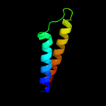
3 d2asra_
55.7
8
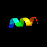
Fold: Four-helical up-and-down bundleSuperfamily: Aspartate receptor, ligand-binding domainFamily: Aspartate receptor, ligand-binding domain4 d1vlta_
54.4
16
Fold: Four-helical up-and-down bundleSuperfamily: Aspartate receptor, ligand-binding domainFamily: Aspartate receptor, ligand-binding domain5 c1p68A_
49.8
14
PDB header: de novo proteinChain: A: PDB Molecule: de novo designed protein s-824;PDBTitle: solution structure of s-824, a de novo designed four helix2 bundle
6 c2d4yA_
49.7
11
PDB header: structural proteinChain: A: PDB Molecule: flagellar hook-associated protein 1;PDBTitle: crystal structure of a 49k fragment of hap1 (flgk)
7 d1d2ta_
49.7
25
Fold: Acid phosphatase/Vanadium-dependent haloperoxidaseSuperfamily: Acid phosphatase/Vanadium-dependent haloperoxidaseFamily: Type 2 phosphatidic acid phosphatase, PAP28 d2j0oa1
48.2
19
Fold: IpaD-likeSuperfamily: IpaD-likeFamily: IpaD-like9 c2j0oA_
48.2
19
PDB header: cell invasionChain: A: PDB Molecule: invasin ipad;PDBTitle: shigella flexneri ipad
10 c2ym0B_
46.3
6
PDB header: cell invasionChain: B: PDB Molecule: cell invasion protein sipd;PDBTitle: truncated sipd from salmonella typhimurium
11 c2d4uA_
43.5
15
PDB header: signaling proteinChain: A: PDB Molecule: methyl-accepting chemotaxis protein i;PDBTitle: crystal structure of the ligand binding domain of the bacterial serine2 chemoreceptor tsr
12 d1q90m_
41.8
50
Fold: Single transmembrane helixSuperfamily: PetM subunit of the cytochrome b6f complexFamily: PetM subunit of the cytochrome b6f complex13 d1oqwa_
41.4
13
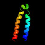
Fold: Pili subunitsSuperfamily: Pili subunitsFamily: Pilin14 d2liga_
39.9
16
Fold: Four-helical up-and-down bundleSuperfamily: Aspartate receptor, ligand-binding domainFamily: Aspartate receptor, ligand-binding domain15 d2pila_
37.6
8
Fold: Pili subunitsSuperfamily: Pili subunitsFamily: Pilin16 c3kdpG_
36.8
38
PDB header: hydrolaseChain: G: PDB Molecule: na+/k+ atpase gamma subunit transcript variant a;PDBTitle: crystal structure of the sodium-potassium pump
17 c3kdpH_
36.8
38
PDB header: hydrolaseChain: H: PDB Molecule: na+/k+ atpase gamma subunit transcript variant a;PDBTitle: crystal structure of the sodium-potassium pump
18 c2zxeG_
34.9
46
PDB header: hydrolase/transport proteinChain: G: PDB Molecule: phospholemman-like protein;PDBTitle: crystal structure of the sodium - potassium pump in the e2.2k+.pi2 state
19 c1gk6B_
33.9
16
PDB header: vimentinChain: B: PDB Molecule: vimentin;PDBTitle: human vimentin coil 2b fragment linked to gcn4 leucine2 zipper (z2b)
20 c3ipdB_
32.2
14
PDB header: exocytosisChain: B: PDB Molecule: syntaxin-1a;PDBTitle: helical extension of the neuronal snare complex into the2 membrane, spacegroup i 21 21 21
21 c1nfoA_
not modelled
30.4
24
PDB header: lipid transportChain: A: PDB Molecule: apolipoprotein e2;PDBTitle: apolipoprotein e2 (apoe2, d154a mutation)
22 c2jo1A_
not modelled
30.0
38
PDB header: hydrolase regulatorChain: A: PDB Molecule: phospholemman;PDBTitle: structure of the na,k-atpase regulatory protein fxyd1 in2 micelles
23 d1rhzb_
not modelled
28.3
20
Fold: Single transmembrane helixSuperfamily: Preprotein translocase SecE subunitFamily: Preprotein translocase SecE subunit24 c1avoD_
not modelled
27.1
11
PDB header: proteasome activatorChain: D: PDB Molecule: 11s regulator;PDBTitle: proteasome activator reg(alpha)
25 c2jp3A_
not modelled
27.0
15
PDB header: transcriptionChain: A: PDB Molecule: fxyd domain-containing ion transport regulator 4;PDBTitle: solution structure of the human fxyd4 (chif) protein in sds2 micelles
26 c2xv5A_
not modelled
26.9
19
PDB header: structural proteinChain: A: PDB Molecule: lamin-a/c;PDBTitle: human lamin a coil 2b fragment
27 c2cpbA_
not modelled
26.1
24
PDB header: viral proteinChain: A: PDB Molecule: m13 major coat protein;PDBTitle: solution nmr structures of the major coat protein of2 filamentous bacteriophage m13 solubilized in3 dodecylphosphocholine micelles, 25 lowest energy structures
28 c1gk4A_
not modelled
25.9
26
PDB header: vimentinChain: A: PDB Molecule: vimentin;PDBTitle: human vimentin coil 2b fragment (cys2)
29 c2k9yB_
not modelled
25.7
22
PDB header: transferaseChain: B: PDB Molecule: ephrin type-a receptor 2;PDBTitle: epha2 dimeric structure in the lipidic bicelle at ph 5.0
30 c2gl2B_
not modelled
25.5
11
PDB header: cell adhesionChain: B: PDB Molecule: adhesion a;PDBTitle: crystal structure of the tetra muntant (t66g,r67g,f68g,2 y69g) of bacterial adhesin fada
31 d2jaaa1
not modelled
25.1
19
Fold: IpaD-likeSuperfamily: IpaD-likeFamily: IpaD-like32 c2p7nA_
not modelled
25.0
6
PDB header: cell invasionChain: A: PDB Molecule: pathogenicity island 1 effector protein;PDBTitle: crystal structure of the pathogenicity island 1 effector2 protein from chromobacterium violaceum. northeast3 structural genomics consortium (nesgc) target cvr69.
33 c3n23E_
not modelled
24.0
38
PDB header: hydrolaseChain: E: PDB Molecule: na+/k+ atpase gamma subunit transcript variant a;PDBTitle: crystal structure of the high affinity complex between ouabain and the2 e2p form of the sodium-potassium pump
34 c2xdjF_
not modelled
23.9
20
PDB header: unknown functionChain: F: PDB Molecule: uncharacterized protein ybgf;PDBTitle: crystal structure of the n-terminal domain of e.coli ybgf
35 d1f6ga_
not modelled
23.8
17
Fold: Voltage-gated potassium channelsSuperfamily: Voltage-gated potassium channelsFamily: Voltage-gated potassium channels36 c2k9yA_
not modelled
22.3
22
PDB header: transferaseChain: A: PDB Molecule: ephrin type-a receptor 2;PDBTitle: epha2 dimeric structure in the lipidic bicelle at ph 5.0
37 c2jwaA_
not modelled
19.3
21
PDB header: transferaseChain: A: PDB Molecule: receptor tyrosine-protein kinase erbb-2;PDBTitle: erbb2 transmembrane segment dimer spatial structure
38 d2axtj1
not modelled
19.1
33
Fold: Single transmembrane helixSuperfamily: Photosystem II reaction center protein J, PsbJFamily: PsbJ-like39 c1ei3E_
not modelled
18.9
16
PDB header: PDB COMPND: 40 c1ei3C_
not modelled
18.5
11
PDB header: PDB COMPND: 41 c1pi7A_
not modelled
17.3
53
PDB header: viral proteinChain: A: PDB Molecule: vpu protein;PDBTitle: structure of the channel-forming trans-membrane domain of2 virus protein "u" (vpu) from hiv-1
42 c2gohA_
not modelled
17.3
53
PDB header: viral proteinChain: A: PDB Molecule: vpu protein;PDBTitle: three-dimensional structure of the trans-membrane domain of2 vpu from hiv-1 in aligned phospholipid bicelles
43 c2gofA_
not modelled
17.3
53
PDB header: viral proteinChain: A: PDB Molecule: vpu protein;PDBTitle: three-dimensional structure of the trans-membrane domain of2 vpu from hiv-1 in aligned phospholipid bicelles
44 c1pi8A_
not modelled
17.3
53
PDB header: viral proteinChain: A: PDB Molecule: vpu protein;PDBTitle: structure of the channel-forming trans-membrane domain of2 virus protein "u" (vpu) from hiv-1
45 c1pjeA_
not modelled
17.3
53
PDB header: viral proteinChain: A: PDB Molecule: vpu protein;PDBTitle: structure of the channel-forming trans-membrane domain of2 virus protein "u"(vpu) from hiv-1
46 c2wvrB_
not modelled
16.7
24
PDB header: replicationChain: B: PDB Molecule: geminin;PDBTitle: human cdt1:geminin complex
47 c3cvfA_
not modelled
16.5
32
PDB header: signaling proteinChain: A: PDB Molecule: homer protein homolog 3;PDBTitle: crystal structure of the carboxy terminus of homer3
48 d2nwwa1
not modelled
16.4
39
Fold: Proton glutamate symport proteinSuperfamily: Proton glutamate symport proteinFamily: Proton glutamate symport protein49 c3sokB_
not modelled
16.3
11
PDB header: cell adhesionChain: B: PDB Molecule: fimbrial protein;PDBTitle: dichelobacter nodosus pilin fima
50 c3b5nF_
not modelled
15.8
16
PDB header: membrane proteinChain: F: PDB Molecule: protein sso1;PDBTitle: structure of the yeast plasma membrane snare complex
51 c2ww9B_
not modelled
15.5
25
PDB header: ribosomeChain: B: PDB Molecule: protein transport protein sss1;PDBTitle: cryo-em structure of the active yeast ssh1 complex bound to the2 yeast 80s ribosome
52 c1n7sB_
not modelled
15.4
14
PDB header: transport proteinChain: B: PDB Molecule: syntaxin 1a;PDBTitle: high resolution structure of a truncated neuronal snare2 complex
53 c2qdqA_
not modelled
15.1
26
PDB header: structural proteinChain: A: PDB Molecule: talin-1;PDBTitle: crystal structure of the talin dimerisation domain
54 c2wwbB_
not modelled
14.5
20
PDB header: ribosomeChain: B: PDB Molecule: protein transport protein sec61 subunit gamma;PDBTitle: cryo-em structure of the mammalian sec61 complex bound to the2 actively translating wheat germ 80s ribosome
55 c3htuB_
not modelled
14.4
15
PDB header: protein transportChain: B: PDB Molecule: vacuolar protein-sorting-associated protein 20;PDBTitle: crystal structure of the human vps25-vps20 subcomplex
56 c2ym9C_
not modelled
13.7
6
PDB header: cell invasionChain: C: PDB Molecule: cell invasion protein sipd;PDBTitle: sipd from salmonella typhimurium
57 d1kpla_
not modelled
13.7
18
Fold: Clc chloride channelSuperfamily: Clc chloride channelFamily: Clc chloride channel58 c2npsB_
not modelled
13.5
16
PDB header: transport proteinChain: B: PDB Molecule: syntaxin 13;PDBTitle: crystal structure of the early endosomal snare complex
59 d1k1fa_
not modelled
12.9
27
Fold: Bcr-Abl oncoprotein oligomerization domainSuperfamily: Bcr-Abl oncoprotein oligomerization domainFamily: Bcr-Abl oncoprotein oligomerization domain60 c2jpxA_
not modelled
12.7
50
PDB header: viral proteinChain: A: PDB Molecule: vpu protein;PDBTitle: a18h vpu tm structure in lipid bilayers
61 c2x2vG_
not modelled
12.6
13
PDB header: membrane proteinChain: G: PDB Molecule: atp synthase subunit c;PDBTitle: structural basis of a novel proton-coordination type in an2 f1fo-atp synthase rotor ring
62 c3cveC_
not modelled
12.6
30
PDB header: signaling proteinChain: C: PDB Molecule: homer protein homolog 1;PDBTitle: crystal structure of the carboxy terminus of homer1
63 c2zt9E_
not modelled
12.3
18
PDB header: photosynthesisChain: E: PDB Molecule: cytochrome b6-f complex subunit 6;PDBTitle: crystal structure of the cytochrome b6f complex from nostoc sp. pcc2 7120
64 c2hg5D_
not modelled
12.0
22
PDB header: membrane proteinChain: D: PDB Molecule: kcsa channel;PDBTitle: cs+ complex of a k channel with an amide to ester substitution in the2 selectivity filter
65 c1sfcJ_
not modelled
11.9
11
PDB header: transport proteinChain: J: PDB Molecule: protein (syntaxin 1a);PDBTitle: neuronal synaptic fusion complex
66 c1lwuH_
not modelled
11.9
10
PDB header: blood clottingChain: H: PDB Molecule: fibrinogen beta chain;PDBTitle: crystal structure of fragment d from lamprey fibrinogen complexed with2 the peptide gly-his-arg-pro-amide
67 c2akcC_
not modelled
11.8
25
PDB header: hydrolaseChain: C: PDB Molecule: class a nonspecific acid phosphatase phon;PDBTitle: crystal structure of tungstate complex of the phon protein2 from s. typhimurium
68 c2vv5D_
not modelled
11.5
15
PDB header: membrane proteinChain: D: PDB Molecule: small-conductance mechanosensitive channel;PDBTitle: the open structure of mscs
69 c2qsgX_
not modelled
11.1
21
PDB header: dna binding protein/dnaChain: X: PDB Molecule: uv excision repair protein rad23;PDBTitle: crystal structure of rad4-rad23 bound to a uv-damaged dna
70 d1x3zb1
not modelled
11.1
21
Fold: XPC-binding domainSuperfamily: XPC-binding domainFamily: XPC-binding domain71 d1otsa_
not modelled
10.9
18
Fold: Clc chloride channelSuperfamily: Clc chloride channelFamily: Clc chloride channel72 d1rh5b_
not modelled
10.7
20
Fold: Single transmembrane helixSuperfamily: Preprotein translocase SecE subunitFamily: Preprotein translocase SecE subunit73 c1x8yA_
not modelled
10.7
18
PDB header: structural proteinChain: A: PDB Molecule: lamin a/c;PDBTitle: human lamin coil 2b
74 c1wu0A_
not modelled
10.6
24
PDB header: hydrolaseChain: A: PDB Molecule: atp synthase c chain;PDBTitle: solution structure of subunit c of f1fo-atp synthase from2 the thermophilic bacillus ps3
75 c2ks1B_
not modelled
10.6
44
PDB header: transferaseChain: B: PDB Molecule: epidermal growth factor receptor;PDBTitle: heterodimeric association of transmembrane domains of erbb1 and erbb22 receptors enabling kinase activation
76 c1deqO_
not modelled
10.5
12
PDB header: PDB COMPND: 77 c3ck6E_
not modelled
10.5
8
PDB header: structural proteinChain: E: PDB Molecule: putative membrane transport protein;PDBTitle: crystal structure of zntb cytoplasmic domain from vibrio2 parahaemolyticus rimd 2210633
78 c2hacA_
not modelled
10.2
18
PDB header: membrane proteinChain: A: PDB Molecule: t-cell surface glycoprotein cd3 zeta chain;PDBTitle: structure of zeta-zeta transmembrane dimer
79 d1zuja1
not modelled
9.4
19
Fold: Ferritin-likeSuperfamily: Ferritin-likeFamily: Ferritin80 d2r6gf2
not modelled
8.9
16
Fold: MetI-likeSuperfamily: MetI-likeFamily: MetI-like81 c1fmhA_
not modelled
8.8
20
PDB header: transcriptionChain: A: PDB Molecule: general control protein gcn4;PDBTitle: nmr solution structure of a designed heterodimeric leucine2 zipper
82 d1q90b_
not modelled
8.4
25
Fold: Heme-binding four-helical bundleSuperfamily: Transmembrane di-heme cytochromesFamily: Cytochrome b of cytochrome bc1 complex (Ubiquinol-cytochrome c reductase)83 c1iflA_
not modelled
8.4
28
PDB header: virusChain: A: PDB Molecule: inovirus;PDBTitle: molecular models and structural comparisons of native and2 mutant class i filamentous bacteriophages ff (fd, f1, m13),3 if1 and ike
84 d1befa_
not modelled
8.3
50
Fold: Trypsin-like serine proteasesSuperfamily: Trypsin-like serine proteasesFamily: Viral proteases85 c2oarA_
not modelled
8.2
20
PDB header: membrane proteinChain: A: PDB Molecule: large-conductance mechanosensitive channel;PDBTitle: mechanosensitive channel of large conductance (mscl)
86 c3jycA_
not modelled
8.1
22
PDB header: metal transportChain: A: PDB Molecule: inward-rectifier k+ channel kir2.2;PDBTitle: crystal structure of the eukaryotic strong inward-rectifier2 k+ channel kir2.2 at 3.1 angstrom resolution
87 c3lkwA_
not modelled
8.0
30
PDB header: viral protein,hydrolaseChain: A: PDB Molecule: fusion protein of nonstructural protein 2b andPDBTitle: crystal structure of dengue virus 1 ns2b/ns3 protease active2 site mutant
88 c2kncB_
not modelled
7.7
18
PDB header: cell adhesionChain: B: PDB Molecule: integrin beta-3;PDBTitle: platelet integrin alfaiib-beta3 transmembrane-cytoplasmic2 heterocomplex
89 d2ijob1
not modelled
7.5
40
Fold: Trypsin-like serine proteasesSuperfamily: Trypsin-like serine proteasesFamily: Viral proteases90 d2fomb1
not modelled
7.5
50
Fold: Trypsin-like serine proteasesSuperfamily: Trypsin-like serine proteasesFamily: Viral proteases91 d1q90l_
not modelled
7.5
24
Fold: Single transmembrane helixSuperfamily: PetL subunit of the cytochrome b6f complexFamily: PetL subunit of the cytochrome b6f complex92 c1q90L_
not modelled
7.5
24
PDB header: photosynthesisChain: L: PDB Molecule: cytochrome b6f complex subunit petl;PDBTitle: structure of the cytochrome b6f (plastohydroquinone : plastocyanin2 oxidoreductase) from chlamydomonas reinhardtii
93 d1c99a_
not modelled
7.4
21
Fold: Transmembrane helix hairpinSuperfamily: F1F0 ATP synthase subunit CFamily: F1F0 ATP synthase subunit C94 d1v54b2
not modelled
7.4
14
Fold: Transmembrane helix hairpinSuperfamily: Cytochrome c oxidase subunit II-like, transmembrane regionFamily: Cytochrome c oxidase subunit II-like, transmembrane region95 d2axti1
not modelled
7.2
15
Fold: Single transmembrane helixSuperfamily: Photosystem II reaction center protein I, PsbIFamily: PsbI-like96 c2e74E_
not modelled
7.1
26
PDB header: photosynthesisChain: E: PDB Molecule: cytochrome b6-f complex subunit 6;PDBTitle: crystal structure of the cytochrome b6f complex from m.laminosus
97 c1vf5E_
not modelled
7.1
26
PDB header: photosynthesisChain: E: PDB Molecule: protein pet l;PDBTitle: crystal structure of cytochrome b6f complex from m.laminosus
98 c1vf5R_
not modelled
7.1
26
PDB header: photosynthesisChain: R: PDB Molecule: protein pet l;PDBTitle: crystal structure of cytochrome b6f complex from m.laminosus
99 c2e75E_
not modelled
7.1
26
PDB header: photosynthesisChain: E: PDB Molecule: cytochrome b6-f complex subunit 6;PDBTitle: crystal structure of the cytochrome b6f complex with 2-nonyl-4-2 hydroxyquinoline n-oxide (nqno) from m.laminosus