1 d2gpfa1
100.0
23
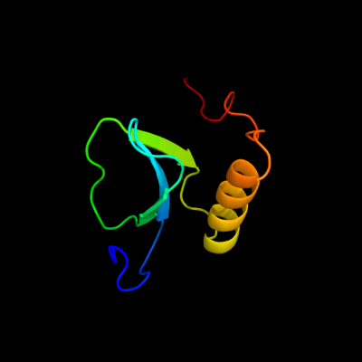
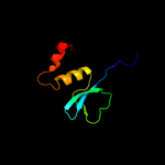
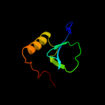
Fold: MbtH/L9 domain-likeSuperfamily: MbtH-likeFamily: MbtH-like2 c2khrA_
100.0
39
PDB header: biosynthetic proteinChain: A: PDB Molecule: protein mbth;PDBTitle: solution structure of rv2377c, a mbth-like protein from mycobacterium2 tuberculosis
3 d2pstx1
100.0
23
Fold: MbtH/L9 domain-likeSuperfamily: MbtH-likeFamily: MbtH-like4 c2jh3C_
21.2
22
PDB header: ribosomal proteinChain: C: PDB Molecule: ribosomal protein s2-related protein;PDBTitle: the crystal structure of dr2241 from deinococcus2 radiodurans at 1.9 a resolution reveals a multi-domain3 protein with structural similarity to chelatases but also4 with two additional novel domains
5 d2b6ca1
19.2
13
Fold: alpha-alpha superhelixSuperfamily: ARM repeatFamily: BC3264-like6 d1j0ha2
18.4
13
Fold: Glycosyl hydrolase domainSuperfamily: Glycosyl hydrolase domainFamily: alpha-Amylases, C-terminal beta-sheet domain7 d1iqqa_
16.0
20
Fold: Ribonuclease Rh-likeSuperfamily: Ribonuclease Rh-likeFamily: Ribonuclease Rh-like8 d1f8ab1
12.2
28
Fold: WW domain-likeSuperfamily: WW domainFamily: WW domain9 d2zjrg1
11.3
38
Fold: Ribosomal protein L13Superfamily: Ribosomal protein L13Family: Ribosomal protein L1310 c3cf5G_
11.3
38
PDB header: ribosome/antibioticChain: G: PDB Molecule: 50s ribosomal protein l13;PDBTitle: thiopeptide antibiotic thiostrepton bound to the large ribosomal2 subunit of deinococcus radiodurans
11 d1k9ra_
10.9
37
Fold: WW domain-likeSuperfamily: WW domainFamily: WW domain12 d1nmva1
10.7
28
Fold: WW domain-likeSuperfamily: WW domainFamily: WW domain13 c2ai4A_
10.6
33
PDB header: structural genomics, unknown functionChain: A: PDB Molecule: hypothetical protein so1698;PDBTitle: structure of protein of unknown function so1698 from shewanella2 oneidensis
14 c3pfyA_
10.5
12
PDB header: hydrolaseChain: A: PDB Molecule: otu domain-containing protein 5;PDBTitle: the catalytic domain of human otud5
15 c1ymzA_
10.4
25
PDB header: unknown functionChain: A: PDB Molecule: cc45;PDBTitle: cc45, an artificial ww domain designed using statistical2 coupling analysis
16 c2zajA_
10.3
30
PDB header: protein bindingChain: A: PDB Molecule: membrane-associated guanylate kinase, ww and pdzPDBTitle: solution structure of the short-isoform of the second ww2 domain from the human membrane-associated guanylate kinase,3 ww and pdz domain-containing protein 1 (magi-1)
17 d2ho2a1
10.0
43
Fold: WW domain-likeSuperfamily: WW domainFamily: WW domain18 d1g0wa2
9.9
10
Fold: Glutamine synthetase/guanido kinaseSuperfamily: Glutamine synthetase/guanido kinaseFamily: Guanido kinase catalytic domain19 c3mc2C_
9.8
19
PDB header: lyase inhibitorChain: C: PDB Molecule: inhibitor of carbonic anhydrase;PDBTitle: crystal structure of the murine inhibitor of carbonic anhydrase
20 d1u6ra2
9.7
10
Fold: Glutamine synthetase/guanido kinaseSuperfamily: Glutamine synthetase/guanido kinaseFamily: Guanido kinase catalytic domain21 c2ftcH_
not modelled
9.6
15
PDB header: ribosomeChain: H: PDB Molecule: 39s ribosomal protein l13, mitochondrial;PDBTitle: structural model for the large subunit of the mammalian mitochondrial2 ribosome
22 c2ysbA_
not modelled
9.2
22
PDB header: protein bindingChain: A: PDB Molecule: salvador homolog 1 protein;PDBTitle: solution structure of the first ww domain from the mouse2 salvador homolog 1 protein (sav1)
23 c3d5bN_
not modelled
8.9
31
PDB header: ribosomeChain: N: PDB Molecule: 50s ribosomal protein l13;PDBTitle: structural basis for translation termination on the 70s ribosome. this2 file contains the 50s subunit of one 70s ribosome. the entire crystal3 structure contains two 70s ribosomes as described in remark 400.
24 d1m15a2
not modelled
8.9
15
Fold: Glutamine synthetase/guanido kinaseSuperfamily: Glutamine synthetase/guanido kinaseFamily: Guanido kinase catalytic domain25 d2ysca1
not modelled
8.9
50
Fold: WW domain-likeSuperfamily: WW domainFamily: WW domain26 d1pina1
not modelled
8.8
36
Fold: WW domain-likeSuperfamily: WW domainFamily: WW domain27 c2kykA_
not modelled
8.5
27
PDB header: ligaseChain: A: PDB Molecule: e3 ubiquitin-protein ligase itchy homolog;PDBTitle: the sandwich region between two lmp2a py motif regulates the2 interaction between aip4ww2domain and py motif
28 c3nicA_
not modelled
8.3
27
PDB header: hydrolase/dnaChain: A: PDB Molecule: eco29kir;PDBTitle: dna binding and cleavage by the giy-yig endonuclease r.eco29ki2 inactive variant y49f
29 c1qk1H_
not modelled
8.2
15
PDB header: transferase (creatine kinase)Chain: H: PDB Molecule: creatine kinase, ubiquitous mitochondrial;PDBTitle: crystal structure of human ubiquitous mitochondrial2 creatine kinase
30 d2j01n1
not modelled
8.2
31
Fold: Ribosomal protein L13Superfamily: Ribosomal protein L13Family: Ribosomal protein L1331 c2lawA_
not modelled
8.2
27
PDB header: signaling protein/transcriptionChain: A: PDB Molecule: yorkie homolog;PDBTitle: structure of the second ww domain from human yap in complex with a2 human smad1 derived peptide
32 d2ilxa1
not modelled
8.1
23
Fold: LigT-likeSuperfamily: LigT-likeFamily: 2',3'-cyclic nucleotide 3'-phosphodiesterase, catalytic domain33 d1qh4a2
not modelled
8.0
10
Fold: Glutamine synthetase/guanido kinaseSuperfamily: Glutamine synthetase/guanido kinaseFamily: Guanido kinase catalytic domain34 c3l4hA_
not modelled
7.8
33
PDB header: protein bindingChain: A: PDB Molecule: e3 ubiquitin-protein ligase hecw1;PDBTitle: helical box domain and second ww domain of the human e3 ubiquitin-2 protein ligase hecw1
35 c3bboL_
not modelled
7.8
46
PDB header: ribosomeChain: L: PDB Molecule: ribosomal protein l13;PDBTitle: homology model for the spinach chloroplast 50s subunit2 fitted to 9.4a cryo-em map of the 70s chlororibosome
36 d1qk1a2
not modelled
7.7
15
Fold: Glutamine synthetase/guanido kinaseSuperfamily: Glutamine synthetase/guanido kinaseFamily: Guanido kinase catalytic domain37 c1tk7A_
not modelled
7.6
19
PDB header: signaling proteinChain: A: PDB Molecule: cg4244-pb;PDBTitle: nmr structure of ww domains (ww3-4) from suppressor of2 deltex
38 c2lb0A_
not modelled
7.6
33
PDB header: signaling protein/transcriptionChain: A: PDB Molecule: e3 ubiquitin-protein ligase smurf1;PDBTitle: structure of the first ww domain of human smurf1 in complex with a di-2 phosphorylated human smad1 derived peptide
39 c2ysgA_
not modelled
7.5
21
PDB header: protein bindingChain: A: PDB Molecule: syntaxin-binding protein 4;PDBTitle: solution structure of the ww domain from the human syntaxin-2 binding protein 4
40 c1e0mA_
not modelled
7.4
31
PDB header: de novo proteinChain: A: PDB Molecule: wwprototype;PDBTitle: prototype ww domain
41 d2jmfa1
not modelled
7.3
33
Fold: WW domain-likeSuperfamily: WW domainFamily: WW domain42 d1vrpa2
not modelled
7.1
10
Fold: Glutamine synthetase/guanido kinaseSuperfamily: Glutamine synthetase/guanido kinaseFamily: Guanido kinase catalytic domain43 d2f21a1
not modelled
7.0
29
Fold: WW domain-likeSuperfamily: WW domainFamily: WW domain44 d1crka2
not modelled
7.0
10
Fold: Glutamine synthetase/guanido kinaseSuperfamily: Glutamine synthetase/guanido kinaseFamily: Guanido kinase catalytic domain45 c1wr4A_
not modelled
7.0
25
PDB header: ligaseChain: A: PDB Molecule: ubiquitin-protein ligase nedd4-2;PDBTitle: solution structure of the second ww domain of nedd4-2
46 c1ce2A_
not modelled
7.0
13
PDB header: metal transportChain: A: PDB Molecule: protein (lactoferrin);PDBTitle: structure of diferric buffalo lactoferrin at 2.5a resolution
47 c2lazA_
not modelled
6.9
33
PDB header: signaling protein/transcriptionChain: A: PDB Molecule: e3 ubiquitin-protein ligase smurf1;PDBTitle: structure of the first ww domain of human smurf1 in complex with a2 mono-phosphorylated human smad1 derived peptide
48 c2dx0B_
not modelled
6.9
16
PDB header: hydrolaseChain: B: PDB Molecule: phospholipase c, gamma 2;PDBTitle: crystal structure of the n-terminal sh2 domain of mouse2 phospholipase c-gamma 2
49 d2itka1
not modelled
6.9
38
Fold: WW domain-likeSuperfamily: WW domainFamily: WW domain50 d1b1xa2
not modelled
6.8
6
Fold: Periplasmic binding protein-like IISuperfamily: Periplasmic binding protein-like IIFamily: Transferrin51 c2djyA_
not modelled
6.8
33
PDB header: ligase/signaling proteinChain: A: PDB Molecule: smad ubiquitination regulatory factor 2;PDBTitle: solution structure of smurf2 ww3 domain-smad7 py peptide2 complex
52 d1dota2
not modelled
6.8
6
Fold: Periplasmic binding protein-like IISuperfamily: Periplasmic binding protein-like IIFamily: Transferrin53 c1yr3A_
not modelled
6.8
18
PDB header: transferaseChain: A: PDB Molecule: xanthosine phosphorylase;PDBTitle: escherichia coli purine nucleoside phosphorylase ii, the2 product of the xapa gene
54 c1lfgA_
not modelled
6.6
19
PDB header: transferrinChain: A: PDB Molecule: lactoferrin;PDBTitle: molecular replacement solution of the structure of apolactoferrin, a2 protein displaying large-scale conformational change
55 c2krxA_
not modelled
6.6
32
PDB header: structural genomics, unknown functionChain: A: PDB Molecule: asl3597 protein;PDBTitle: solution nmr structure of asl3597 from nostoc sp. pcc7120. northeast2 structural genomics consortium target id nsr244.
56 c1rl9A_
not modelled
6.6
15
PDB header: transferaseChain: A: PDB Molecule: arginine kinase;PDBTitle: crystal structure of creatine-adp arginine kinase ternary2 complex
57 c2vncB_
not modelled
6.5
25
PDB header: hydrolaseChain: B: PDB Molecule: glycogen operon protein glgx;PDBTitle: crystal structure of glycogen debranching enzyme trex from2 sulfolobus solfataricus
58 c3l2eB_
not modelled
6.5
10
PDB header: transferaseChain: B: PDB Molecule: glycocyamine kinase beta chain;PDBTitle: glycocyamine kinase, alpha-beta heterodimer from marine worm2 namalycastis sp.
59 c2kxqA_
not modelled
6.4
26
PDB header: protein bindingChain: A: PDB Molecule: e3 ubiquitin-protein ligase smurf2;PDBTitle: solution structure of smurf2 ww2 and ww3 bound to smad7 py motif2 containing peptide
60 c1yiuA_
not modelled
6.4
27
PDB header: ligaseChain: A: PDB Molecule: itchy e3 ubiquitin protein ligase;PDBTitle: itch e3 ubiquitin ligase ww3 domain
61 c2l4jA_
not modelled
6.4
32
PDB header: transcriptionChain: A: PDB Molecule: yes-associated protein 2 (yap2);PDBTitle: yap ww2
62 c3bvsA_
not modelled
6.3
12
PDB header: hydrolaseChain: A: PDB Molecule: alkylpurine dna glycosylase alkd;PDBTitle: crystal structure of bacillus cereus alkylpurine dna glycosylase alkd
63 d1i8gb_
not modelled
6.3
36
Fold: WW domain-likeSuperfamily: WW domainFamily: WW domain64 d1iama2
not modelled
6.3
43
Fold: Immunoglobulin-like beta-sandwichSuperfamily: ImmunoglobulinFamily: I set domains65 d2e45a1
not modelled
6.2
67
Fold: WW domain-likeSuperfamily: WW domainFamily: WW domain66 c1ryxA_
not modelled
6.1
6
PDB header: metal transportChain: A: PDB Molecule: ovotransferrin;PDBTitle: crystal structure of hen serum transferrin in apo- form
67 d1g3pa1
not modelled
6.0
29
Fold: N-terminal domains of the minor coat protein g3pSuperfamily: N-terminal domains of the minor coat protein g3pFamily: N-terminal domains of the minor coat protein g3p68 c3ju6A_
not modelled
5.9
10
PDB header: transferaseChain: A: PDB Molecule: arginine kinase;PDBTitle: crystal structure of dimeric arginine kinase in complex with2 amppnp and arginine
69 c2yscA_
not modelled
5.6
25
PDB header: protein bindingChain: A: PDB Molecule: amyloid beta a4 precursor protein-binding familyPDBTitle: solution structure of the ww domain from the human amyloid2 beta a4 precursor protein-binding family b member 3, apbb3
70 c2jmfA_
not modelled
5.6
28
PDB header: ligase/signaling proteinChain: A: PDB Molecule: e3 ubiquitin-protein ligase suppressor of deltex;PDBTitle: solution structure of the su(dx) ww4- notch py peptide2 complex
71 d1dota1
not modelled
5.5
6
Fold: Periplasmic binding protein-like IISuperfamily: Periplasmic binding protein-like IIFamily: Transferrin72 c2yshA_
not modelled
5.5
32
PDB header: protein bindingChain: A: PDB Molecule: growth-arrest-specific protein 7;PDBTitle: solution structure of the ww domain from the human growth-2 arrest-specific protein 7, gas-7
73 c2hauA_
not modelled
5.4
19
PDB header: metal transportChain: A: PDB Molecule: serotransferrin;PDBTitle: apo-human serum transferrin (non-glycosylated)
74 c1i0eD_
not modelled
5.3
10
PDB header: transferaseChain: D: PDB Molecule: creatine kinase,m chain;PDBTitle: crystal structure of creatine kinase from human muscle
75 c2ysfA_
not modelled
5.2
27
PDB header: protein bindingChain: A: PDB Molecule: e3 ubiquitin-protein ligase itchy homolog;PDBTitle: solution structure of the fourth ww domain from the human2 e3 ubiquitin-protein ligase itchy homolog, itch
76 c2kieA_
not modelled
5.2
33
PDB header: hydrolaseChain: A: PDB Molecule: inositol polyphosphate 5-phosphatase ocrl-1;PDBTitle: a ph domain within ocrl bridges clathrin mediated membrane2 trafficking to phosphoinositide metabolis
77 d2c5ra1
not modelled
5.2
23
Fold: Phage replication organizer domainSuperfamily: Phage replication organizer domainFamily: Phage replication organizer domain78 c2kq0A_
not modelled
5.2
36
PDB header: ligaseChain: A: PDB Molecule: e3 ubiquitin-protein ligase nedd4;PDBTitle: human nedd4 3rd ww domain complex with ebola zaire virus matrix2 protein vp40 derived peptide ilptappeymea
79 d1ejxb_
not modelled
5.1
27
Fold: beta-clipSuperfamily: Urease, beta-subunitFamily: Urease, beta-subunit