1 c3nb2B_
100.0
20
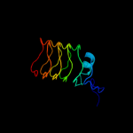
PDB header: ligaseChain: B: PDB Molecule: secreted effector protein;PDBTitle: crystal structure of e. coli o157:h7 effector protein nlel
2 c2qzaA_
100.0
17
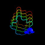
PDB header: ligaseChain: A: PDB Molecule: secreted effector protein;PDBTitle: crystal structure of salmonella effector protein sopa
3 c3du1X_
99.9
13
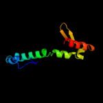
PDB header: structural proteinChain: X: PDB Molecule: all3740 protein;PDBTitle: the 2.0 angstrom resolution crystal structure of hetl, a pentapeptide2 repeat protein involved in heterocyst differentiation regulation from3 the cyanobacterium nostoc sp. strain pcc 7120
4 c2o6wA_
99.9
15
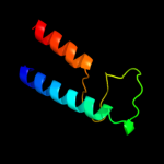
PDB header: unknown functionChain: A: PDB Molecule: repeat five residue (rfr) protein orPDBTitle: crystal structure of a pentapeptide repeat protein (rfr23)2 from the cyanobacterium cyanothece 51142
5 c2xt4B_
99.9
19
PDB header: cell cycleChain: B: PDB Molecule: mcbg-like protein;PDBTitle: structure of the pentapeptide repeat protein albg, a2 resistance factor for the topoisomerase poison albicidin.
6 c3pssB_
99.9
11
PDB header: cell cycleChain: B: PDB Molecule: qnr;PDBTitle: crystal structure of ahqnr, the qnr protein from aeromonas hydrophila2 (p21 crystal form)
7 d2j8ka1
99.9
17
Fold: Single-stranded right-handed beta-helixSuperfamily: Pentapeptide repeat-likeFamily: Pentapeptide repeats8 c2j8kA_
99.9
17
PDB header: toxinChain: A: PDB Molecule: np275-np276;PDBTitle: structure of the fusion of np275 and np276, pentapeptide2 repeat proteins from nostoc punctiforme
9 c2w7zB_
99.8
16
PDB header: inhibitorChain: B: PDB Molecule: pentapeptide repeat family protein;PDBTitle: structure of the pentapeptide repeat protein efsqnr, a dna2 gyrase inhibitor. free amines modified by cyclic3 pentylation with glutaraldehyde.
10 c2xtwB_
99.8
15
PDB header: cell cycleChain: B: PDB Molecule: qnrb1;PDBTitle: structure of qnrb1 (full length), a plasmid-mediated2 fluoroquinolone resistance protein
11 c2xt4A_
99.8
14
PDB header: cell cycleChain: A: PDB Molecule: mcbg-like protein;PDBTitle: structure of the pentapeptide repeat protein albg, a2 resistance factor for the topoisomerase poison albicidin.
12 c3n90A_
99.8
12
PDB header: unknown functionChain: A: PDB Molecule: thylakoid lumenal 15 kda protein 1, chloroplastic;PDBTitle: the 1.7 angstrom resolution crystal structure of at2g44920, a2 pentapeptide repeat protein from arabidopsis thaliana thylakoid3 lumen.
13 c2g0yA_
99.8
11
PDB header: unknown functionChain: A: PDB Molecule: pentapeptide repeat protein;PDBTitle: crystal structure of a lumenal pentapeptide repeat protein from2 cyanothece sp 51142 at 2.3 angstrom resolution. tetragonal crystal3 form
14 d2bm5a1
99.8
9
Fold: Single-stranded right-handed beta-helixSuperfamily: Pentapeptide repeat-likeFamily: Pentapeptide repeats15 d2f3la1
99.8
11
Fold: Single-stranded right-handed beta-helixSuperfamily: Pentapeptide repeat-likeFamily: Pentapeptide repeats16 c2j8iB_
99.8
15
PDB header: toxinChain: B: PDB Molecule: np275;PDBTitle: structure of np275, a pentapeptide repeat protein from2 nostoc punctiforme
17 d2j8ia1
99.5
14
Fold: Single-stranded right-handed beta-helixSuperfamily: Pentapeptide repeat-likeFamily: Pentapeptide repeats18 c3etvA_
78.7
15
PDB header: transport proteinChain: A: PDB Molecule: protein transport protein tip20,linker,proteinPDBTitle: crystal structure of a tip20p-dsl1p fusion protein
19 d3buxb2
52.8
13
Fold: N-cbl likeSuperfamily: N-terminal domain of cbl (N-cbl)Family: N-terminal domain of cbl (N-cbl)20 c3bunB_
47.1
13
PDB header: ligase/signaling proteinChain: B: PDB Molecule: e3 ubiquitin-protein ligase cbl;PDBTitle: crystal structure of c-cbl-tkb domain complexed with its2 binding motif in sprouty4
21 c2cblA_
not modelled
42.8
13
PDB header: complex (proto-oncogene/peptide)Chain: A: PDB Molecule: proto-oncogene cbl;PDBTitle: n-terminal domain of cbl in complex with its binding site2 on zap-70
22 d1td6a_
not modelled
39.9
16
Fold: Hypothetical protein MPN330Superfamily: Hypothetical protein MPN330Family: Hypothetical protein MPN33023 d1h8ba_
not modelled
37.4
19
Fold: EF Hand-likeSuperfamily: EF-handFamily: EF-hand modules in multidomain proteins24 c3ddrC_
not modelled
36.0
11
PDB header: membrane protein/heme binding proteinChain: C: PDB Molecule: hemophore hasa;PDBTitle: structure of the serratia marcescens hemophore receptor hasr-ile671gly2 mutant in complex with its hemophore hasa and heme
25 c1fbvA_
not modelled
35.1
13
PDB header: ligaseChain: A: PDB Molecule: signal transduction protein cbl;PDBTitle: structure of a cbl-ubch7 complex: ring domain function in2 ubiquitin-protein ligases
26 d1dlya_
not modelled
33.4
22
Fold: Globin-likeSuperfamily: Globin-likeFamily: Truncated hemoglobin27 c1dlyA_
not modelled
33.4
22
PDB header: oxygen storage/transportChain: A: PDB Molecule: hemoglobin;PDBTitle: x-ray crystal structure of hemoglobin from the green2 unicellular alga chlamydomonas eugametos
28 d1idra_
not modelled
30.7
23
Fold: Globin-likeSuperfamily: Globin-likeFamily: Truncated hemoglobin29 c2kscA_
not modelled
28.9
16
PDB header: unknown functionChain: A: PDB Molecule: cyanoglobin;PDBTitle: solution structure of synechococcus sp. pcc 7002 hemoglobin
30 d1dk0a_
not modelled
26.9
12
Fold: Heme-binding protein A (HasA)Superfamily: Heme-binding protein A (HasA)Family: Heme-binding protein A (HasA)31 c3aq8A_
not modelled
25.6
26
PDB header: oxygen bindingChain: A: PDB Molecule: group 1 truncated hemoglobin;PDBTitle: crystal structure of truncated hemoglobin from tetrahymena pyriformis,2 q46e mutant, fe(iii) form
32 d1s69a_
not modelled
23.4
13
Fold: Globin-likeSuperfamily: Globin-likeFamily: Truncated hemoglobin33 d1m8na_
not modelled
23.3
11
Fold: Single-stranded left-handed beta-helixSuperfamily: An insect antifreeze proteinFamily: An insect antifreeze protein34 d1dlwa_
not modelled
21.5
13
Fold: Globin-likeSuperfamily: Globin-likeFamily: Truncated hemoglobin35 d1ngka_
not modelled
20.6
13
Fold: Globin-likeSuperfamily: Globin-likeFamily: Truncated hemoglobin36 c3op0B_
not modelled
17.2
8
PDB header: signaling protein/signaling protein reguChain: B: PDB Molecule: signal transduction protein cbl-c;PDBTitle: crystal structure of cbl-c (cbl-3) tkb domain in complex with egfr2 py1069 peptide
37 c1z9eA_
not modelled
16.0
11
PDB header: protein binding/transcriptionChain: A: PDB Molecule: pc4 and sfrs1 interacting protein 2;PDBTitle: solution structure of the hiv-1 integrase-binding domain in2 ledgf/p75
38 d1ogmx2
not modelled
15.1
18
Fold: Single-stranded right-handed beta-helixSuperfamily: Pectin lyase-likeFamily: Dextranase, catalytic domain39 d2b4jc1
not modelled
15.0
11
Fold: N-cbl likeSuperfamily: HIV integrase-binding domainFamily: HIV integrase-binding domain40 d1fqia_
not modelled
14.1
14
Fold: Regulator of G-protein signaling, RGSSuperfamily: Regulator of G-protein signaling, RGSFamily: Regulator of G-protein signaling, RGS41 c2crpA_
not modelled
12.3
13
PDB header: signaling proteinChain: A: PDB Molecule: regulator of g-protein signaling 5;PDBTitle: solution structure of the rgs domain of regulator of g-2 protein signalling 5 (rgs 5)
42 c2p90B_
not modelled
11.8
12
PDB header: structural genomics, unknown functionChain: B: PDB Molecule: hypothetical protein cgl1923;PDBTitle: the crystal structure of a protein of unknown function from2 corynebacterium glutamicum atcc 13032
43 d1ko7a1
not modelled
11.5
20
Fold: MurF and HprK N-domain-likeSuperfamily: HprK N-terminal domain-likeFamily: HPr kinase/phoshatase HprK N-terminal domain44 c2bmmA_
not modelled
11.3
9
PDB header: oxygen storage/transportChain: A: PDB Molecule: thermostable hemoglobin from thermobifida fusca;PDBTitle: x-ray structure of a novel thermostable hemoglobin from the2 actinobacterium thermobifida fusca
45 c2gtpD_
not modelled
10.4
19
PDB header: signaling proteinChain: D: PDB Molecule: regulator of g-protein signaling 1;PDBTitle: crystal structure of the heterodimeric complex of human rgs1 and2 activated gi alpha 1
46 c2pbiA_
not modelled
10.0
13
PDB header: signaling proteinChain: A: PDB Molecule: regulator of g-protein signaling 9;PDBTitle: the multifunctional nature of gbeta5/rgs9 revealed from its crystal2 structure
47 c2d9jA_
not modelled
8.7
13
PDB header: signaling proteinChain: A: PDB Molecule: regulator of g-protein signalling 7;PDBTitle: solution structure of the rgs domain of regulator of g-2 protein signaling 7
48 c1d0rA_
not modelled
8.7
0
PDB header: hormone/growth factorChain: A: PDB Molecule: glucagon-like peptide-1-(7-36)-amide;PDBTitle: solution structure of glucagon-like peptide-1-(7-36)-amide2 in trifluoroethanol/water
49 d3buxb3
not modelled
8.2
17
Fold: SH2-likeSuperfamily: SH2 domainFamily: SH2 domain50 d1jg5a_
not modelled
7.6
31
Fold: GTP cyclohydrolase I feedback regulatory protein, GFRPSuperfamily: GTP cyclohydrolase I feedback regulatory protein, GFRPFamily: GTP cyclohydrolase I feedback regulatory protein, GFRP51 c2xykB_
not modelled
7.1
11
PDB header: oxygen storage/transportChain: B: PDB Molecule: 2-on-2 hemoglobin;PDBTitle: group ii 2-on-2 hemoglobin from the plant pathogen2 agrobacterium tumefaciens
52 d2etda1
not modelled
6.3
24
Fold: Bromodomain-likeSuperfamily: LemA-likeFamily: LemA-like53 c2af0A_
not modelled
6.1
10
PDB header: signaling proteinChain: A: PDB Molecule: regulator of g-protein signaling 2;PDBTitle: structure of the regulator of g-protein signaling domain of2 rgs2
54 d7pcka_
not modelled
6.1
11
Fold: Cysteine proteinasesSuperfamily: Cysteine proteinasesFamily: Papain-like55 c1jrjA_
not modelled
5.8
6
PDB header: hormone/growth factorChain: A: PDB Molecule: exendin-4;PDBTitle: solution structure of exendin-4 in 30-vol% trifluoroethanol
56 c2ebzA_
not modelled
5.6
6
PDB header: signaling proteinChain: A: PDB Molecule: regulator of g-protein signaling 12;PDBTitle: solution structure of the rgs domain from human regulator2 of g-protein signaling 12 (rgs12)