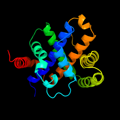
Phyre Home Retrieve Phyre Job Id
Emaill.a.kelley@imperial.ac.uk
DescriptionP25746
DateThu Jan 5 11:42:34 GMT 2012
Unique Job IDfc1afae978af8daf

Summary 

Top model
Image coloured by rainbow N → C terminus
Model (left) based on template d1sdia_
Top template information
Fold:YcfC-like
Superfamily:YcfC-like
Family:YcfC-like
Confidence and coverage
Confidence:100.0% Coverage:100%
212 residues (100% of your sequence) have been modelled with 100.0% confidence by the single highest scoring template.
3D viewing
Interactive 3D view in Jmol

Sequence analysis 

Secondary structure and disorder prediction 

   1........10.........20.........30.........40.........50.........60
Sequence  MAKNYYDITLALAGICQSARLVQQLAHQGHCDADALHVSLNSIIDMNPSSTLAVFGGSEA
Secondary structure 


SS confidence 


Disorder  ????


?


Disorder confidence 


   .........70.........80.........90.........100.........110.........120
Sequence  NLRVGLETLLGVLNASSRQGLNAELTRYTLSLMVLERKLSSAKGALDTLGNRINGLQRQL
Secondary structure 


SS confidence 


Disorder 


??????????


???


Disorder confidence 


   .........130.........140.........150.........160.........170.........180
Sequence  EHFDLQSETLMSAMAAIYVDVISPLGPRIQVTGSPAVLQSPQVQAKVRATLLAGIRAAVL
Secondary structure 


SS confidence 


Disorder 


??

?


?


???


Disorder confidence 


   .........190.........200.........210...
Sequence  WHQVGGGRLQLMFSRNRLTTQAKQILAHLTPEL
Secondary structure 


SS confidence 


Disorder 


??????
Disorder confidence 


Confidence Key
High(9)                    Low (0)
?Disordered
Alpha helix
Beta strand

Domain analysis 

Hover over an aligned region to see model and summary info

Please note, only up to the top 20 hits are modelled to reduce computer load

RankAligned region

PDB 1sdi chain A

3D model

Region: 2 - 213
Aligned: 212
Modelled: 212
Confidence: 100.0%
Identity: 100%
Fold: YcfC-like
Superfamily: YcfC-like
Family: YcfC-like

Phyre2

PDB 2xr1 chain A

3D model

Region: 127 - 167
Aligned: 41
Modelled: 41
Confidence: 39.1%
Identity: 20%
PDB header:hydrolase
Chain: A: PDB Molecule:cleavage and polyadenylation specificity factor 100 kd
PDBTitle: dimeric archaeal cleavage and polyadenylation specificity2 factor with n-terminal kh domains (kh-cpsf) from methanosarcina3 mazei

Phyre2

PDB 2gej chain A

3D model

Region: 97 - 152
Aligned: 50
Modelled: 51
Confidence: 33.1%
Identity: 12%
PDB header:transferase
Chain: A: PDB Molecule:phosphatidylinositol mannosyltransferase (pima);
PDBTitle: crystal structure of phosphatidylinositol mannosyltransferase (pima)2 from mycobacterium smegmatis in complex with gdp-man

Phyre2

PDB 2ycb chain A

3D model

Region: 127 - 167
Aligned: 41
Modelled: 41
Confidence: 30.0%
Identity: 15%
PDB header:hydrolase
Chain: A: PDB Molecule:cleavage and polyadenylation specificity factor;
PDBTitle: structure of the archaeal beta-casp protein with n-terminal2 kh domains from methanothermobacter thermautotrophicus

Phyre2

PDB 2rdc chain A

3D model

Region: 86 - 178
Aligned: 87
Modelled: 93
Confidence: 27.0%
Identity: 22%
PDB header:lipid binding protein
Chain: A: PDB Molecule:uncharacterized protein;
PDBTitle: crystal structure of a putative lipid binding protein (gsu0061) from2 geobacter sulfurreducens pca at 1.80 a resolution

Phyre2

PDB 3af5 chain A

3D model

Region: 139 - 167
Aligned: 29
Modelled: 29
Confidence: 22.9%
Identity: 34%
PDB header:hydrolase
Chain: A: PDB Molecule:putative uncharacterized protein ph1404;
PDBTitle: the crystal structure of an archaeal cpsf subunit, ph1404 from2 pyrococcus horikoshii

Phyre2

PDB 2fge chain A domain 1

3D model

Region: 59 - 159
Aligned: 99
Modelled: 101
Confidence: 21.5%
Identity: 17%
Fold: LuxS/MPP-like metallohydrolase
Superfamily: LuxS/MPP-like metallohydrolase
Family: MPP-like

Phyre2

PDB 2k1v chain B

3D model

Region: 175 - 189
Aligned: 13
Modelled: 15
Confidence: 15.7%
Identity: 46%
PDB header:hormone
Chain: B: PDB Molecule:relaxin-3;
PDBTitle: r3/i5 relaxin chimera

Phyre2

PDB 2rpa chain A

3D model

Region: 9 - 33
Aligned: 25
Modelled: 25
Confidence: 15.1%
Identity: 28%
PDB header:hydrolase
Chain: A: PDB Molecule:katanin p60 atpase-containing subunit a1;
PDBTitle: the solution structure of n-terminal domain of microtubule severing2 enzyme

Phyre2

PDB 3fxd chain D

3D model

Region: 134 - 144
Aligned: 11
Modelled: 11
Confidence: 14.0%
Identity: 36%
PDB header:unknown function
Chain: D: PDB Molecule:protein icmr;
PDBTitle: crystal structure of interacting domains of icmr and icmq

Phyre2

PDB 3cx3 chain A

3D model

Region: 104 - 157
Aligned: 54
Modelled: 54
Confidence: 11.7%
Identity: 7%
PDB header:metal binding protein
Chain: A: PDB Molecule:lipoprotein;
PDBTitle: crystal structure analysis of the streptococcus pneumoniae2 adcaii protein

Phyre2

PDB 3mzk chain C

3D model

Region: 160 - 193
Aligned: 34
Modelled: 34
Confidence: 11.7%
Identity: 18%
PDB header:protein transport
Chain: C: PDB Molecule:protein transport protein sec16;
PDBTitle: sec13/sec16 complex, s.cerevisiae

Phyre2

PDB 2rms chain B

3D model

Region: 9 - 17
Aligned: 9
Modelled: 9
Confidence: 11.6%
Identity: 56%
PDB header:transcription
Chain: B: PDB Molecule:msin3a-binding protein;
PDBTitle: solution structure of the msin3a pah1-sap25 sid complex

Phyre2

PDB 2xdj chain F

3D model

Region: 92 - 140
Aligned: 49
Modelled: 48
Confidence: 10.4%
Identity: 22%
PDB header:unknown function
Chain: F: PDB Molecule:uncharacterized protein ybgf;
PDBTitle: crystal structure of the n-terminal domain of e.coli ybgf

Phyre2

PDB 2h8b chain B

3D model

Region: 180 - 189
Aligned: 10
Modelled: 10
Confidence: 9.8%
Identity: 40%
PDB header:hormone/growth factor
Chain: B: PDB Molecule:insulin-like 3;
PDBTitle: solution structure of insl3

Phyre2

PDB 2ogw chain B

3D model

Region: 103 - 157
Aligned: 55
Modelled: 55
Confidence: 9.0%
Identity: 13%
PDB header:transport protein
Chain: B: PDB Molecule:high-affinity zinc uptake system protein znua
PDBTitle: structure of abc type zinc transporter from e. coli

Phyre2

PDB 2r5u chain D

3D model

Region: 80 - 140
Aligned: 50
Modelled: 61
Confidence: 8.9%
Identity: 20%
PDB header:hydrolase
Chain: D: PDB Molecule:replicative dna helicase;
PDBTitle: crystal structure of the n-terminal domain of dnab helicase from2 mycobacterium tuberculosis

Phyre2

PDB 2juf chain A

3D model

Region: 179 - 189
Aligned: 11
Modelled: 11
Confidence: 8.5%
Identity: 27%
PDB header:gene regulation
Chain: A: PDB Molecule:p53-associated parkin-like cytoplasmic protein;
PDBTitle: nmr solution structure of parc cph domain. nesg target2 hr3443b/sgc-toronto

Phyre2

PDB 1qi9 chain A

3D model

Region: 27 - 54
Aligned: 28
Modelled: 28
Confidence: 8.3%
Identity: 29%
Fold: Acid phosphatase/Vanadium-dependent haloperoxidase
Superfamily: Acid phosphatase/Vanadium-dependent haloperoxidase
Family: Haloperoxidase (bromoperoxidase)

Phyre2

PDB 2fhd chain A

3D model

Region: 178 - 187
Aligned: 10
Modelled: 10
Confidence: 8.3%
Identity: 20%
PDB header:cell cycle
Chain: A: PDB Molecule:dna repair protein rhp9/crb2;
PDBTitle: crystal structure of crb2 tandem tudor domains

Phyre2
1

d1sdia_
2

c2xr1A_
3

c2gejA_
4

c2ycbA_
5

c2rdcA_
6

c3af5A_
7

d2fgea1
8

c2k1vB_
9

c2rpaA_
10

c3fxdD_
11

c3cx3A_
12

c3mzkC_
13

c2rmsB_
14

c2xdjF_
15

c2h8bB_
16

c2ogwB_
17

c2r5uD_
18

c2jufA_
19

d1qi9a_
20

c2fhdA_
21


22


23


24


25


26


27


28


29


30


31


32


33


34


35


36


37


38


39


40


41


42


43


44


45


46


47


Detailed template information 

#
Template Alignment Coverage3D Model Confidence
% i.d. Template Information
1d1sdia_


100.0 100 Fold:YcfC-like
Superfamily:YcfC-like
Family:YcfC-like
2c2xr1A_


39.1 20 PDB header:hydrolase
Chain: A: PDB Molecule:cleavage and polyadenylation specificity factor 100 kd
PDBTitle: dimeric archaeal cleavage and polyadenylation specificity2 factor with n-terminal kh domains (kh-cpsf) from methanosarcina3 mazei
3c2gejA_


33.1 12 PDB header:transferase
Chain: A: PDB Molecule:phosphatidylinositol mannosyltransferase (pima);
PDBTitle: crystal structure of phosphatidylinositol mannosyltransferase (pima)2 from mycobacterium smegmatis in complex with gdp-man
4c2ycbA_


30.0 15 PDB header:hydrolase
Chain: A: PDB Molecule:cleavage and polyadenylation specificity factor;
PDBTitle: structure of the archaeal beta-casp protein with n-terminal2 kh domains from methanothermobacter thermautotrophicus
5c2rdcA_


27.0 22 PDB header:lipid binding protein
Chain: A: PDB Molecule:uncharacterized protein;
PDBTitle: crystal structure of a putative lipid binding protein (gsu0061) from2 geobacter sulfurreducens pca at 1.80 a resolution
6c3af5A_


22.9 34 PDB header:hydrolase
Chain: A: PDB Molecule:putative uncharacterized protein ph1404;
PDBTitle: the crystal structure of an archaeal cpsf subunit, ph1404 from2 pyrococcus horikoshii
7d2fgea1


21.5 17 Fold:LuxS/MPP-like metallohydrolase
Superfamily:LuxS/MPP-like metallohydrolase
Family:MPP-like
8c2k1vB_


15.7 46 PDB header:hormone
Chain: B: PDB Molecule:relaxin-3;
PDBTitle: r3/i5 relaxin chimera
9c2rpaA_


15.1 28 PDB header:hydrolase
Chain: A: PDB Molecule:katanin p60 atpase-containing subunit a1;
PDBTitle: the solution structure of n-terminal domain of microtubule severing2 enzyme
10c3fxdD_


14.0 36 PDB header:unknown function
Chain: D: PDB Molecule:protein icmr;
PDBTitle: crystal structure of interacting domains of icmr and icmq
11c3cx3A_


11.7 7 PDB header:metal binding protein
Chain: A: PDB Molecule:lipoprotein;
PDBTitle: crystal structure analysis of the streptococcus pneumoniae2 adcaii protein
12c3mzkC_


11.7 18 PDB header:protein transport
Chain: C: PDB Molecule:protein transport protein sec16;
PDBTitle: sec13/sec16 complex, s.cerevisiae
13c2rmsB_


11.6 56 PDB header:transcription
Chain: B: PDB Molecule:msin3a-binding protein;
PDBTitle: solution structure of the msin3a pah1-sap25 sid complex
14c2xdjF_


10.4 22 PDB header:unknown function
Chain: F: PDB Molecule:uncharacterized protein ybgf;
PDBTitle: crystal structure of the n-terminal domain of e.coli ybgf
15c2h8bB_


9.8 40 PDB header:hormone/growth factor
Chain: B: PDB Molecule:insulin-like 3;
PDBTitle: solution structure of insl3
16c2ogwB_


9.0 13 PDB header:transport protein
Chain: B: PDB Molecule:high-affinity zinc uptake system protein znua
PDBTitle: structure of abc type zinc transporter from e. coli
17c2r5uD_


8.9 20 PDB header:hydrolase
Chain: D: PDB Molecule:replicative dna helicase;
PDBTitle: crystal structure of the n-terminal domain of dnab helicase from2 mycobacterium tuberculosis
18c2jufA_


8.5 27 PDB header:gene regulation
Chain: A: PDB Molecule:p53-associated parkin-like cytoplasmic protein;
PDBTitle: nmr solution structure of parc cph domain. nesg target2 hr3443b/sgc-toronto
19d1qi9a_


8.3 29 Fold:Acid phosphatase/Vanadium-dependent haloperoxidase
Superfamily:Acid phosphatase/Vanadium-dependent haloperoxidase
Family:Haloperoxidase (bromoperoxidase)
20c2fhdA_


8.3 20 PDB header:cell cycle
Chain: A: PDB Molecule:dna repair protein rhp9/crb2;
PDBTitle: crystal structure of crb2 tandem tudor domains
21c3h25A_


not modelled 8.2 32 PDB header:replication/dna
Chain: A: PDB Molecule:replication protein b;
PDBTitle: crystal structure of the catalytic domain of primase repb' in complex2 with initiator dna
22c3hgkE_


not modelled 8.2 20 PDB header:transferase
Chain: E: PDB Molecule:effector protein hopab2;
PDBTitle: crystal structure of effect protein avrptob complexed with2 kinase pto
23d2jnga1


not modelled 7.9 27 Fold:SH3-like barrel
Superfamily:Tudor/PWWP/MBT
Family:CPH domain
24d1qhba_


not modelled 7.7 18 Fold:Acid phosphatase/Vanadium-dependent haloperoxidase
Superfamily:Acid phosphatase/Vanadium-dependent haloperoxidase
Family:Haloperoxidase (bromoperoxidase)
25d1vkea_


not modelled 7.6 13 Fold:AhpD-like
Superfamily:AhpD-like
Family:CMD-like
26d1up8a_


not modelled 7.5 14 Fold:Acid phosphatase/Vanadium-dependent haloperoxidase
Superfamily:Acid phosphatase/Vanadium-dependent haloperoxidase
Family:Haloperoxidase (bromoperoxidase)
27c2o1eB_


not modelled 7.5 11 PDB header:structural genomics, unknown function
Chain: B: PDB Molecule:ycdh;
PDBTitle: crystal structure of the metal-dependent lipoprotein ycdh2 from bacillus subtilis, northeast structural genomics3 target sr583
28d1u7ka_


not modelled 7.2 33 Fold:Retrovirus capsid protein, N-terminal core domain
Superfamily:Retrovirus capsid protein, N-terminal core domain
Family:Retrovirus capsid protein, N-terminal core domain
29d2jeka1


not modelled 7.1 20 Fold:Rv1873-like
Superfamily:Rv1873-like
Family:Rv1873-like
30c1yv0T_


not modelled 7.0 27 PDB header:contractile protein
Chain: T: PDB Molecule:troponin t, fast skeletal muscle isoforms;
PDBTitle: crystal structure of skeletal muscle troponin in the ca2+-2 free state
31c1xouA_


not modelled 7.0 24 PDB header:structural protein/chaperone
Chain: A: PDB Molecule:espa;
PDBTitle: crystal structure of the cesa-espa complex
32d1xoua_


not modelled 7.0 24 Fold:EspA/CesA-like
Superfamily:EspA/CesA-like
Family:EspA-like
33d2v7qi1


not modelled 6.7 60 Fold:Non-globular all-alpha subunits of globular proteins
Superfamily:Epsilon subunit of mitochondrial F1F0-ATP synthase
Family:Epsilon subunit of mitochondrial F1F0-ATP synthase
34c2w9zA_


not modelled 6.5 17 PDB header:transferase
Chain: A: PDB Molecule:g1/s-specific cyclin-d1;
PDBTitle: crystal structure of cdk4 in complex with a d-type cyclin
35c3nrhA_


not modelled 6.4 13 PDB header:structural genomics, unknown function
Chain: A: PDB Molecule:uncharacterized protein bf1032;
PDBTitle: crystal structure of protein bf1032 from bacteroides fragilis,2 northeast structural genomics consortium target bfr309
36d1jwea_


not modelled 6.2 18 Fold:N-terminal domain of DnaB helicase
Superfamily:N-terminal domain of DnaB helicase
Family:N-terminal domain of DnaB helicase
37c1cosA_


not modelled 6.2 29 PDB header:alpha-helical bundle
Chain: A: PDB Molecule:coiled serine;
PDBTitle: crystal structure of a synthetic triple-stranded alpha-2 helical bundle
38c1cosB_


not modelled 6.2 29 PDB header:alpha-helical bundle
Chain: B: PDB Molecule:coiled serine;
PDBTitle: crystal structure of a synthetic triple-stranded alpha-2 helical bundle
39c1cosC_


not modelled 6.2 29 PDB header:alpha-helical bundle
Chain: C: PDB Molecule:coiled serine;
PDBTitle: crystal structure of a synthetic triple-stranded alpha-2 helical bundle
40c3imoC_


not modelled 6.1 21 PDB header:unknown function
Chain: C: PDB Molecule:integron cassette protein;
PDBTitle: structure from the mobile metagenome of vibrio cholerae.2 integron cassette protein vch_cass14
41d1hula_


not modelled 5.7 19 Fold:4-helical cytokines
Superfamily:4-helical cytokines
Family:Short-chain cytokines
42c2f59B_


not modelled 5.5 20 PDB header:transferase
Chain: B: PDB Molecule:6,7-dimethyl-8-ribityllumazine synthase 1;
PDBTitle: lumazine synthase ribh1 from brucella abortus (gene bruab1_0785,2 swiss-prot entry q57dy1) complexed with inhibitor 5-nitro-6-(d-3 ribitylamino)-2,4(1h,3h) pyrimidinedione
43d1b8za_


not modelled 5.5 14 Fold:IHF-like DNA-binding proteins
Superfamily:IHF-like DNA-binding proteins
Family:Prokaryotic DNA-bending protein
44d2bs2b2


not modelled 5.4 24 Fold:beta-Grasp (ubiquitin-like)
Superfamily:2Fe-2S ferredoxin-like
Family:2Fe-2S ferredoxin domains from multidomain proteins
45c3dlaD_


not modelled 5.3 19 PDB header:ligase
Chain: D: PDB Molecule:glutamine-dependent nad(+) synthetase;
PDBTitle: x-ray crystal structure of glutamine-dependent nad+ synthetase from2 mycobacterium tuberculosis bound to naad+ and don
46c3c4vB_


not modelled 5.2 14 PDB header:transferase
Chain: B: PDB Molecule:predicted glycosyltransferases;
PDBTitle: structure of the retaining glycosyltransferase msha:the2 first step in mycothiol biosynthesis. organism:3 corynebacterium glutamicum : complex with udp and 1l-ins-1-4 p.
47c3lk2B_


not modelled 5.0 18 PDB header:protein binding
Chain: B: PDB Molecule:f-actin-capping protein subunit beta isoforms 1 and 2;
PDBTitle: crystal structure of capz bound to the uncapping motif from carmil

Binding site prediction 

Due to computational demand, binding site predictions are not run for batch jobs

If you want to predict binding sites, please manually submit your model of choice to 3DLigandSite


Phyre is for academic use only

Please cite: Protein structure prediction on the web: a case study using the Phyre server
Kelley LA and Sternberg MJE. Nature Protocols 4, 363 - 371 (2009) [pdf] [Import into BibTeX]
 
If you use the binding site predictions from 3DLigandSite, please also cite:
3DLigandSite: predicting ligand-binding sites using similar structures.
Wass MN, Kelley LA and Sternberg MJ Nucleic Acids Research 38, W469-73 (2010) [PubMed]
 
© Structural Bioinformatics Group
Imperial College London
Lawrence Kelley, Benjamin Jefferys
Disclaimer
Terms and Conditions
Component software
Template detection: HHpred 1.51
Secondary structure prediction: Psi-pred 2.5
Disorder prediction: Disopred 2.4
Transmembrane prediction: Memsat_SVM
Multi-template modelling and ab initio: Poing 1.0