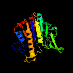
1 c3m8yC_
100.0
26
PDB header: isomeraseChain: C: PDB Molecule: phosphopentomutase;PDBTitle: phosphopentomutase from bacillus cereus after glucose-1,6-bisphosphate2 activation
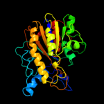
2 c2i09A_
100.0
28
PDB header: isomeraseChain: A: PDB Molecule: phosphopentomutase;PDBTitle: crystal structure of putative phosphopentomutase from streptococcus2 mutans
3 d1p49a_
100.0
11
Fold: Alkaline phosphatase-likeSuperfamily: Alkaline phosphatase-likeFamily: Arylsulfatase4 c3b5qB_
100.0
12
PDB header: hydrolaseChain: B: PDB Molecule: putative sulfatase yidj;PDBTitle: crystal structure of a putative sulfatase (np_810509.1)2 from bacteroides thetaiotaomicron vpi-5482 at 2.40 a3 resolution
5 d1auka_
100.0
15
Fold: Alkaline phosphatase-likeSuperfamily: Alkaline phosphatase-likeFamily: Arylsulfatase6 c2zktB_
100.0
13
PDB header: isomeraseChain: B: PDB Molecule: 2,3-bisphosphoglycerate-independent phosphoglyceratePDBTitle: structure of ph0037 protein from pyrococcus horikoshii
7 c3ed4A_
100.0
12
PDB header: transferaseChain: A: PDB Molecule: arylsulfatase;PDBTitle: crystal structure of putative arylsulfatase from escherichia coli
8 d1hdha_
100.0
16
Fold: Alkaline phosphatase-likeSuperfamily: Alkaline phosphatase-likeFamily: Arylsulfatase9 c2vqrA_
100.0
13
PDB header: hydrolaseChain: A: PDB Molecule: putative sulfatase;PDBTitle: crystal structure of a phosphonate monoester hydrolase2 from rhizobium leguminosarum: a new member of the3 alkaline phosphatase superfamily
10 c2qzuA_
100.0
13
PDB header: hydrolaseChain: A: PDB Molecule: putative sulfatase yidj;PDBTitle: crystal structure of the putative sulfatase yidj from bacteroides2 fragilis. northeast structural genomics consortium target bfr123
11 d1fsua_
100.0
14
Fold: Alkaline phosphatase-likeSuperfamily: Alkaline phosphatase-likeFamily: Arylsulfatase12 d2i09a1
100.0
28
Fold: Alkaline phosphatase-likeSuperfamily: Alkaline phosphatase-likeFamily: DeoB catalytic domain-like13 c3lxqB_
100.0
15
PDB header: structural genomics, unknown functionChain: B: PDB Molecule: uncharacterized protein vp1736;PDBTitle: the crystal structure of a protein in the alkaline2 phosphatase superfamily from vibrio parahaemolyticus to3 1.95a
14 c2w8dB_
100.0
11
PDB header: transferaseChain: B: PDB Molecule: processed glycerol phosphate lipoteichoic acid synthase 2;PDBTitle: distinct and essential morphogenic functions for wall- and2 lipo-teichoic acids in bacillus subtilis
15 c2w5tA_
100.0
11
PDB header: transferaseChain: A: PDB Molecule: processed glycerol phosphate lipoteichoic acidPDBTitle: structure-based mechanism of lipoteichoic acid synthesis by2 staphylococcus aureus ltas.
16 d1o98a2
100.0
21
Fold: Alkaline phosphatase-likeSuperfamily: Alkaline phosphatase-likeFamily: 2,3-Bisphosphoglycerate-independent phosphoglycerate mutase, catalytic domain17 c3q3qA_
100.0
18
PDB header: hydrolaseChain: A: PDB Molecule: alkaline phosphatase;PDBTitle: crystal structure of spap: an novel alkaline phosphatase from2 bacterium sphingomonas sp. strain bsar-1
18 c2gsoB_
100.0
25
PDB header: hydrolaseChain: B: PDB Molecule: phosphodiesterase-nucleotide pyrophosphatase;PDBTitle: structure of xac nucleotide2 pyrophosphatase/phosphodiesterase in complex with vanadate
19 c3szzA_
100.0
19
PDB header: hydrolaseChain: A: PDB Molecule: phosphonoacetate hydrolase;PDBTitle: crystal structure of phosphonoacetate hydrolase from sinorhizobium2 meliloti 1021 in complex with acetate
20 d1ei6a_
100.0
18
Fold: Alkaline phosphatase-likeSuperfamily: Alkaline phosphatase-likeFamily: Phosphonoacetate hydrolase21 c2xrgA_
not modelled
100.0
19
PDB header: hydrolaseChain: A: PDB Molecule: ectonucleotide pyrophosphatase/phosphodiesterase familyPDBTitle: crystal structure of autotaxin (enpp2) in complex with the2 ha155 boronic acid inhibitor
22 c2xr9A_
not modelled
100.0
15
PDB header: hydrolaseChain: A: PDB Molecule: ectonucleotide pyrophosphatase/phosphodiesterase familyPDBTitle: crystal structure of autotaxin (enpp2)
23 d1y6va1
not modelled
100.0
13
Fold: Alkaline phosphatase-likeSuperfamily: Alkaline phosphatase-likeFamily: Alkaline phosphatase24 c1ew2A_
not modelled
100.0
15
PDB header: hydrolaseChain: A: PDB Molecule: phosphatase;PDBTitle: crystal structure of a human phosphatase
25 d1zeda1
not modelled
100.0
15
Fold: Alkaline phosphatase-likeSuperfamily: Alkaline phosphatase-likeFamily: Alkaline phosphatase26 d1k7ha_
not modelled
100.0
15
Fold: Alkaline phosphatase-likeSuperfamily: Alkaline phosphatase-likeFamily: Alkaline phosphatase27 c2iucB_
not modelled
100.0
13
PDB header: hydrolaseChain: B: PDB Molecule: alkaline phosphatase;PDBTitle: structure of alkaline phosphatase from the antarctic2 bacterium tab5
28 c3e2dB_
not modelled
100.0
15
PDB header: hydrolaseChain: B: PDB Molecule: alkaline phosphatase;PDBTitle: the 1.4 a crystal structure of the large and cold-active2 vibrio sp. alkaline phosphatase
29 c1o98A_
not modelled
100.0
24
PDB header: isomeraseChain: A: PDB Molecule: 2,3-bisphosphoglycerate-independentPDBTitle: 1.4a crystal structure of phosphoglycerate mutase from2 bacillus stearothermophilus complexed with3 2-phosphoglycerate
30 c3a52A_
not modelled
100.0
15
PDB header: hydrolaseChain: A: PDB Molecule: cold-active alkaline phosphatase;PDBTitle: crystal structure of cold-active alkailne phosphatase from2 psychrophile shewanella sp.
31 c2w0yB_
not modelled
100.0
16
PDB header: hydrolaseChain: B: PDB Molecule: alkaline phosphatase;PDBTitle: h.salinarum alkaline phosphatase
32 c2x98A_
not modelled
100.0
14
PDB header: hydrolaseChain: A: PDB Molecule: alkaline phosphatase;PDBTitle: h.salinarum alkaline phosphatase
33 c3igzB_
not modelled
99.9
24
PDB header: isomeraseChain: B: PDB Molecule: cofactor-independent phosphoglycerate mutase;PDBTitle: crystal structures of leishmania mexicana phosphoglycerate2 mutase at low cobalt concentration
34 c3iddA_
not modelled
99.9
15
PDB header: isomeraseChain: A: PDB Molecule: 2,3-bisphosphoglycerate-independentPDBTitle: cofactor-independent phosphoglycerate mutase from2 thermoplasma acidophilum dsm 1728
35 c2d1gB_
not modelled
99.4
11
PDB header: hydrolaseChain: B: PDB Molecule: acid phosphatase;PDBTitle: structure of francisella tularensis acid phosphatase a (acpa) bound to2 orthovanadate
36 d2i09a2
not modelled
87.2
7
Fold: DeoB insert domain-likeSuperfamily: DeoB insert domain-likeFamily: DeoB insert domain-like37 d1b4ub_
not modelled
68.0
16
Fold: Phosphorylase/hydrolase-likeSuperfamily: LigB-likeFamily: LigB-like38 d1j33a_
not modelled
46.4
32
Fold: Nicotinate mononucleotide:5,6-dimethylbenzimidazole phosphoribosyltransferase (CobT)Superfamily: Nicotinate mononucleotide:5,6-dimethylbenzimidazole phosphoribosyltransferase (CobT)Family: Nicotinate mononucleotide:5,6-dimethylbenzimidazole phosphoribosyltransferase (CobT)39 c3oaaO_
not modelled
40.4
20
PDB header: hydrolase/transport proteinChain: O: PDB Molecule: atp synthase gamma chain;PDBTitle: structure of the e.coli f1-atp synthase inhibited by subunit epsilon
40 c3f5dA_
not modelled
39.4
17
PDB header: structural genomics, unknown functionChain: A: PDB Molecule: protein ydea;PDBTitle: crystal structure of a protein of unknown function from2 bacillus subtilis
41 c3cagF_
not modelled
34.9
33
PDB header: dna binding proteinChain: F: PDB Molecule: arginine repressor;PDBTitle: crystal structure of the oligomerization domain hexamer of the2 arginine repressor protein from mycobacterium tuberculosis in complex3 with 9 arginines.
42 d2p5ma1
not modelled
33.0
14
Fold: DCoH-likeSuperfamily: C-terminal domain of arginine repressorFamily: C-terminal domain of arginine repressor43 d1b4ba_
not modelled
31.6
19
Fold: DCoH-likeSuperfamily: C-terminal domain of arginine repressorFamily: C-terminal domain of arginine repressor44 c2xokG_
not modelled
31.4
17
PDB header: hydrolaseChain: G: PDB Molecule: atp synthase subunit gamma, mitochondrial;PDBTitle: refined structure of yeast f1c10 atpase complex to 3 a2 resolution
45 c3ereD_
not modelled
29.7
33
PDB header: dna binding protein/dnaChain: D: PDB Molecule: arginine repressor;PDBTitle: crystal structure of the arginine repressor protein from mycobacterium2 tuberculosis in complex with the dna operator
46 c1b4aA_
not modelled
23.3
19
PDB header: repressorChain: A: PDB Molecule: arginine repressor;PDBTitle: structure of the arginine repressor from bacillus stearothermophilus
47 d1l5oa_
not modelled
20.9
35
Fold: Nicotinate mononucleotide:5,6-dimethylbenzimidazole phosphoribosyltransferase (CobT)Superfamily: Nicotinate mononucleotide:5,6-dimethylbenzimidazole phosphoribosyltransferase (CobT)Family: Nicotinate mononucleotide:5,6-dimethylbenzimidazole phosphoribosyltransferase (CobT)48 d1xxaa_
not modelled
20.4
24
Fold: DCoH-likeSuperfamily: C-terminal domain of arginine repressorFamily: C-terminal domain of arginine repressor49 d1a05a_
not modelled
19.4
23
Fold: Isocitrate/Isopropylmalate dehydrogenase-likeSuperfamily: Isocitrate/Isopropylmalate dehydrogenase-likeFamily: Dimeric isocitrate & isopropylmalate dehydrogenases50 d2nlya1
not modelled
19.3
40
Fold: 7-stranded beta/alpha barrelSuperfamily: Glycoside hydrolase/deacetylaseFamily: Divergent polysaccharide deacetylase51 d1w0da_
not modelled
16.2
23
Fold: Isocitrate/Isopropylmalate dehydrogenase-likeSuperfamily: Isocitrate/Isopropylmalate dehydrogenase-likeFamily: Dimeric isocitrate & isopropylmalate dehydrogenases52 d1fs0g_
not modelled
15.7
16
Fold: Pyruvate kinase C-terminal domain-likeSuperfamily: ATP synthase (F1-ATPase), gamma subunitFamily: ATP synthase (F1-ATPase), gamma subunit53 c3v4gA_
not modelled
15.1
14
PDB header: dna binding proteinChain: A: PDB Molecule: arginine repressor;PDBTitle: 1.60 angstrom resolution crystal structure of an arginine repressor2 from vibrio vulnificus cmcp6
54 c3e20C_
not modelled
14.4
8
PDB header: translationChain: C: PDB Molecule: eukaryotic peptide chain release factor subunit 1;PDBTitle: crystal structure of s.pombe erf1/erf3 complex
55 c3pnuA_
not modelled
13.6
17
PDB header: hydrolaseChain: A: PDB Molecule: dihydroorotase;PDBTitle: 2.4 angstrom crystal structure of dihydroorotase (pyrc) from2 campylobacter jejuni.
56 d1dc1a_
not modelled
11.9
10
Fold: Restriction endonuclease-likeSuperfamily: Restriction endonuclease-likeFamily: Restriction endonuclease BsobI57 d1dt9a3
not modelled
11.2
13
Fold: N-terminal domain of eukaryotic peptide chain release factor subunit 1, ERF1Superfamily: N-terminal domain of eukaryotic peptide chain release factor subunit 1, ERF1Family: N-terminal domain of eukaryotic peptide chain release factor subunit 1, ERF158 c1zzgB_
not modelled
11.2
12
PDB header: isomeraseChain: B: PDB Molecule: glucose-6-phosphate isomerase;PDBTitle: crystal structure of hypothetical protein tt0462 from thermus2 thermophilus hb8
59 d1t6la1
not modelled
11.1
10
Fold: DNA clampSuperfamily: DNA clampFamily: DNA polymerase processivity factor60 c2w6jG_
not modelled
10.8
33
PDB header: hydrolaseChain: G: PDB Molecule: atp synthase subunit gamma, mitochondrial;PDBTitle: low resolution structures of bovine mitochondrial f1-atpase2 during controlled dehydration: hydration state 5.
61 c3p19A_
not modelled
10.4
40
PDB header: oxidoreductaseChain: A: PDB Molecule: putative blue fluorescent protein;PDBTitle: improved nadph-dependent blue fluorescent protein
62 d1xo1a2
not modelled
9.9
21
Fold: PIN domain-likeSuperfamily: PIN domain-likeFamily: 5' to 3' exonuclease catalytic domain63 c3ff1B_
not modelled
9.7
20
PDB header: isomeraseChain: B: PDB Molecule: glucose-6-phosphate isomerase;PDBTitle: structure of glucose 6-phosphate isomerase from staphylococcus aureus
64 c1yx3A_
not modelled
9.5
29
PDB header: structural genomics, unknown functionChain: A: PDB Molecule: hypothetical protein dsrc;PDBTitle: nmr structure of allochromatium vinosum dsrc: northeast2 structural genomics consortium target op4
65 d1nfga2
not modelled
9.5
23
Fold: TIM beta/alpha-barrelSuperfamily: Metallo-dependent hydrolasesFamily: Hydantoinase (dihydropyrimidinase), catalytic domain66 c2qe7G_
not modelled
9.2
33
PDB header: hydrolaseChain: G: PDB Molecule: atp synthase subunit gamma;PDBTitle: crystal structure of the f1-atpase from the thermoalkaliphilic2 bacterium bacillus sp. ta2.a1
67 d1tfra2
not modelled
9.1
25
Fold: PIN domain-likeSuperfamily: PIN domain-likeFamily: 5' to 3' exonuclease catalytic domain68 d2jdig1
not modelled
9.0
33
Fold: Pyruvate kinase C-terminal domain-likeSuperfamily: ATP synthase (F1-ATPase), gamma subunitFamily: ATP synthase (F1-ATPase), gamma subunit69 d1szpb1
not modelled
9.0
14
Fold: SAM domain-likeSuperfamily: Rad51 N-terminal domain-likeFamily: DNA repair protein Rad51, N-terminal domain70 c3ajfA_
not modelled
8.9
16
PDB header: viral proteinChain: A: PDB Molecule: non-structural protein 3;PDBTitle: structural insigths into dsrna binding and rna silencing suppression2 by ns3 protein of rice hoja blanca tenuivirus
71 d1gkra2
not modelled
8.8
23
Fold: TIM beta/alpha-barrelSuperfamily: Metallo-dependent hydrolasesFamily: Hydantoinase (dihydropyrimidinase), catalytic domain72 d1uqra_
not modelled
8.8
56
Fold: Flavodoxin-likeSuperfamily: Type II 3-dehydroquinate dehydrataseFamily: Type II 3-dehydroquinate dehydratase73 d1k1da2
not modelled
8.4
17
Fold: TIM beta/alpha-barrelSuperfamily: Metallo-dependent hydrolasesFamily: Hydantoinase (dihydropyrimidinase), catalytic domain74 c2q8nB_
not modelled
8.3
8
PDB header: isomeraseChain: B: PDB Molecule: glucose-6-phosphate isomerase;PDBTitle: crystal structure of glucose-6-phosphate isomerase (ec2 5.3.1.9) (tm1385) from thermotoga maritima at 1.82 a3 resolution
75 d1fe0a_
not modelled
8.3
25
Fold: Ferredoxin-likeSuperfamily: HMA, heavy metal-associated domainFamily: HMA, heavy metal-associated domain76 d1p94a_
not modelled
8.3
17
Fold: Ribbon-helix-helixSuperfamily: Ribbon-helix-helixFamily: CopG-like77 d1yzfa1
not modelled
8.3
50
Fold: Flavodoxin-likeSuperfamily: SGNH hydrolaseFamily: TAP-like78 d1ji8a_
not modelled
8.2
24
Fold: DsrC, the gamma subunit of dissimilatory sulfite reductaseSuperfamily: DsrC, the gamma subunit of dissimilatory sulfite reductaseFamily: DsrC, the gamma subunit of dissimilatory sulfite reductase79 d1c7qa_
not modelled
8.2
16
Fold: SIS domainSuperfamily: SIS domainFamily: Phosphoglucose isomerase, PGI80 d1dzfa1
not modelled
8.1
15
Fold: Restriction endonuclease-likeSuperfamily: Eukaryotic RPB5 N-terminal domainFamily: Eukaryotic RPB5 N-terminal domain81 d2i5ia1
not modelled
7.9
14
Fold: 7-stranded beta/alpha barrelSuperfamily: Glycoside hydrolase/deacetylaseFamily: YdjC-like82 c2o2cB_
not modelled
7.8
8
PDB header: isomeraseChain: B: PDB Molecule: glucose-6-phosphate isomerase, glycosomal;PDBTitle: crystal structure of phosphoglucose isomerase from t. brucei2 containing glucose-6-phosphate in the active site
83 c1ly1A_
not modelled
7.6
11
PDB header: transferaseChain: A: PDB Molecule: polynucleotide kinase;PDBTitle: structure and mechanism of t4 polynucleotide kinase
84 d1ly1a_
not modelled
7.6
11
Fold: P-loop containing nucleoside triphosphate hydrolasesSuperfamily: P-loop containing nucleoside triphosphate hydrolasesFamily: Nucleotide and nucleoside kinases85 d1qrda_
not modelled
7.4
11
Fold: Flavodoxin-likeSuperfamily: FlavoproteinsFamily: Quinone reductase86 c2e67D_
not modelled
7.2
27
PDB header: structural genomics, unknown functionChain: D: PDB Molecule: hypothetical protein tthb029;PDBTitle: crystal structure of the hypothetical protein tthb029 from thermus2 thermophilus hb8
87 d2v4jc1
not modelled
7.2
18
Fold: DsrC, the gamma subunit of dissimilatory sulfite reductaseSuperfamily: DsrC, the gamma subunit of dissimilatory sulfite reductaseFamily: DsrC, the gamma subunit of dissimilatory sulfite reductase88 d2eg6a1
not modelled
7.1
17
Fold: TIM beta/alpha-barrelSuperfamily: Metallo-dependent hydrolasesFamily: Dihydroorotase89 c3jzeC_
not modelled
7.0
14
PDB header: hydrolaseChain: C: PDB Molecule: dihydroorotase;PDBTitle: 1.8 angstrom resolution crystal structure of dihydroorotase (pyrc)2 from salmonella enterica subsp. enterica serovar typhimurium str. lt2
90 d1ypra_
not modelled
7.0
10
Fold: Profilin-likeSuperfamily: Profilin (actin-binding protein)Family: Profilin (actin-binding protein)91 d1ynya2
not modelled
6.9
27
Fold: TIM beta/alpha-barrelSuperfamily: Metallo-dependent hydrolasesFamily: Hydantoinase (dihydropyrimidinase), catalytic domain92 d1hm5a_
not modelled
6.8
4
Fold: SIS domainSuperfamily: SIS domainFamily: Phosphoglucose isomerase, PGI93 c3nbuC_
not modelled
6.8
8
PDB header: isomeraseChain: C: PDB Molecule: glucose-6-phosphate isomerase;PDBTitle: crystal structure of pgi glucosephosphate isomerase
94 c3c9pA_
not modelled
6.7
3
PDB header: structural genomics, unknown functionChain: A: PDB Molecule: uncharacterized protein sp1917;PDBTitle: crystal structure of uncharacterized protein sp1917
95 c3hjbA_
not modelled
6.5
8
PDB header: isomeraseChain: A: PDB Molecule: glucose-6-phosphate isomerase;PDBTitle: 1.5 angstrom crystal structure of glucose-6-phosphate isomerase from2 vibrio cholerae.
96 d1u0fa_
not modelled
6.5
4
Fold: SIS domainSuperfamily: SIS domainFamily: Phosphoglucose isomerase, PGI97 d1f2ka_
not modelled
6.5
13
Fold: Profilin-likeSuperfamily: Profilin (actin-binding protein)Family: Profilin (actin-binding protein)98 c1yypA_
not modelled
6.3
11
PDB header: replication/transferaseChain: A: PDB Molecule: dna polymerase processivity factor;PDBTitle: crystal structure of cytomegalovirus ul44 bound to c-terminal peptide2 from cmv ul54
99 d1iata_
not modelled
6.1
4
Fold: SIS domainSuperfamily: SIS domainFamily: Phosphoglucose isomerase, PGI