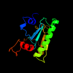
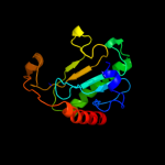
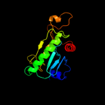
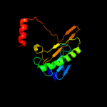
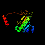
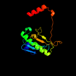
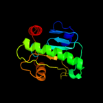
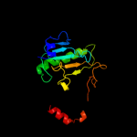
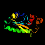
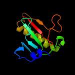
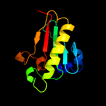
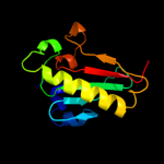
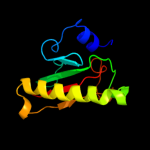
1 c1gupC_
100.0
100
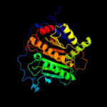
PDB header: nucleotidyltransferaseChain: C: PDB Molecule: galactose-1-phosphate uridylyltransferase;PDBTitle: structure of nucleotidyltransferase complexed with udp-2 galactose
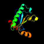
2 c1zwjA_
100.0
25
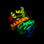
PDB header: structural genomics, unknown functionChain: A: PDB Molecule: putative galactose-1-phosphate uridyl transferase;PDBTitle: x-ray structure of galt-like protein from arabidopsis thaliana2 at5g18200
3 d1guqa1
100.0
99
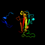
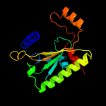
Fold: HIT-likeSuperfamily: HIT-likeFamily: Hexose-1-phosphate uridylyltransferase4 d1guqa2
100.0
100
Fold: HIT-likeSuperfamily: HIT-likeFamily: Hexose-1-phosphate uridylyltransferase5 d1z84a1
100.0
25
Fold: HIT-likeSuperfamily: HIT-likeFamily: Hexose-1-phosphate uridylyltransferase6 d1z84a2
100.0
24
Fold: HIT-likeSuperfamily: HIT-likeFamily: Hexose-1-phosphate uridylyltransferase7 c3lb5B_
99.9
19
PDB header: cell cycleChain: B: PDB Molecule: hit-like protein involved in cell-cycle regulation;PDBTitle: crystal structure of hit-like protein involved in cell-cycle2 regulation from bartonella henselae with unknown ligand
8 c3anoA_
99.9
18
PDB header: transferaseChain: A: PDB Molecule: ap-4-a phosphorylase;PDBTitle: crystal structure of a novel diadenosine 5',5'''-p1,p4-tetraphosphate2 phosphorylase from mycobacterium tuberculosis h37rv
9 c3o0mB_
99.9
17
PDB header: hydrolaseChain: B: PDB Molecule: hit family protein;PDBTitle: crystal structure of a zn-bound histidine triad family protein from2 mycobacterium smegmatis
10 c3l7xA_
99.9
19
PDB header: cell cycleChain: A: PDB Molecule: putative hit-like protein involved in cell-cyclePDBTitle: the crystal structure of smu.412c from streptococcus mutans ua159
11 c3ksvA_
99.9
15
PDB header: unknown functionChain: A: PDB Molecule: uncharacterized protein;PDBTitle: hypothetical protein from leishmania major
12 c3imiB_
99.9
17
PDB header: structural genomics, unknown functionChain: B: PDB Molecule: hit family protein;PDBTitle: 2.01 angstrom resolution crystal structure of a hit family protein2 from bacillus anthracis str. 'ames ancestor'
13 c3p0tB_
99.9
18
PDB header: unknown functionChain: B: PDB Molecule: uncharacterized protein;PDBTitle: crystal structure of an hit-like protein from mycobacterium2 paratuberculosis
14 d1y23a_
99.9
18
Fold: HIT-likeSuperfamily: HIT-likeFamily: HIT (HINT, histidine triad) family of protein kinase-interacting proteins15 c2eo4A_
99.9
18
PDB header: hydrolaseChain: A: PDB Molecule: 150aa long hypothetical histidine triad nucleotide-bindingPDBTitle: crystal structure of hypothetical histidine triad nucleotide-binding2 protein st2152 from sulfolobus tokodaii strain7
16 c3n1tE_
99.9
13
PDB header: hydrolaseChain: E: PDB Molecule: hit-like protein hint;PDBTitle: crystal structure of the h101a mutant echint gmp complex
17 d1xqua_
99.9
19
Fold: HIT-likeSuperfamily: HIT-likeFamily: HIT (HINT, histidine triad) family of protein kinase-interacting proteins18 c1xquA_
99.9
19
PDB header: hydrolaseChain: A: PDB Molecule: hit family hydrolase;PDBTitle: hit family hydrolase from clostridium thermocellum cth-393
19 d1rzya_
99.9
19
Fold: HIT-likeSuperfamily: HIT-likeFamily: HIT (HINT, histidine triad) family of protein kinase-interacting proteins20 d1fita_
99.9
21
Fold: HIT-likeSuperfamily: HIT-likeFamily: HIT (HINT, histidine triad) family of protein kinase-interacting proteins21 d1kpfa_
not modelled
99.9
18
Fold: HIT-likeSuperfamily: HIT-likeFamily: HIT (HINT, histidine triad) family of protein kinase-interacting proteins22 d2oika1
not modelled
99.9
16
Fold: HIT-likeSuperfamily: HIT-likeFamily: HIT (HINT, histidine triad) family of protein kinase-interacting proteins23 d1emsa1
not modelled
99.9
20
Fold: HIT-likeSuperfamily: HIT-likeFamily: HIT (HINT, histidine triad) family of protein kinase-interacting proteins24 c3r6fA_
not modelled
99.9
16
PDB header: hydrolaseChain: A: PDB Molecule: hit family protein;PDBTitle: crystal structure of a zinc-containing hit family protein from2 encephalitozoon cuniculi
25 c3oj7A_
not modelled
99.8
17
PDB header: metal binding proteinChain: A: PDB Molecule: putative histidine triad family protein;PDBTitle: crystal structure of a histidine triad family protein from entamoeba2 histolytica, bound to sulfate
26 c1emsB_
not modelled
99.8
19
PDB header: antitumor proteinChain: B: PDB Molecule: nit-fragile histidine triad fusion protein;PDBTitle: crystal structure of the c. elegans nitfhit protein
27 c3i24B_
not modelled
99.6
15
PDB header: hydrolaseChain: B: PDB Molecule: hit family hydrolase;PDBTitle: crystal structure of a hit family hydrolase protein from2 vibrio fischeri. northeast structural genomics consortium3 target id vfr176
28 c3nrdB_
not modelled
99.6
16
PDB header: nucleotide binding proteinChain: B: PDB Molecule: histidine triad (hit) protein;PDBTitle: crystal structure of a histidine triad (hit) protein (smc02904) from2 sinorhizobium meliloti 1021 at 2.06 a resolution
29 c3i4sB_
not modelled
99.6
17
PDB header: hydrolaseChain: B: PDB Molecule: histidine triad protein;PDBTitle: crystal structure of histidine triad protein blr8122 from2 bradyrhizobium japonicum
30 c3oheA_
not modelled
99.6
17
PDB header: hydrolaseChain: A: PDB Molecule: histidine triad (hit) protein;PDBTitle: crystal structure of a histidine triad protein (maqu_1709) from2 marinobacter aquaeolei vt8 at 1.20 a resolution
31 d3bl9a1
not modelled
98.7
18
Fold: HIT-likeSuperfamily: HIT-likeFamily: mRNA decapping enzyme DcpS C-terminal domain32 c3bl9B_
not modelled
98.6
18
PDB header: hydrolaseChain: B: PDB Molecule: scavenger mrna-decapping enzyme dcps;PDBTitle: synthetic gene encoded dcps bound to inhibitor dg157493
33 d1vlra1
not modelled
98.4
21
Fold: HIT-likeSuperfamily: HIT-likeFamily: mRNA decapping enzyme DcpS C-terminal domain34 c1xmlA_
not modelled
98.4
18
PDB header: chaperoneChain: A: PDB Molecule: heat shock-like protein 1;PDBTitle: structure of human dcps
35 d2pofa1
not modelled
85.2
14
Fold: HIT-likeSuperfamily: HIT-likeFamily: CDH-like36 c1m6vE_
not modelled
34.9
13
PDB header: ligaseChain: E: PDB Molecule: carbamoyl phosphate synthetase large chain;PDBTitle: crystal structure of the g359f (small subunit) point mutant of2 carbamoyl phosphate synthetase
37 d1vpra1
not modelled
22.3
60
Fold: LipocalinsSuperfamily: LipocalinsFamily: Dinoflagellate luciferase repeat38 c3bddD_
not modelled
22.3
27
PDB header: transcriptionChain: D: PDB Molecule: regulatory protein marr;PDBTitle: crystal structure of a putative multiple antibiotic-resistance2 repressor (ssu05_1136) from streptococcus suis 89/1591 at 2.20 a3 resolution
39 c3g2bA_
not modelled
19.5
28
PDB header: biosynthetic proteinChain: A: PDB Molecule: coenzyme pqq synthesis protein d;PDBTitle: crystal structure of pqqd from xanthomonas campestris
40 c3ohmB_
not modelled
18.2
25
PDB header: signaling protein / hydrolaseChain: B: PDB Molecule: 1-phosphatidylinositol-4,5-bisphosphate phosphodiesterasePDBTitle: crystal structure of activated g alpha q bound to its effector2 phospholipase c beta 3
41 d1zaka2
not modelled
15.2
56
Fold: Rubredoxin-likeSuperfamily: Microbial and mitochondrial ADK, insert "zinc finger" domainFamily: Microbial and mitochondrial ADK, insert "zinc finger" domain42 d1ukfa_
not modelled
14.7
19
Fold: Cysteine proteinasesSuperfamily: Cysteine proteinasesFamily: Avirulence protein Avrpph343 d1v58a1
not modelled
14.0
50
Fold: Thioredoxin foldSuperfamily: Thioredoxin-likeFamily: DsbC/DsbG C-terminal domain-like44 c1t3bA_
not modelled
13.8
38
PDB header: isomeraseChain: A: PDB Molecule: thiol:disulfide interchange protein dsbc;PDBTitle: x-ray structure of dsbc from haemophilus influenzae
45 d1qasa3
not modelled
13.5
27
Fold: TIM beta/alpha-barrelSuperfamily: PLC-like phosphodiesterasesFamily: Mammalian PLC46 c1v57A_
not modelled
13.0
50
PDB header: isomeraseChain: A: PDB Molecule: thiol:disulfide interchange protein dsbg;PDBTitle: crystal structure of the disulfide bond isomerase dsbg
47 c1jzdA_
not modelled
12.4
18
PDB header: oxidoreductaseChain: A: PDB Molecule: thiol:disulfide interchange protein dsbc;PDBTitle: dsbc-dsbdalpha complex
48 d2nvna1
not modelled
11.6
18
Fold: ssDNA-binding transcriptional regulator domainSuperfamily: ssDNA-binding transcriptional regulator domainFamily: PMN2A0962/syc2379c-like49 c2rodB_
not modelled
10.9
24
PDB header: apoptosisChain: B: PDB Molecule: noxa;PDBTitle: solution structure of mcl-1 complexed with noxaa
50 d1rk4a2
not modelled
10.7
17
Fold: Thioredoxin foldSuperfamily: Thioredoxin-likeFamily: Glutathione S-transferase (GST), N-terminal domain51 d2psba1
not modelled
10.6
33
Fold: YerB-likeSuperfamily: YerB-likeFamily: YerB-like52 c2psbA_
not modelled
10.6
33
PDB header: structural genomics, unknown functionChain: A: PDB Molecule: yerb protein;PDBTitle: crystal structure of yerb protein from bacillus subtilis.2 northeast structural genomics target sr586
53 d1eeja1
not modelled
10.6
38
Fold: Thioredoxin foldSuperfamily: Thioredoxin-likeFamily: DsbC/DsbG C-terminal domain-like54 c1hyuA_
not modelled
10.5
25
PDB header: oxidoreductaseChain: A: PDB Molecule: alkyl hydroperoxide reductase subunit f;PDBTitle: crystal structure of intact ahpf
55 c2hwtA_
not modelled
10.4
19
PDB header: replication, hydrolaseChain: A: PDB Molecule: putative replicase-associated protein;PDBTitle: nmr solution structure of the master-rep protein nuclease2 domain (2-95) from the faba bean necrotic yellows virus
56 d1ji8a_
not modelled
10.4
11
Fold: DsrC, the gamma subunit of dissimilatory sulfite reductaseSuperfamily: DsrC, the gamma subunit of dissimilatory sulfite reductaseFamily: DsrC, the gamma subunit of dissimilatory sulfite reductase57 d2v4jc1
not modelled
9.7
11
Fold: DsrC, the gamma subunit of dissimilatory sulfite reductaseSuperfamily: DsrC, the gamma subunit of dissimilatory sulfite reductaseFamily: DsrC, the gamma subunit of dissimilatory sulfite reductase58 d2drpa2
not modelled
9.6
50
Fold: beta-beta-alpha zinc fingersSuperfamily: beta-beta-alpha zinc fingersFamily: Classic zinc finger, C2H259 c2elpA_
not modelled
9.0
22
PDB header: transcriptionChain: A: PDB Molecule: zinc finger protein 406;PDBTitle: solution structure of the 13th c2h2 zinc finger of human2 zinc finger protein 406
60 d1t3ba1
not modelled
8.4
38
Fold: Thioredoxin foldSuperfamily: Thioredoxin-likeFamily: DsbC/DsbG C-terminal domain-like61 c3ghaA_
not modelled
8.1
26
PDB header: oxidoreductaseChain: A: PDB Molecule: disulfide bond formation protein d;PDBTitle: crystal structure of etda-treated bdbd (reduced)
62 c3gv1A_
not modelled
8.1
63
PDB header: structural genomics, unknown functionChain: A: PDB Molecule: disulfide interchange protein;PDBTitle: crystal structure of disulfide interchange protein from neisseria2 gonorrhoeae
63 d1dqwa_
not modelled
8.0
20
Fold: TIM beta/alpha-barrelSuperfamily: Ribulose-phoshate binding barrelFamily: Decarboxylase64 d1ee8a1
not modelled
7.9
21
Fold: S13-like H2TH domainSuperfamily: S13-like H2TH domainFamily: Middle domain of MutM-like DNA repair proteins65 d1igna2
not modelled
7.9
11
Fold: DNA/RNA-binding 3-helical bundleSuperfamily: Homeodomain-likeFamily: DNA-binding domain of rap166 d1k3sa_
not modelled
7.7
20
Fold: Secretion chaperone-likeSuperfamily: Type III secretory system chaperone-likeFamily: Type III secretory system chaperone67 c3c6cA_
not modelled
7.7
42
PDB header: hydrolaseChain: A: PDB Molecule: 3-keto-5-aminohexanoate cleavage enzyme;PDBTitle: crystal structure of a putative 3-keto-5-aminohexanoate cleavage2 enzyme (reut_c6226) from ralstonia eutropha jmp134 at 1.72 a3 resolution
68 c3lotC_
not modelled
7.7
36
PDB header: structure genomics, unknown functionChain: C: PDB Molecule: uncharacterized protein;PDBTitle: crystal structure of protein of unknown function (np_070038.1) from2 archaeoglobus fulgidus at 1.89 a resolution
69 c3e49A_
not modelled
7.1
36
PDB header: metal binding proteinChain: A: PDB Molecule: uncharacterized protein duf849 with a tim barrel fold;PDBTitle: crystal structure of a prokaryotic domain of unknown function (duf849)2 with a tim barrel fold (bxe_c0966) from burkholderia xenovorans lb4003 at 1.75 a resolution
70 c3e02A_
not modelled
6.9
33
PDB header: metal binding proteinChain: A: PDB Molecule: uncharacterized protein duf849;PDBTitle: crystal structure of a duf849 family protein (bxe_c0271) from2 burkholderia xenovorans lb400 at 1.90 a resolution
71 c3no5C_
not modelled
6.8
27
PDB header: structural genomics, unknown functionChain: C: PDB Molecule: uncharacterized protein;PDBTitle: crystal structure of a pfam duf849 domain containing protein2 (reut_a1631) from ralstonia eutropha jmp134 at 1.90 a resolution
72 d2fbka1
not modelled
6.2
8
Fold: DNA/RNA-binding 3-helical bundleSuperfamily: "Winged helix" DNA-binding domainFamily: MarR-like transcriptional regulators73 c3chvA_
not modelled
6.2
27
PDB header: metal binding proteinChain: A: PDB Molecule: prokaryotic domain of unknown function (duf849) with a timPDBTitle: crystal structure of a prokaryotic domain of unknown function (duf849)2 member (spoa0042) from silicibacter pomeroyi dss-3 at 1.45 a3 resolution
74 c3gn3B_
not modelled
6.2
25
PDB header: structural genomics, unknown functionChain: B: PDB Molecule: putative protein-disulfide isomerase;PDBTitle: crystal structure of a putative protein-disulfide isomerase from2 pseudomonas syringae to 2.5a resolution.
75 d1aisa1
not modelled
6.1
15
Fold: TBP-likeSuperfamily: TATA-box binding protein-likeFamily: TATA-box binding protein (TBP), C-terminal domain76 c3nznA_
not modelled
6.1
25
PDB header: oxidoreductaseChain: A: PDB Molecule: glutaredoxin;PDBTitle: the crystal structure of the glutaredoxin from methanosarcina mazei2 go1
77 c2fd4A_
not modelled
6.1
25
PDB header: ligaseChain: A: PDB Molecule: avirulence protein avrptob;PDBTitle: crystal structure of avrptob (436-553)
78 c3nqnB_
not modelled
6.0
47
PDB header: structural genomics, unknown functionChain: B: PDB Molecule: uncharacterized protein;PDBTitle: crystal structure of a protein with unknown function. (dr_2006) from2 deinococcus radiodurans at 1.88 a resolution
79 c2ab3A_
not modelled
5.9
45
PDB header: rna binding proteinChain: A: PDB Molecule: znf29;PDBTitle: solution structures and characterization of hiv rre iib rna2 targeting zinc finger proteins
80 d1wika_
not modelled
5.9
11
Fold: Thioredoxin foldSuperfamily: Thioredoxin-likeFamily: Thioltransferase81 d2csta_
not modelled
5.7
7
Fold: PLP-dependent transferase-likeSuperfamily: PLP-dependent transferasesFamily: AAT-like82 c3oopA_
not modelled
5.7
13
PDB header: structural genomics, unknown functionChain: A: PDB Molecule: lin2960 protein;PDBTitle: the structure of a protein with unknown function from listeria innocua2 clip11262
83 d2fiya1
not modelled
5.6
22
Fold: FdhE-likeSuperfamily: FdhE-likeFamily: FdhE-like84 c2y7eA_
not modelled
5.6
27
PDB header: lyaseChain: A: PDB Molecule: 3-keto-5-aminohexanoate cleavage enzyme;PDBTitle: crystal structure of the 3-keto-5-aminohexanoate cleavage enzyme2 (kce) from candidatus cloacamonas acidaminovorans (tetragonal form)
85 d1dl0a_
not modelled
5.5
50
Fold: Knottins (small inhibitors, toxins, lectins)Superfamily: omega toxin-likeFamily: Spider toxins86 c1dl0A_
not modelled
5.5
50
PDB header: toxinChain: A: PDB Molecule: j-atracotoxin-hv1c;PDBTitle: solution structure of the insecticidal neurotoxin j-2 atracotoxin-hv1c
87 d1fm4a_
not modelled
5.5
16
Fold: TBP-likeSuperfamily: Bet v1-likeFamily: Pathogenesis-related protein 10 (PR10)-like88 c2hw0A_
not modelled
5.4
28
PDB header: hydrolase, replicationChain: A: PDB Molecule: replicase;PDBTitle: nmr solution structure of the nuclease domain from the2 replicator initiator protein from porcine circovirus pcv2
89 d1s6la1
not modelled
5.4
26
Fold: DNA/RNA-binding 3-helical bundleSuperfamily: "Winged helix" DNA-binding domainFamily: MerB N-terminal domain-like90 d1qnaa1
not modelled
5.4
12
Fold: TBP-likeSuperfamily: TATA-box binding protein-likeFamily: TATA-box binding protein (TBP), C-terminal domain91 c1zypB_
not modelled
5.3
25
PDB header: oxidoreductaseChain: B: PDB Molecule: alkyl hydroperoxide reductase subunit f;PDBTitle: synchrotron reduced form of the n-terminal domain of2 salmonella typhimurium ahpf
92 d1egoa_
not modelled
5.3
25
Fold: Thioredoxin foldSuperfamily: Thioredoxin-likeFamily: Thioltransferase93 d1k0ma2
not modelled
5.1
17
Fold: Thioredoxin foldSuperfamily: Thioredoxin-likeFamily: Glutathione S-transferase (GST), N-terminal domain94 d1t3ca_
not modelled
5.1
25
Fold: Zincin-likeSuperfamily: Metalloproteases ("zincins"), catalytic domainFamily: Clostridium neurotoxins, catalytic domain95 d1vr7a1
not modelled
5.1
16
Fold: S-adenosylmethionine decarboxylaseSuperfamily: S-adenosylmethionine decarboxylaseFamily: Bacterial S-adenosylmethionine decarboxylase


