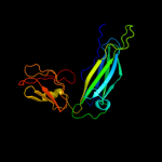
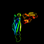
1 c1z9sA_
100.0
29
PDB header: chaperone/immune systemChain: A: PDB Molecule: chaperone protein caf1m;PDBTitle: crystal structure of the native chaperone:subunit:subunit2 caf1m:caf1:caf1 complex
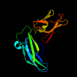
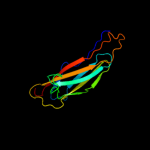
2 c1qunA_
100.0
30
PDB header: chaperone/structural proteinChain: A: PDB Molecule: papd-like chaperone fimc;PDBTitle: x-ray structure of the fimc-fimh chaperone adhesin complex2 from uropathogenic e.coli
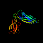
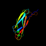
3 c2co7B_
100.0
21
PDB header: fibril proteinChain: B: PDB Molecule: putative fimbriae assembly chaperone;PDBTitle: salmonella enterica safa pilin in complex with the safb2 chaperone (type ii)
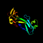
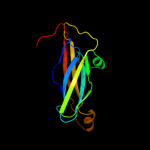
4 c3q48B_
100.0
33
PDB header: chaperoneChain: B: PDB Molecule: chaperone cupb2;PDBTitle: crystal structure of pseudomonas aeruginosa cupb2 chaperone
5 c1qpxA_
100.0
31
PDB header: chaperoneChain: A: PDB Molecule: papd chaperone;PDBTitle: crystal structures of self-capping papd chaperone homodimers
6 c1l4iA_
100.0
32
PDB header: chaperoneChain: A: PDB Molecule: sfae protein;PDBTitle: crystal structure of the periplasmic chaperone sfae
7 c3f6iB_
100.0
24
PDB header: chaperoneChain: B: PDB Molecule: chaperone protein faee;PDBTitle: structure of the semet labeled f4 fibrial chaperone faee
8 d2co7b1
100.0
26
Fold: Immunoglobulin-like beta-sandwichSuperfamily: PapD-likeFamily: Pilus chaperone9 d1p5va1
100.0
35
Fold: Immunoglobulin-like beta-sandwichSuperfamily: PapD-likeFamily: Pilus chaperone10 d2j2za1
100.0
36
Fold: Immunoglobulin-like beta-sandwichSuperfamily: PapD-likeFamily: Pilus chaperone11 d3bwuc1
100.0
38
Fold: Immunoglobulin-like beta-sandwichSuperfamily: PapD-likeFamily: Pilus chaperone12 d1l4ia1
100.0
36
Fold: Immunoglobulin-like beta-sandwichSuperfamily: PapD-likeFamily: Pilus chaperone13 d3bwuc2
99.7
22
Fold: C2 domain-likeSuperfamily: Periplasmic chaperone C-domainFamily: Periplasmic chaperone C-domain14 d1p5va2
99.7
18
Fold: C2 domain-likeSuperfamily: Periplasmic chaperone C-domainFamily: Periplasmic chaperone C-domain15 d2co7b2
99.6
15
Fold: C2 domain-likeSuperfamily: Periplasmic chaperone C-domainFamily: Periplasmic chaperone C-domain16 d1l4ia2
99.6
25
Fold: C2 domain-likeSuperfamily: Periplasmic chaperone C-domainFamily: Periplasmic chaperone C-domain17 d2j2za2
99.6
27
Fold: C2 domain-likeSuperfamily: Periplasmic chaperone C-domainFamily: Periplasmic chaperone C-domain18 d1m1sa_
97.8
13
Fold: Immunoglobulin-like beta-sandwichSuperfamily: PapD-likeFamily: MSP-like19 d1rowa_
97.3
13
Fold: Immunoglobulin-like beta-sandwichSuperfamily: PapD-likeFamily: MSP-like20 d1grwa_
97.2
9
Fold: Immunoglobulin-like beta-sandwichSuperfamily: PapD-likeFamily: MSP-like21 c2qsvA_
not modelled
96.8
13
PDB header: structural genomics, unknown functionChain: A: PDB Molecule: uncharacterized protein;PDBTitle: crystal structure of protein of unknown function from porphyromonas2 gingivalis w83
22 c1z9oB_
not modelled
96.5
15
PDB header: protein binding/lipid binding proteinChain: B: PDB Molecule: vesicle-associated membrane protein-associated protein a;PDBTitle: 1.9 angstrom crystal structure of the rat vap-a msp homology domain in2 complex with the rat orp1 ffat motif
23 d1mspa_
not modelled
95.8
11
Fold: Immunoglobulin-like beta-sandwichSuperfamily: PapD-likeFamily: MSP-like24 c2ys4A_
not modelled
95.0
16
PDB header: structural genomics, unknown functionChain: A: PDB Molecule: hydrocephalus-inducing protein homolog;PDBTitle: solution structure of the n-terminal papd-like domain of2 hydin protein from human
25 c2e6jA_
not modelled
94.5
20
PDB header: structural genomics, unknown functionChain: A: PDB Molecule: hydin protein;PDBTitle: solution structure of the c-terminal papd-like domain from2 human hydin protein
26 d1wica_
not modelled
93.6
9
Fold: Immunoglobulin-like beta-sandwichSuperfamily: PapD-likeFamily: MSP-like27 c3qisA_
not modelled
92.3
9
PDB header: hydrolase/protein bindingChain: A: PDB Molecule: inositol polyphosphate 5-phosphatase ocrl-1;PDBTitle: recognition of the f&h motif by the lowe syndrome protein ocrl
28 c3o0lB_
not modelled
91.2
13
PDB header: structural genomics, unknown functionChain: B: PDB Molecule: uncharacterized protein;PDBTitle: crystal structure of a pfam duf1425 family member (shew_1734) from2 shewanella sp. pv-4 at 1.81 a resolution
29 c3qbtH_
not modelled
90.9
9
PDB header: protein transport/hydrolaseChain: H: PDB Molecule: inositol polyphosphate 5-phosphatase ocrl-1;PDBTitle: crystal structure of ocrl1 540-678 in complex with rab8a:gppnhp
30 d2vzsa2
not modelled
90.8
12
Fold: Immunoglobulin-like beta-sandwichSuperfamily: beta-Galactosidase/glucuronidase domainFamily: beta-Galactosidase/glucuronidase domain31 c3ac0B_
not modelled
88.8
11
PDB header: hydrolaseChain: B: PDB Molecule: beta-glucosidase i;PDBTitle: crystal structure of beta-glucosidase from kluyveromyces marxianus in2 complex with glucose
32 c2x41A_
not modelled
87.2
11
PDB header: hydrolaseChain: A: PDB Molecule: beta-glucosidase;PDBTitle: structure of beta-glucosidase 3b from thermotoga neapolitana2 in complex with glucose
33 c2je8B_
not modelled
83.2
12
PDB header: hydrolaseChain: B: PDB Molecule: beta-mannosidase;PDBTitle: structure of a beta-mannosidase from bacteroides2 thetaiotaomicron
34 d4ubpb_
not modelled
76.9
11
Fold: beta-clipSuperfamily: Urease, beta-subunitFamily: Urease, beta-subunit35 c3ginB_
not modelled
75.6
9
PDB header: metal binding proteinChain: B: PDB Molecule: sodium/calcium exchanger 1;PDBTitle: crystal structure of e454k-cbd1
36 d1ejxb_
not modelled
72.8
8
Fold: beta-clipSuperfamily: Urease, beta-subunitFamily: Urease, beta-subunit37 d1e9ya1
not modelled
71.0
11
Fold: beta-clipSuperfamily: Urease, beta-subunitFamily: Urease, beta-subunit38 c3qgaD_
not modelled
65.2
15
PDB header: hydrolaseChain: D: PDB Molecule: fusion of urease beta and gamma subunits;PDBTitle: 3.0 a model of iron containing urease urea2b2 from helicobacter2 mustelae
39 d2dpka1
not modelled
62.8
10
Fold: Immunoglobulin-like beta-sandwichSuperfamily: CalX-likeFamily: CalX-beta domain40 c2qvkA_
not modelled
58.6
11
PDB header: metal binding proteinChain: A: PDB Molecule: sodium/calcium exchanger 1;PDBTitle: the second ca2+-binding domain of the na+-ca2+ exchanger is2 essential for regulation: crystal structures and3 mutational analysis
41 d1ufga_
not modelled
56.4
16
Fold: Immunoglobulin-like beta-sandwichSuperfamily: Lamin A/C globular tail domainFamily: Lamin A/C globular tail domain42 d1e42a1
not modelled
55.9
25
Fold: Immunoglobulin-like beta-sandwichSuperfamily: Clathrin adaptor appendage domainFamily: Alpha-adaptin ear subdomain-like43 c1e9zA_
not modelled
55.9
11
PDB header: hydrolaseChain: A: PDB Molecule: urease subunit alpha;PDBTitle: crystal structure of helicobacter pylori urease
44 d1k3ra1
not modelled
49.1
18
Fold: OB-foldSuperfamily: Nucleic acid-binding proteinsFamily: Hypothetical protein MTH1 (MT0001), insert domain45 c2lllA_
not modelled
49.0
21
PDB header: structural proteinChain: A: PDB Molecule: lamin-b2;PDBTitle: solution nmr structure of c-terminal globular domain of human lamin-2 b2, northeast structural genomics consortium target hr8546a
46 c2f1eA_
not modelled
48.5
17
PDB header: structural genomics, unknown functionChain: A: PDB Molecule: protein apag;PDBTitle: solution structure of apag protein
47 c3jt0B_
not modelled
48.2
11
PDB header: structural proteinChain: B: PDB Molecule: lamin-b1;PDBTitle: crystal structure of the c-terminal fragment (426-558)2 lamin-b1 from homo sapiens, northeast structural genomics3 consortium target hr5546a
48 c3eujB_
not modelled
45.5
20
PDB header: cell cycleChain: B: PDB Molecule: chromosome partition protein mukf;PDBTitle: crystal structure of muke-mukf(residues 292-443)-mukb(head2 domain)-atpgammas complex, symmetric dimer
49 c3rb7E_
not modelled
45.2
10
PDB header: metal binding proteinChain: E: PDB Molecule: na/ca exchange protein;PDBTitle: crystal structure of cbd12 from calx1.2
50 c1e42A_
not modelled
37.6
23
PDB header: endocytosisChain: A: PDB Molecule: ap-2 complex subunit beta;PDBTitle: beta2-adaptin appendage domain, from clathrin adaptor ap2
51 d1aoza2
not modelled
36.3
6
Fold: Cupredoxin-likeSuperfamily: CupredoxinsFamily: Multidomain cupredoxins52 d1w8oa1
not modelled
33.3
12
Fold: Immunoglobulin-like beta-sandwichSuperfamily: E set domainsFamily: E-set domains of sugar-utilizing enzymes53 d1tzaa_
not modelled
31.2
17
Fold: Immunoglobulin-like beta-sandwichSuperfamily: ApaG-likeFamily: ApaG-like54 d1ifra_
not modelled
30.7
16
Fold: Immunoglobulin-like beta-sandwichSuperfamily: Lamin A/C globular tail domainFamily: Lamin A/C globular tail domain55 d1hfua2
not modelled
30.4
6
Fold: Cupredoxin-likeSuperfamily: CupredoxinsFamily: Multidomain cupredoxins56 c2r39A_
not modelled
30.3
3
PDB header: structural genomics, unknown functionChain: A: PDB Molecule: fixg-related protein;PDBTitle: crystal structure of fixg-related protein from vibrio parahaemolyticus
57 d2fwua1
not modelled
26.2
10
Fold: Immunoglobulin-like beta-sandwichSuperfamily: CalX-likeFamily: CalX-beta domain58 c1yycA_
not modelled
25.7
14
PDB header: structural genomics, unknown functionChain: A: PDB Molecule: putative late embryogenesis abundant protein;PDBTitle: solution structure of a putative late embryogenesis2 abundant (lea) protein at2g46140.1
59 c3isyA_
not modelled
24.0
12
PDB header: protein bindingChain: A: PDB Molecule: intracellular proteinase inhibitor;PDBTitle: crystal structure of an intracellular proteinase inhibitor (ipi,2 bsu11130) from bacillus subtilis at 2.61 a resolution
60 d1kyaa2
not modelled
23.7
4
Fold: Cupredoxin-likeSuperfamily: CupredoxinsFamily: Multidomain cupredoxins61 c3cfuA_
not modelled
23.2
11
PDB header: lipoproteinChain: A: PDB Molecule: uncharacterized lipoprotein yjha;PDBTitle: crystal structure of the yjha protein from bacillus2 subtilis. northeast structural genomics consortium target3 sr562
62 d1xo8a_
not modelled
22.4
7
Fold: Immunoglobulin-like beta-sandwichSuperfamily: LEA14-likeFamily: LEA14-like63 d1v8ha1
not modelled
22.2
19
Fold: Immunoglobulin-like beta-sandwichSuperfamily: E set domainsFamily: SoxZ-like64 d1v10a2
not modelled
21.0
10
Fold: Cupredoxin-likeSuperfamily: CupredoxinsFamily: Multidomain cupredoxins65 c3butA_
not modelled
20.0
19
PDB header: structural genomics, unknown functionChain: A: PDB Molecule: uncharacterized protein af_0446;PDBTitle: crystal structure of protein af_0446 from archaeoglobus fulgidus
66 c2vzvB_
not modelled
17.8
13
PDB header: hydrolaseChain: B: PDB Molecule: exo-beta-d-glucosaminidase;PDBTitle: substrate complex of amycolatopsis orientalis exo-2 chitosanase csxa e541a with chitosan
67 d1ivta_
not modelled
17.1
16
Fold: Immunoglobulin-like beta-sandwichSuperfamily: Lamin A/C globular tail domainFamily: Lamin A/C globular tail domain68 d1gyca2
not modelled
16.1
8
Fold: Cupredoxin-likeSuperfamily: CupredoxinsFamily: Multidomain cupredoxins69 d1xq4a_
not modelled
16.0
11
Fold: Immunoglobulin-like beta-sandwichSuperfamily: ApaG-likeFamily: ApaG-like70 d1jz8a2
not modelled
15.7
12
Fold: Immunoglobulin-like beta-sandwichSuperfamily: beta-Galactosidase/glucuronidase domainFamily: beta-Galactosidase/glucuronidase domain71 d1v7wa1
not modelled
15.1
12
Fold: alpha/alpha toroidSuperfamily: Six-hairpin glycosidasesFamily: Glycosyltransferase family 36 C-terminal domain72 d1ahsa_
not modelled
14.8
25
Fold: Viral protein domainSuperfamily: Viral protein domainFamily: Top domain of virus capsid protein73 c2l02B_
not modelled
14.6
17
PDB header: structural genomics, unknown functionChain: B: PDB Molecule: uncharacterized protein;PDBTitle: solution nmr structure of protein bt2368 from bacteroides2 thetaiotaomicron, northeast structural genomics consortium target3 btr375
74 c1l9mB_
not modelled
14.4
10
PDB header: transferaseChain: B: PDB Molecule: protein-glutamine glutamyltransferase e3;PDBTitle: three-dimensional structure of the human transglutaminase 32 enzyme: binding of calcium ions change structure for3 activation
75 c1wkwB_
not modelled
13.7
67
PDB header: translation/protein bindingChain: B: PDB Molecule: eukaryotic translation initiation factor 4ePDBTitle: crystal structure of the ternary complex of eif4e-m7gpppa-2 4ebp1 peptide
76 d1ex0a2
not modelled
12.8
9
Fold: Immunoglobulin-like beta-sandwichSuperfamily: Transglutaminase, two C-terminal domainsFamily: Transglutaminase, two C-terminal domains77 c2l0dA_
not modelled
12.4
21
PDB header: cell adhesionChain: A: PDB Molecule: cell surface protein;PDBTitle: solution nmr structure of putative cell surface protein ma_4588 (272-2 376 domain) from methanosarcina acetivorans, northeast structural3 genomics consortium target mvr254a
78 d2pwwa1
not modelled
11.9
17
Fold: TBP-likeSuperfamily: YugN-likeFamily: YugN-like79 c3h6aB_
not modelled
11.8
6
PDB header: cell adhesionChain: B: PDB Molecule: integrin beta-4;PDBTitle: structure of the calx-beta domain of integrin beta42 crystallized in the presence of calcium
80 d1cuoa_
not modelled
11.5
21
Fold: Cupredoxin-likeSuperfamily: CupredoxinsFamily: Plastocyanin/azurin-like81 c3k6sB_
not modelled
11.1
11
PDB header: cell adhesionChain: B: PDB Molecule: integrin beta-2;PDBTitle: structure of integrin alphaxbeta2 ectodomain
82 d1xvsa_
not modelled
10.5
19
Fold: Immunoglobulin-like beta-sandwichSuperfamily: ApaG-likeFamily: ApaG-like83 d1hmja_
not modelled
9.8
23
Fold: RPB5-like RNA polymerase subunitSuperfamily: RPB5-like RNA polymerase subunitFamily: RPB584 d2ccwa1
not modelled
9.7
14
Fold: Cupredoxin-likeSuperfamily: CupredoxinsFamily: Plastocyanin/azurin-like85 c3qe5A_
not modelled
9.3
17
PDB header: cell adhesionChain: A: PDB Molecule: major cell-surface adhesin pac;PDBTitle: complete structure of streptococcus mutans antigen i/ii carboxy-2 terminus
86 c2h47C_
not modelled
9.3
17
PDB header: oxidoreductase/electron transportChain: C: PDB Molecule: azurin;PDBTitle: crystal structure of an electron transfer complex between2 aromatic amine dephydrogenase and azurin from alcaligenes3 faecalis (form 1)
87 d1vjja2
not modelled
9.2
5
Fold: Immunoglobulin-like beta-sandwichSuperfamily: Transglutaminase, two C-terminal domainsFamily: Transglutaminase, two C-terminal domains88 c2f7fA_
not modelled
9.2
13
PDB header: transferaseChain: A: PDB Molecule: nicotinate phosphoribosyltransferase, putative;PDBTitle: crystal structure of enterococcus faecalis putative nicotinate2 phosphoribosyltransferase, new york structural genomics consortium
89 d1cc3a_
not modelled
9.1
21
Fold: Cupredoxin-likeSuperfamily: CupredoxinsFamily: Plastocyanin/azurin-like90 c2kl6A_
not modelled
8.9
12
PDB header: structural genomics, unknown functionChain: A: PDB Molecule: uncharacterized protein;PDBTitle: solution nmr structure of the cardb domain of pf1109 from2 pyrococcus furiosus. northeast structural genomics3 consortium target pfr193a
91 c1v7wA_
not modelled
8.8
12
PDB header: transferaseChain: A: PDB Molecule: chitobiose phosphorylase;PDBTitle: crystal structure of vibrio proteolyticus chitobiose phosphorylase in2 complex with glcnac
92 d1g0da2
not modelled
8.7
15
Fold: Immunoglobulin-like beta-sandwichSuperfamily: Transglutaminase, two C-terminal domainsFamily: Transglutaminase, two C-terminal domains93 c2qziA_
not modelled
8.5
13
PDB header: structural genomics, unknown functionChain: A: PDB Molecule: uncharacterized protein;PDBTitle: the crystal structure of a conserved protein of unknown function from2 streptococcus thermophilus lmg 18311.
94 d2q9oa2
not modelled
8.5
9
Fold: Cupredoxin-likeSuperfamily: CupredoxinsFamily: Multidomain cupredoxins95 c2ii8F_
not modelled
8.0
16
PDB header: signaling proteinChain: F: PDB Molecule: anabaena sensory rhodopsin transducer protein;PDBTitle: anabaena sensory rhodopsin transducer
96 c3rfrI_
not modelled
7.9
18
PDB header: oxidoreductaseChain: I: PDB Molecule: pmob;PDBTitle: crystal structure of particulate methane monooxygenase (pmmo) from2 methylocystis sp. strain m
97 d1dd1a_
not modelled
7.9
15
Fold: SMAD/FHA domainSuperfamily: SMAD/FHA domainFamily: SMAD domain98 c3tqzA_
not modelled
7.8
18
PDB header: hydrolaseChain: A: PDB Molecule: deoxyuridine 5'-triphosphate nucleotidohydrolase;PDBTitle: structure of a deoxyuridine 5'-triphosphate nucleotidohydrolase (dut)2 from coxiella burnetii
99 d1jzga_
not modelled
7.8
21
Fold: Cupredoxin-likeSuperfamily: CupredoxinsFamily: Plastocyanin/azurin-like