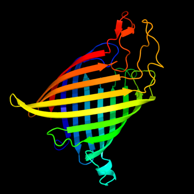
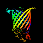
Phyre Home Retrieve Phyre Job Id
Emaill.a.kelley@imperial.ac.uk
DescriptionP0A927
DateThu Jan 5 11:09:14 GMT 2012
Unique Job IDb8a24512c3d908f0

Summary 

Top model
Image coloured by rainbow N → C terminus
Model (left) based on template d1tlya_
Top template information
Fold:Transmembrane beta-barrels
Superfamily:Tsx-like channel
Family:Tsx-like channel
Confidence and coverage
Confidence:100.0% Coverage: 84%
248 residues ( 84% of your sequence) have been modelled with 100.0% confidence by the single highest scoring template.
3D viewing
Interactive 3D view in Jmol

Sequence analysis 

Secondary structure and disorder prediction 

   1........10.........20.........30.........40.........50.........60
Sequence  MKKTLLAAGAVLALSSSFTVNAAENDKPQYLSDWWHQSVNVVGSYHTRFGPQIRNDTYLE
Secondary structure 


SS confidence 


Disorder  ??


??????????????


?????


?


Disorder confidence 


   .........70.........80.........90.........100.........110.........120
Sequence  YEAFAKKDWFDFYGYADAPVFFGGNSDAKGIWNHGSPLFMEIEPRFSIDKLTNTDLSFGP
Secondary structure 


SS confidence 


Disorder 


????????????


?


Disorder confidence 


   .........130.........140.........150.........160.........170.........180
Sequence  FKEWYFANNYIYDMGRNKDGRQSTWYMGLGTDIDTGLPMSLSMNVYAKYQWQNYGAANEN
Secondary structure 


SS confidence 


Disorder 


??

????
?


??????????
Disorder confidence 


   .........190.........200.........210.........220.........230.........240
Sequence  EWDGYRFKIKYFVPITDLWGGQLSYIGFTNFDWGSDLGDDSGNAINGIKTRTNNSIASSH
Secondary structure 


SS confidence 


Disorder  ???


???????????
?
????


Disorder confidence 


   .........250.........260.........270.........280.........290....
Sequence  ILALNYDHWHYSVVARYWHDGGQWNDDAELNFGNGNFNVRSTGWGGYLVVGYNF
Secondary structure 


SS confidence 


Disorder 


????


?

?

??


??
Disorder confidence 


Confidence Key
High(9)                    Low (0)
?Disordered
Alpha helix
Beta strand

Domain analysis 

Hover over an aligned region to see model and summary info

Please note, only up to the top 20 hits are modelled to reduce computer load

RankAligned region

PDB 1tly chain A

3D model

Region: 31 - 294
Aligned: 248
Modelled: 248
Confidence: 100.0%
Identity: 100%
Fold: Transmembrane beta-barrels
Superfamily: Tsx-like channel
Family: Tsx-like channel

Phyre2

PDB 2e9x chain B domain 2

3D model

Region: 101 - 122
Aligned: 22
Modelled: 22
Confidence: 20.4%
Identity: 41%
Fold: GINS/PriA/YqbF domain
Superfamily: PriA/YqbF domain
Family: PSF2 N-terminal domain-like

Phyre2

PDB 2jna chain A domain 1

3D model

Region: 1 - 42
Aligned: 41
Modelled: 42
Confidence: 20.3%
Identity: 39%
Fold: Dodecin subunit-like
Superfamily: YdgH-like
Family: YdgH-like

Phyre2

PDB 3epz chain A

3D model

Region: 116 - 127
Aligned: 12
Modelled: 12
Confidence: 20.2%
Identity: 33%
PDB header:transferase
Chain: A: PDB Molecule:dna (cytosine-5)-methyltransferase 1;
PDBTitle: structure of the replication foci-targeting sequence of human dna2 cytosine methyltransferase dnmt1

Phyre2

PDB 3f09 chain B

3D model

Region: 143 - 153
Aligned: 11
Modelled: 11
Confidence: 18.9%
Identity: 27%
PDB header:transferase
Chain: B: PDB Molecule:holo-[acyl-carrier-protein] synthase;
PDBTitle: 1.82 angstrom resolution crystal structure of holo-(acyl-carrier-2 protein) synthase (acps) from staphylococcus aureus

Phyre2

PDB 1u8c chain B

3D model

Region: 36 - 88
Aligned: 45
Modelled: 53
Confidence: 17.7%
Identity: 16%
PDB header:cell adhesion
Chain: B: PDB Molecule:integrin beta-3;
PDBTitle: a novel adaptation of the integrin psi domain revealed from its2 crystal structure

Phyre2

PDB 3n6r chain K

3D model

Region: 93 - 116
Aligned: 24
Modelled: 24
Confidence: 16.7%
Identity: 25%
PDB header:ligase
Chain: K: PDB Molecule:propionyl-coa carboxylase, alpha subunit;
PDBTitle: crystal structure of the holoenzyme of propionyl-coa carboxylase (pcc)

Phyre2

PDB 2e9x chain F

3D model

Region: 101 - 122
Aligned: 22
Modelled: 22
Confidence: 15.5%
Identity: 41%
PDB header:replication
Chain: F: PDB Molecule:dna replication complex gins protein psf2;
PDBTitle: the crystal structure of human gins core complex

Phyre2

PDB 2j9g chain A domain 3

3D model

Region: 93 - 116
Aligned: 24
Modelled: 24
Confidence: 14.0%
Identity: 29%
Fold: ATP-grasp
Superfamily: Glutathione synthetase ATP-binding domain-like
Family: BC ATP-binding domain-like

Phyre2

PDB 3u9s chain E

3D model

Region: 93 - 116
Aligned: 24
Modelled: 24
Confidence: 13.7%
Identity: 33%
PDB header:ligase
Chain: E: PDB Molecule:methylcrotonyl-coa carboxylase, alpha-subunit;
PDBTitle: crystal structure of p. aeruginosa 3-methylcrotonyl-coa carboxylase2 (mcc) 750 kd holoenzyme, coa complex

Phyre2

PDB 2x27 chain X

3D model

Region: 241 - 294
Aligned: 54
Modelled: 54
Confidence: 13.1%
Identity: 17%
PDB header:membrane protein
Chain: X: PDB Molecule:outer membrane protein oprg;
PDBTitle: crystal structure of the outer membrane protein oprg from2 pseudomonas aeruginosa

Phyre2

PDB 3g8c chain B

3D model

Region: 91 - 116
Aligned: 26
Modelled: 26
Confidence: 12.5%
Identity: 27%
PDB header:ligase
Chain: B: PDB Molecule:biotin carboxylase;
PDBTitle: crystal stucture of biotin carboxylase in complex with2 biotin, bicarbonate, adp and mg ion

Phyre2

PDB 2k3a chain A

3D model

Region: 1 - 34
Aligned: 34
Modelled: 34
Confidence: 12.5%
Identity: 15%
PDB header:hydrolase
Chain: A: PDB Molecule:chap domain protein;
PDBTitle: nmr solution structure of staphylococcus saprophyticus chap2 (cysteine, histidine-dependent amidohydrolases/peptidases)3 domain protein. northeast structural genomics consortium4 target syr11

Phyre2

PDB 1w96 chain A domain 3

3D model

Region: 92 - 116
Aligned: 25
Modelled: 25
Confidence: 12.0%
Identity: 24%
Fold: ATP-grasp
Superfamily: Glutathione synthetase ATP-binding domain-like
Family: BC ATP-binding domain-like

Phyre2

PDB 1gso chain A domain 3

3D model

Region: 94 - 116
Aligned: 23
Modelled: 23
Confidence: 10.0%
Identity: 35%
Fold: ATP-grasp
Superfamily: Glutathione synthetase ATP-binding domain-like
Family: BC ATP-binding domain-like

Phyre2

PDB 2qdz chain A

3D model

Region: 182 - 294
Aligned: 93
Modelled: 101
Confidence: 9.3%
Identity: 17%
PDB header:protein transport
Chain: A: PDB Molecule:tpsb transporter fhac;
PDBTitle: structure of the membrane protein fhac: a member of the2 omp85/tpsb transporter family

Phyre2

PDB 2vpq chain A

3D model

Region: 92 - 116
Aligned: 25
Modelled: 25
Confidence: 7.6%
Identity: 32%
PDB header:ligase
Chain: A: PDB Molecule:acetyl-coa carboxylase;
PDBTitle: crystal structure of biotin carboxylase from s. aureus2 complexed with amppnp

Phyre2

PDB 3bg5 chain C

3D model

Region: 93 - 116
Aligned: 24
Modelled: 24
Confidence: 7.6%
Identity: 25%
PDB header:ligase
Chain: C: PDB Molecule:pyruvate carboxylase;
PDBTitle: crystal structure of staphylococcus aureus pyruvate2 carboxylase

Phyre2

PDB 2pvp chain B

3D model

Region: 203 - 236
Aligned: 34
Modelled: 34
Confidence: 7.5%
Identity: 21%
PDB header:ligase
Chain: B: PDB Molecule:d-alanine-d-alanine ligase;
PDBTitle: crystal structure of d-alanine-d-alanine ligase from helicobacter2 pylori

Phyre2

PDB 2gpw chain C

3D model

Region: 91 - 116
Aligned: 26
Modelled: 26
Confidence: 7.3%
Identity: 27%
PDB header:ligase
Chain: C: PDB Molecule:biotin carboxylase;
PDBTitle: crystal structure of the biotin carboxylase subunit, f363a2 mutant, of acetyl-coa carboxylase from escherichia coli.

Phyre2
1

d1tlya_
2

d2e9xb2
3

d2jnaa1
4

c3epzA_
5

c3f09B_
6

c1u8cB_
7

c3n6rK_
8

c2e9xF_
9

d2j9ga3
10

c3u9sE_
11

c2x27X_
12

c3g8cB_
13

c2k3aA_
14

d1w96a3
15

d1gsoa3
16

c2qdzA_
17

c2vpqA_
18

c3bg5C_
19

c2pvpB_
20

c2gpwC_
21


22


23


24


25


26


27


28


29


30


31


32


33


34


Detailed template information 

#
Template Alignment Coverage3D Model Confidence
% i.d. Template Information
1d1tlya_


100.0 100 Fold:Transmembrane beta-barrels
Superfamily:Tsx-like channel
Family:Tsx-like channel
2d2e9xb2


20.4 41 Fold:GINS/PriA/YqbF domain
Superfamily:PriA/YqbF domain
Family:PSF2 N-terminal domain-like
3d2jnaa1


20.3 39 Fold:Dodecin subunit-like
Superfamily:YdgH-like
Family:YdgH-like
4c3epzA_


20.2 33 PDB header:transferase
Chain: A: PDB Molecule:dna (cytosine-5)-methyltransferase 1;
PDBTitle: structure of the replication foci-targeting sequence of human dna2 cytosine methyltransferase dnmt1
5c3f09B_


18.9 27 PDB header:transferase
Chain: B: PDB Molecule:holo-[acyl-carrier-protein] synthase;
PDBTitle: 1.82 angstrom resolution crystal structure of holo-(acyl-carrier-2 protein) synthase (acps) from staphylococcus aureus
6c1u8cB_


17.7 16 PDB header:cell adhesion
Chain: B: PDB Molecule:integrin beta-3;
PDBTitle: a novel adaptation of the integrin psi domain revealed from its2 crystal structure
7c3n6rK_


16.7 25 PDB header:ligase
Chain: K: PDB Molecule:propionyl-coa carboxylase, alpha subunit;
PDBTitle: crystal structure of the holoenzyme of propionyl-coa carboxylase (pcc)
8c2e9xF_


15.5 41 PDB header:replication
Chain: F: PDB Molecule:dna replication complex gins protein psf2;
PDBTitle: the crystal structure of human gins core complex
9d2j9ga3


14.0 29 Fold:ATP-grasp
Superfamily:Glutathione synthetase ATP-binding domain-like
Family:BC ATP-binding domain-like
10c3u9sE_


13.7 33 PDB header:ligase
Chain: E: PDB Molecule:methylcrotonyl-coa carboxylase, alpha-subunit;
PDBTitle: crystal structure of p. aeruginosa 3-methylcrotonyl-coa carboxylase2 (mcc) 750 kd holoenzyme, coa complex
11c2x27X_


13.1 17 PDB header:membrane protein
Chain: X: PDB Molecule:outer membrane protein oprg;
PDBTitle: crystal structure of the outer membrane protein oprg from2 pseudomonas aeruginosa
12c3g8cB_


12.5 27 PDB header:ligase
Chain: B: PDB Molecule:biotin carboxylase;
PDBTitle: crystal stucture of biotin carboxylase in complex with2 biotin, bicarbonate, adp and mg ion
13c2k3aA_


12.5 15 PDB header:hydrolase
Chain: A: PDB Molecule:chap domain protein;
PDBTitle: nmr solution structure of staphylococcus saprophyticus chap2 (cysteine, histidine-dependent amidohydrolases/peptidases)3 domain protein. northeast structural genomics consortium4 target syr11
14d1w96a3


12.0 24 Fold:ATP-grasp
Superfamily:Glutathione synthetase ATP-binding domain-like
Family:BC ATP-binding domain-like
15d1gsoa3


10.0 35 Fold:ATP-grasp
Superfamily:Glutathione synthetase ATP-binding domain-like
Family:BC ATP-binding domain-like
16c2qdzA_


9.3 17 PDB header:protein transport
Chain: A: PDB Molecule:tpsb transporter fhac;
PDBTitle: structure of the membrane protein fhac: a member of the2 omp85/tpsb transporter family
17c2vpqA_


7.6 32 PDB header:ligase
Chain: A: PDB Molecule:acetyl-coa carboxylase;
PDBTitle: crystal structure of biotin carboxylase from s. aureus2 complexed with amppnp
18c3bg5C_


7.6 25 PDB header:ligase
Chain: C: PDB Molecule:pyruvate carboxylase;
PDBTitle: crystal structure of staphylococcus aureus pyruvate2 carboxylase
19c2pvpB_


7.5 21 PDB header:ligase
Chain: B: PDB Molecule:d-alanine-d-alanine ligase;
PDBTitle: crystal structure of d-alanine-d-alanine ligase from helicobacter2 pylori
20c2gpwC_


7.3 27 PDB header:ligase
Chain: C: PDB Molecule:biotin carboxylase;
PDBTitle: crystal structure of the biotin carboxylase subunit, f363a2 mutant, of acetyl-coa carboxylase from escherichia coli.
21c2hjwA_


not modelled 7.2 29 PDB header:ligase
Chain: A: PDB Molecule:acetyl-coa carboxylase 2;
PDBTitle: crystal structure of the bc domain of acc2
22c1w96B_


not modelled 7.0 24 PDB header:ligase
Chain: B: PDB Molecule:acetyl-coenzyme a carboxylase;
PDBTitle: crystal structure of biotin carboxylase domain of acetyl-2 coenzyme a carboxylase from saccharomyces cerevisiae in3 complex with soraphen a
23d1iu4a_


not modelled 6.5 21 Fold:Cysteine proteinases
Superfamily:Cysteine proteinases
Family:Microbial transglutaminase
24d1ev11_


not modelled 6.4 19 Fold:Nucleoplasmin-like/VP (viral coat and capsid proteins)
Superfamily:Positive stranded ssRNA viruses
Family:Picornaviridae-like VP (VP1, VP2, VP3 and VP4)
25c3iu0A_


not modelled 6.1 21 PDB header:transferase
Chain: A: PDB Molecule:protein-glutamine gamma-glutamyltransferase;
PDBTitle: structural basis for zymogen activation and substrate binding of2 transglutaminase from streptomyces mobaraense
26c3lyvF_


not modelled 5.8 24 PDB header:chaperone
Chain: F: PDB Molecule:ribosome-associated factor y;
PDBTitle: crystal structure of a domain of ribosome-associated factor y from2 streptococcus pyogenes serotype m6. northeast structural genomics3 consortium target id dr64a
27c3k6sB_


not modelled 5.8 15 PDB header:cell adhesion
Chain: B: PDB Molecule:integrin beta-2;
PDBTitle: structure of integrin alphaxbeta2 ectodomain
28c2ktlA_


not modelled 5.7 30 PDB header:ligase
Chain: A: PDB Molecule:tyrosyl-trna synthetase;
PDBTitle: structure of c-terminal domain from mttyrrs of a. nidulans
29d1vkza3


not modelled 5.6 25 Fold:ATP-grasp
Superfamily:Glutathione synthetase ATP-binding domain-like
Family:BC ATP-binding domain-like
30c2k0lA_


not modelled 5.6 12 PDB header:membrane protein
Chain: A: PDB Molecule:outer membrane protein a;
PDBTitle: nmr structure of the transmembrane domain of the outer2 membrane protein a from klebsiella pneumoniae in dhpc3 micelles.
31c3ouzA_


not modelled 5.5 15 PDB header:ligase
Chain: A: PDB Molecule:biotin carboxylase;
PDBTitle: crystal structure of biotin carboxylase-adp complex from campylobacter2 jejuni
32c2q5xA_


not modelled 5.5 29 PDB header:protein transport
Chain: A: PDB Molecule:nuclear pore complex protein nup98;
PDBTitle: crystal structure of the c-terminal domain of hnup98
33c2jmmA_


not modelled 5.3 20 PDB header:membrane protein
Chain: A: PDB Molecule:outer membrane protein a;
PDBTitle: nmr solution structure of a minimal transmembrane beta-2 barrel platform protein
34d1jyaa_


not modelled 5.2 19 Fold:Secretion chaperone-like
Superfamily:Type III secretory system chaperone-like
Family:Type III secretory system chaperone

Binding site prediction 

Due to computational demand, binding site predictions are not run for batch jobs

If you want to predict binding sites, please manually submit your model of choice to 3DLigandSite


Phyre is for academic use only

Please cite: Protein structure prediction on the web: a case study using the Phyre server
Kelley LA and Sternberg MJE. Nature Protocols 4, 363 - 371 (2009) [pdf] [Import into BibTeX]
 
If you use the binding site predictions from 3DLigandSite, please also cite:
3DLigandSite: predicting ligand-binding sites using similar structures.
Wass MN, Kelley LA and Sternberg MJ Nucleic Acids Research 38, W469-73 (2010) [PubMed]
 
© Structural Bioinformatics Group
Imperial College London
Lawrence Kelley, Benjamin Jefferys
Disclaimer
Terms and Conditions
Component software
Template detection: HHpred 1.51
Secondary structure prediction: Psi-pred 2.5
Disorder prediction: Disopred 2.4
Transmembrane prediction: Memsat_SVM
Multi-template modelling and ab initio: Poing 1.0